

Search Count: 47
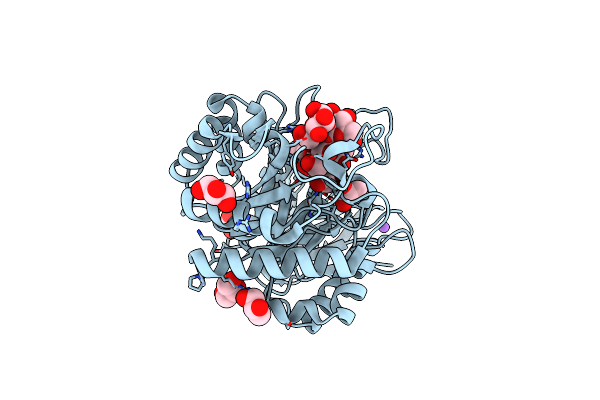 |
Structure Of Pslg With A Covalently- Bound Pentasaccharide
Organism: Pseudomonas aeruginosa
Method: X-RAY DIFFRACTION Resolution:1.55 Å Release Date: 2025-03-05 Classification: HYDROLASE Ligands: CL, NA, GOL, YLL |
Organism: Pseudomonas aeruginosa
Method: X-RAY DIFFRACTION
Release Date: 2025-03-05
Ligands: CL, NA, GOL, YLL
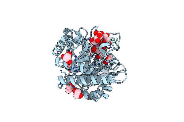 |
Structure Of Pslg With An Iminosugar Inhibitor
Organism: Pseudomonas aeruginosa
Method: X-RAY DIFFRACTION Resolution:1.50 Å Release Date: 2025-03-05 Classification: HYDROLASE Ligands: GOL, CL |
Organism: Pseudomonas aeruginosa
Method: X-RAY DIFFRACTION
Release Date: 2025-03-05
Ligands: GOL, CL
 |
Crystal Structure Of Susa Amylase From Bacteroides Thetaiotaomicron Covalently Bound To Alpha-1,6 Branched Pseudo-Trisaccharide Activity-Based Probe
Organism: Bacteroides thetaiotaomicron
Method: X-RAY DIFFRACTION Resolution:2.43 Å Release Date: 2024-12-11 Classification: HYDROLASE Ligands: PBW, OC9, IMD, CA |
Organism: Bacteroides thetaiotaomicron
Method: X-RAY DIFFRACTION
Release Date: 2024-12-11
Ligands: PBW, OC9, IMD, CA
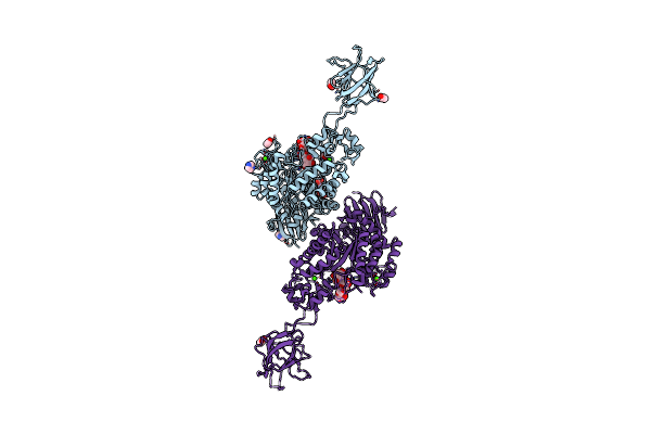 |
Crystal Structure Of Susg From Bacteroides Thetaiotaomicron Covalently Bound To Alpha-1,6 Branched Pseudo-Trisaccharide Activity-Based Probe
Organism: Bacteroides thetaiotaomicron
Method: X-RAY DIFFRACTION Resolution:2.65 Å Release Date: 2024-12-11 Classification: HYDROLASE Ligands: PBW, OC9, A1ILG, CA, IMD, ACT |
Organism: Bacteroides thetaiotaomicron
Method: X-RAY DIFFRACTION
Release Date: 2024-12-11
Ligands: PBW, OC9, A1ILG, CA, IMD, ACT
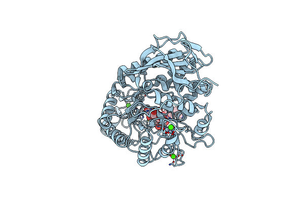 |
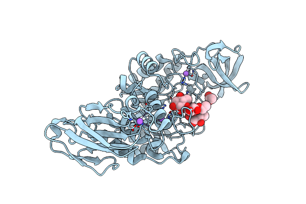
Crystal Structure Of Amylase 5 (Amy5) From Ruminococcus Bromii Covalently Bound To Alpha-1,6 Branched Pseudo-Trisaccharide Activity-Based Probe
Organism: Ruminococcus bromii
Method: X-RAY DIFFRACTION Resolution:1.40 Å Release Date: 2024-12-11 Classification: HYDROLASE Ligands: PBW, A1IHI, CA |
Organism: Ruminococcus bromii
Method: X-RAY DIFFRACTION
Release Date: 2024-12-11
Ligands: PBW, A1IHI, CA
 |
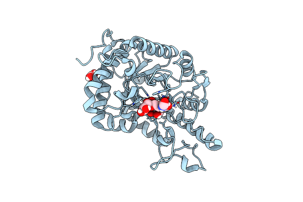
Crystal Structure Of K38 Amylase From Bacillus Sp. Strain Ksm-K38 Covalently Bound To Alpha-1,6 Branched Pseudo-Trisaccharide Activity-Based Probe
Organism: Bacillus sp. ksm-k38
Method: X-RAY DIFFRACTION Resolution:2.02 Å Release Date: 2024-12-11 Classification: HYDROLASE Ligands: PBW, OC9, ACT, NA |
Organism: Bacillus sp. ksm-k38
Method: X-RAY DIFFRACTION
Release Date: 2024-12-11
Ligands: PBW, OC9, ACT, NA
 |
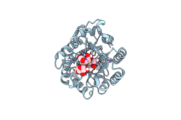
Cjcel5D Endo-Xyloglucanase Bounc To Cb665 Covalent Inhibitor
Organism: Cellvibrio japonicus ueda107
Method: X-RAY DIFFRACTION Resolution:1.30 Å Release Date: 2024-02-28 Classification: HYDROLASE Ligands: BO3, RBH, YLL, XYS |
Organism: Cellvibrio japonicus ueda107
Method: X-RAY DIFFRACTION
Release Date: 2024-02-28
Ligands: BO3, RBH, YLL, XYS
 |
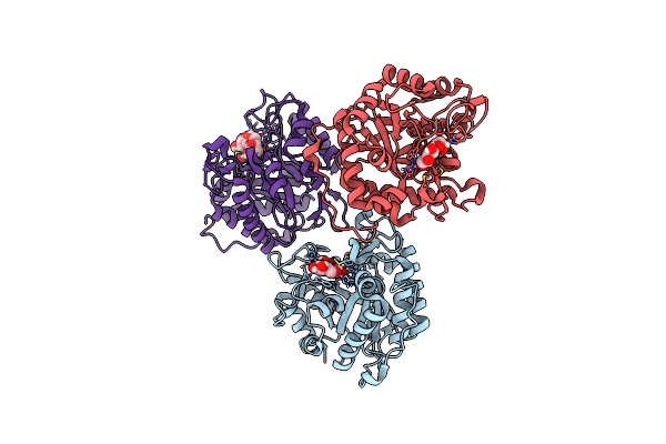
Cjcel5B Endo-Glucanase Bound To Cb665 Covalent Inhibitor
Organism: Cellvibrio japonicus
Method: X-RAY DIFFRACTION Resolution:1.67 Å Release Date: 2023-11-29 Classification: HYDROLASE Ligands: RBH, XYS, YLL |
Organism: Cellvibrio japonicus
Method: X-RAY DIFFRACTION
Release Date: 2023-11-29
Ligands: RBH, XYS, YLL
 |
Cjcel5C Endo-Glucanase Bound To Cb396 Covalent Inhibitor
Organism: Cellvibrio japonicus ueda107
Method: X-RAY DIFFRACTION Resolution:2.70 Å Release Date: 2023-11-29 Classification: HYDROLASE Ligands: YLL, BGC |
Organism: Cellvibrio japonicus ueda107
Method: X-RAY DIFFRACTION
Release Date: 2023-11-29
Ligands: YLL, BGC
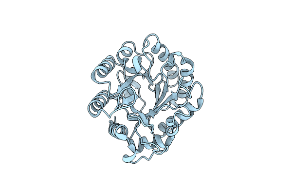 |
Cjcel5B Endo-Glucanase
Organism: Cellvibrio japonicus
Method: X-RAY DIFFRACTION Resolution:1.57 Å Release Date: 2023-11-29 Classification: HYDROLASE |
Organism: Cellvibrio japonicus
Method: X-RAY DIFFRACTION
Release Date: 2023-11-29
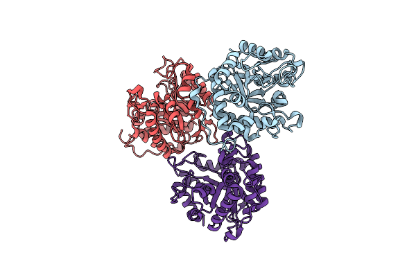 |
Cjcel5C Endo-Glucanase
Organism: Cellvibrio japonicus
Method: X-RAY DIFFRACTION Resolution:2.18 Å Release Date: 2023-11-22 Classification: HYDROLASE |
Organism: Cellvibrio japonicus
Method: X-RAY DIFFRACTION
Release Date: 2023-11-22
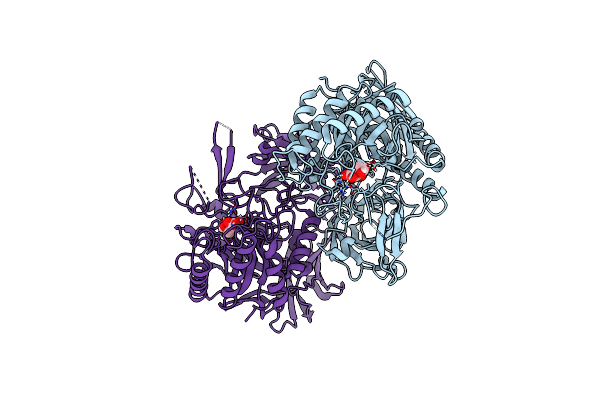 |
Crystal Structure Of E. Coli Beta-Glucuronidase In Complex With Covalent Inhibitor Me727
Organism: Escherichia coli (strain k12)
Method: X-RAY DIFFRACTION Resolution:1.99 Å Release Date: 2022-08-03 Classification: CARBOHYDRATE Ligands: 8I4 |
Organism: Escherichia coli (strain k12)
Method: X-RAY DIFFRACTION
Release Date: 2022-08-03
Ligands: 8I4
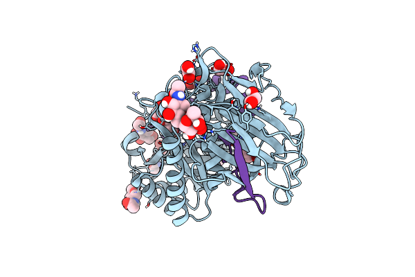 |
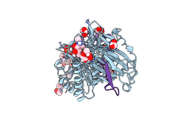
Crystal Structure Of Human Heparanase In Complex With Covalent Inhibitor Vl166
Organism: Homo sapiens
Method: X-RAY DIFFRACTION Resolution:1.52 Å Release Date: 2022-08-03 Classification: CARBOHYDRATE Ligands: NAG, EDO, CL |
Organism: Homo sapiens
Method: X-RAY DIFFRACTION
Release Date: 2022-08-03
Ligands: NAG, EDO, CL
 |
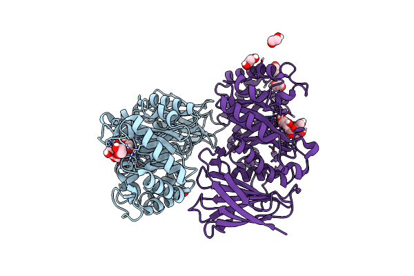
Crystal Structure Of Human Heparanase In Complex With Covalent Inhibitor Gr109
Organism: Homo sapiens
Method: X-RAY DIFFRACTION Resolution:1.66 Å Release Date: 2022-08-03 Classification: CARBOHYDRATE Ligands: NAG, EDO, CL |
Organism: Homo sapiens
Method: X-RAY DIFFRACTION
Release Date: 2022-08-03
Ligands: NAG, EDO, CL
 |
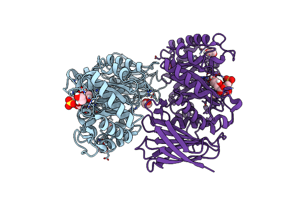
Crystal Structure Of Burkholderia Pseudomallei Heparanase In Complex With Covalent Inhibitor Vl166
Organism: Burkholderia pseudomallei (strain k96243)
Method: X-RAY DIFFRACTION Resolution:1.34 Å Release Date: 2022-08-03 Classification: HYDROLASE Ligands: EDO |
Organism: Burkholderia pseudomallei (strain k96243)
Method: X-RAY DIFFRACTION
Release Date: 2022-08-03
Ligands: EDO
 |
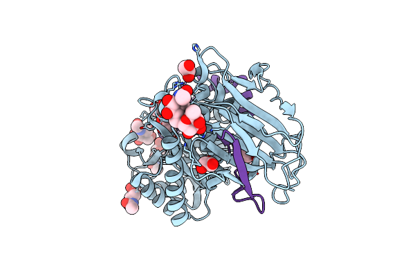
Crystal Structure Of Burkholderia Pseudomallei Heparanase In Complex With Covalent Inhibitor Gr109
Organism: Burkholderia pseudomallei (strain k96243)
Method: X-RAY DIFFRACTION Resolution:1.31 Å Release Date: 2022-08-03 Classification: HYDROLASE Ligands: EDO |
Organism: Burkholderia pseudomallei (strain k96243)
Method: X-RAY DIFFRACTION
Release Date: 2022-08-03
Ligands: EDO
 |
Crystal Structure Of Human Heparanase In Complex With Covalent Inhibitor Cb678
Organism: Homo sapiens
Method: X-RAY DIFFRACTION Resolution:1.70 Å Release Date: 2022-08-03 Classification: CARBOHYDRATE Ligands: NAG, EDO, CL |
Organism: Homo sapiens
Method: X-RAY DIFFRACTION
Release Date: 2022-08-03
Ligands: NAG, EDO, CL
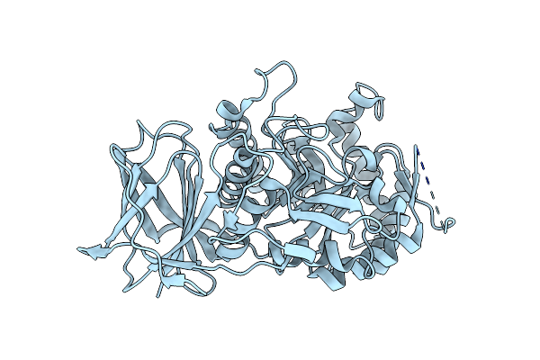 |
Crystal Structure Of Beta-Glucuronidase From Acidobacterium Capsulatum At 1.24 Angstrom Resolution
Organism: Acidobacterium capsulatum (strain atcc 51196 / dsm 11244 / jcm 7670 / nbrc 15755 / ncimb 13165 / 161)
Method: X-RAY DIFFRACTION Resolution:1.24 Å Release Date: 2022-08-03 Classification: CARBOHYDRATE |
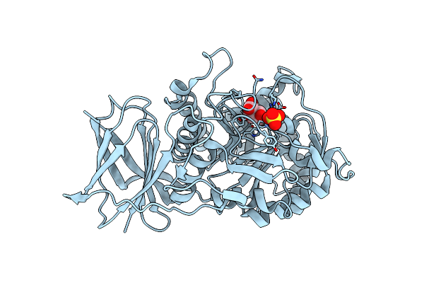 |
Crystal Structure Of Beta-Glucuronidase From Acidobacterium Capsulatum In Complex With Covalent Inhibitor Me727
Organism: Acidobacterium capsulatum (strain atcc 51196 / dsm 11244 / jcm 7670 / nbrc 15755 / ncimb 13165 / 161)
Method: X-RAY DIFFRACTION Resolution:1.25 Å Release Date: 2022-08-03 Classification: CARBOHYDRATE Ligands: 8I4, SO4 |
Organism: Acidobacterium capsulatum (strain atcc 51196 / dsm 11244 / jcm 7670 / nbrc 15755 / ncimb 13165 / 161)
Method: X-RAY DIFFRACTION
Release Date: 2022-08-03
Ligands: 8I4, SO4
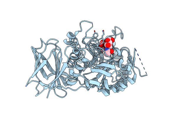 |
Crystal Structure Of Beta-Glucuronidase From Acidobacterium Capsulatum In Complex With Covalent Inhibitor Vl166
Organism: Acidobacterium capsulatum (strain atcc 51196 / dsm 11244 / jcm 7670 / nbrc 15755 / ncimb 13165 / 161)
Method: X-RAY DIFFRACTION Resolution:1.55 Å Release Date: 2022-08-03 Classification: CARBOHYDRATE |