

Search Count: 25
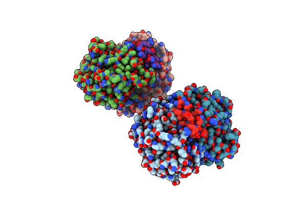 |
2.6A Resolution Structure Of Mind Complexed With Mine (12-31) Peptide
Organism: Escherichia coli
Method: X-RAY DIFFRACTION Resolution:2.60 Å Release Date: 2011-08-17 Classification: CELL CYCLE,HYDROLASE/CELL CYCLE Ligands: ADP |
Organism: Escherichia coli
Method: X-RAY DIFFRACTION
Release Date: 2011-08-17
Ligands: ADP
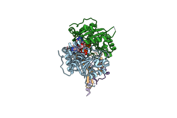 |
4.3A Resolution Structure Of A Mind-Mine(I24N) Protein Complex
Organism: Escherichia coli
Method: X-RAY DIFFRACTION Resolution:4.30 Å Release Date: 2011-08-17 Classification: CELL CYCLE,HYDROLASE/CELL CYCLE Ligands: ADP |
Organism: Escherichia coli
Method: X-RAY DIFFRACTION
Release Date: 2011-08-17
Ligands: ADP
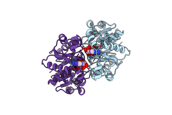 |
The Structure Of The Dimeric E.Coli Mind-Atp Complex
Organism: Escherichia coli
Method: X-RAY DIFFRACTION Resolution:2.34 Å Release Date: 2011-01-26 Classification: CELL CYCLE, HYDROLASE Ligands: MG, ATP |
Organism: Escherichia coli
Method: X-RAY DIFFRACTION
Release Date: 2011-01-26
Ligands: MG, ATP
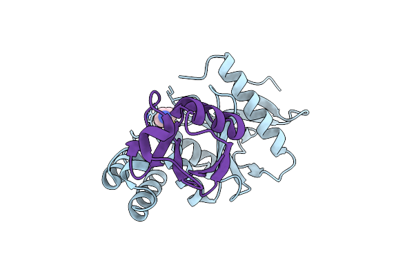 |
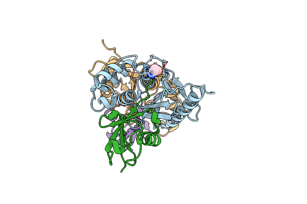
Structure Of Ovarian Tumor (Otu) Domain In Complex With Ubiquitin
Organism: Saccharomyces cerevisiae, Homo sapiens
Method: X-RAY DIFFRACTION Resolution:1.55 Å Release Date: 2008-02-19 Classification: CELL CYCLE, HYDROLASE Ligands: 3CN |
Organism: Saccharomyces cerevisiae, Homo sapiens
Method: X-RAY DIFFRACTION
Release Date: 2008-02-19
Ligands: 3CN
 |
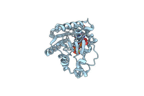
Structure Of Ovarian Tumor (Otu) Domain In Complex With Ubiquitin
Organism: Saccharomyces cerevisiae, Homo sapiens
Method: X-RAY DIFFRACTION Resolution:2.32 Å Release Date: 2008-02-19 Classification: CELL CYCLE, HYDROLASE Ligands: 3CN |
Organism: Saccharomyces cerevisiae, Homo sapiens
Method: X-RAY DIFFRACTION
Release Date: 2008-02-19
Ligands: 3CN
 |
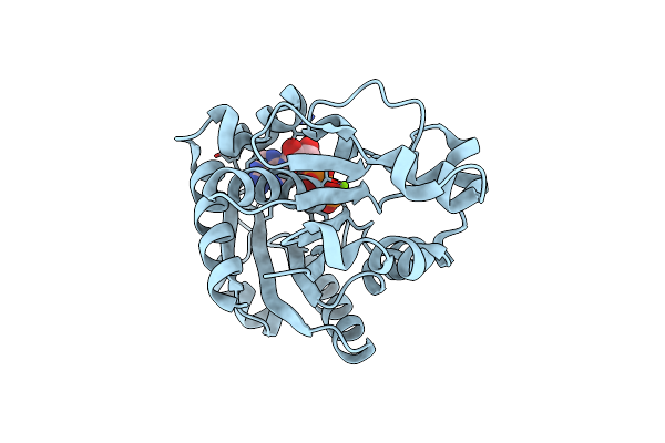
Crystal Structure Analysis Of Pyrococcus Furiosus Cell Division Atpase Mind
Organism: Pyrococcus furiosus
Method: X-RAY DIFFRACTION Resolution:2.70 Å Release Date: 2001-04-21 Classification: cell cycle, hydrolase Ligands: MG, ACP |
Organism: Pyrococcus furiosus
Method: X-RAY DIFFRACTION
Release Date: 2001-04-21
Ligands: MG, ACP
 |
Crystal Structure Analysis Of Pyrococcus Furiosus Cell Division Atpase Mind
Organism: Pyrococcus furiosus
Method: X-RAY DIFFRACTION Resolution:2.00 Å Release Date: 2001-04-21 Classification: cell cycle, hydrolase Ligands: MG, ADP |
Organism: Pyrococcus furiosus
Method: X-RAY DIFFRACTION
Release Date: 2001-04-21
Ligands: MG, ADP
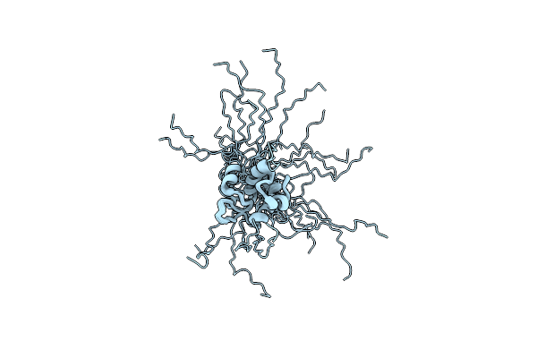 |
Solution Structure Of A Resuscitation Promoting Factor Domain From Mycobacterium Tuberculosis
Organism: Mycobacterium tuberculosis
Method: SOLUTION NMR Release Date: 2005-02-15 Classification: CELL CYCLE, HYDROLASE |
Organism: Mycobacterium tuberculosis
Method: SOLUTION NMR
Release Date: 2005-02-15
 |
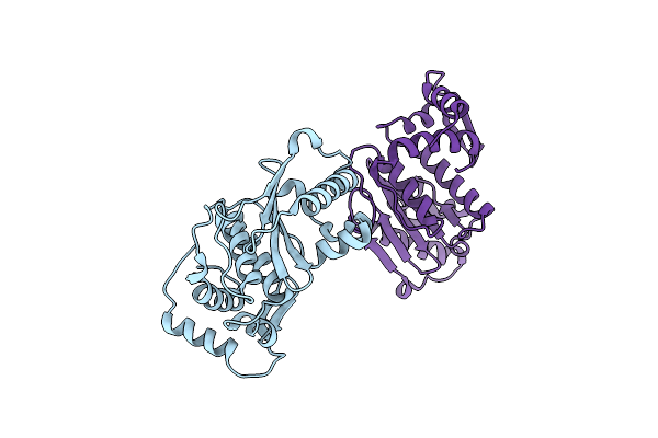
Structure Of Human Senp2 In Complex With Sumo-1
Organism: Homo sapiens
Method: X-RAY DIFFRACTION Resolution:2.80 Å Release Date: 2004-09-14 Classification: CELL CYCLE, HYDROLASE Ligands: SO4 |
Organism: Homo sapiens
Method: X-RAY DIFFRACTION
Release Date: 2004-09-14
Ligands: SO4
 |
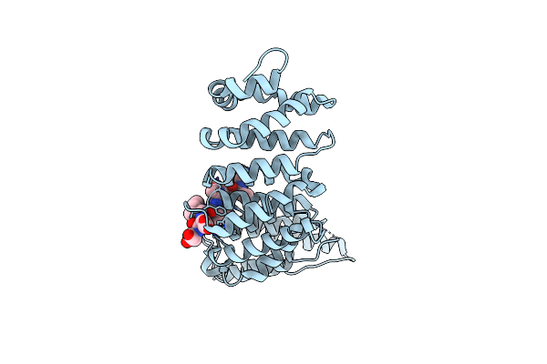
Structure Of Human Senp2
Organism: Homo sapiens
Method: X-RAY DIFFRACTION Resolution:2.20 Å Release Date: 2004-09-14 Classification: CELL CYCLE, HYDROLASE |
Organism: Homo sapiens
Method: X-RAY DIFFRACTION
Release Date: 2004-09-14
 |
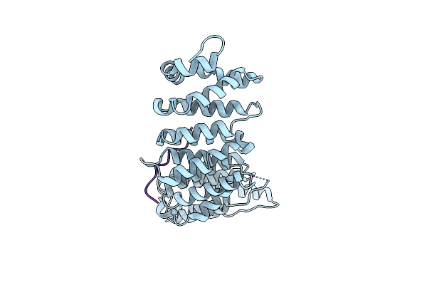
The Structure Of The Pp2A B56 Subunit Double Phosphorylated Bubr1 Complex
Organism: Homo sapiens
Method: X-RAY DIFFRACTION Resolution:2.82 Å Release Date: 2016-11-30 Classification: Hydrolase, cell cycle |
Organism: Homo sapiens
Method: X-RAY DIFFRACTION
Release Date: 2016-11-30
 |
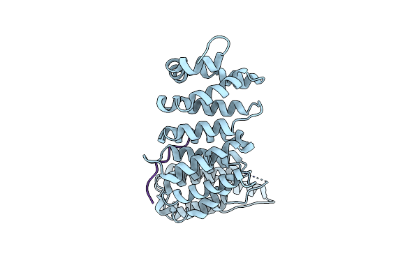
The Structure Of The Pp2A B56 Subunit Repoman Complex
Organism: Homo sapiens
Method: X-RAY DIFFRACTION Resolution:2.85 Å Release Date: 2016-11-30 Classification: Hydrolase, cell cycle |
Organism: Homo sapiens
Method: X-RAY DIFFRACTION
Release Date: 2016-11-30
 |
The Structure Of The Pp2A B56 Subunit Bubr1 Complex
Organism: Homo sapiens
Method: X-RAY DIFFRACTION Resolution:2.79 Å Release Date: 2016-12-07 Classification: Hydrolase, cell cycle |
Organism: Homo sapiens
Method: X-RAY DIFFRACTION
Release Date: 2016-12-07
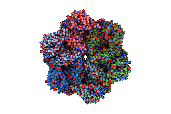 |
Human P97 Double Hexamer Conformer I With D1-Atpgammas And D2-Adp Bound
Organism: Homo sapiens
Method: ELECTRON MICROSCOPY Release Date: 2022-03-02 Classification: HYDROLASE Ligands: AGS, MG, ADP |
Organism: Homo sapiens
Method: ELECTRON MICROSCOPY
Release Date: 2022-03-02
Ligands: AGS, MG, ADP
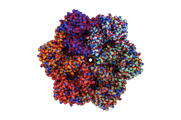 |
Human P97 Single Hexamer Conformer Ii With Atpgammas Bound
Organism: Homo sapiens
Method: ELECTRON MICROSCOPY Release Date: 2022-03-02 Classification: HYDROLASE Ligands: AGS, MG |
Organism: Homo sapiens
Method: ELECTRON MICROSCOPY
Release Date: 2022-03-02
Ligands: AGS, MG
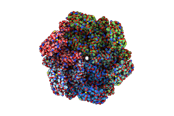 |
Human P97 Double Hexamer Conformer Ii With Atpgammas Bound
Organism: Homo sapiens
Method: ELECTRON MICROSCOPY Release Date: 2022-03-02 Classification: HYDROLASE Ligands: AGS, MG |
Organism: Homo sapiens
Method: ELECTRON MICROSCOPY
Release Date: 2022-03-02
Ligands: AGS, MG
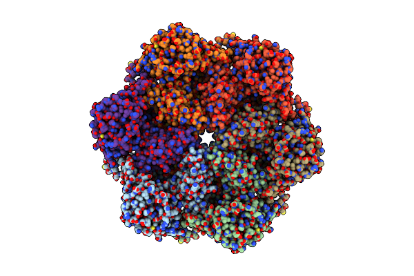 |
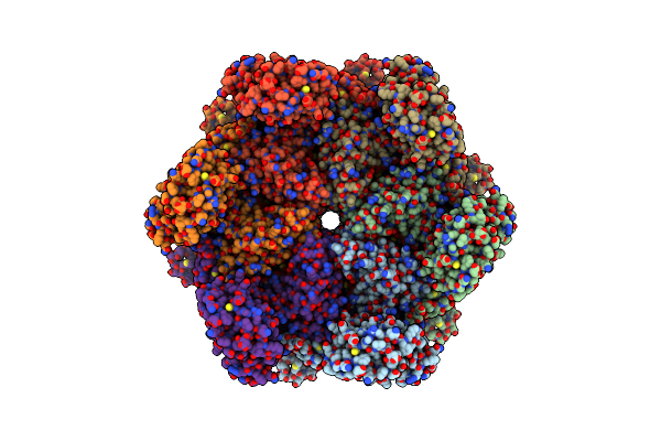
Human P97 Single Hexamer Conformer Iii With D1-Atpgammas And D2-Adp Bound
Organism: Homo sapiens
Method: ELECTRON MICROSCOPY Release Date: 2022-03-02 Classification: HYDROLASE Ligands: AGS, MG, ADP |
Organism: Homo sapiens
Method: ELECTRON MICROSCOPY
Release Date: 2022-03-02
Ligands: AGS, MG, ADP
 |
Human P97 Single Hexamer Conformer I With Atpgammas Bound
Organism: Homo sapiens
Method: ELECTRON MICROSCOPY Release Date: 2022-03-02 Classification: HYDROLASE Ligands: AGS, MG |
Organism: Homo sapiens
Method: ELECTRON MICROSCOPY
Release Date: 2022-03-02
Ligands: AGS, MG
 |
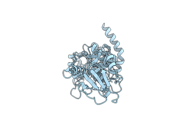
Pyridinylpyrimidines Selectively Inhibit Human Methionine Aminopeptidase-1
Organism: Homo sapiens
Method: X-RAY DIFFRACTION Resolution:2.09 Å Release Date: 2013-04-03 Classification: hydrolase/hydrolase inhibitor Ligands: CO, K, 1AY, SO4 |
Organism: Homo sapiens
Method: X-RAY DIFFRACTION
Release Date: 2013-04-03
Ligands: CO, K, 1AY, SO4
 |
Structure Of Cdc14 In Complex With Tungstate
Organism: Homo sapiens
Method: X-RAY DIFFRACTION Resolution:2.60 Å Release Date: 2003-07-24 Classification: HYDROLASE Ligands: WO4 |
Organism: Homo sapiens
Method: X-RAY DIFFRACTION
Release Date: 2003-07-24
Ligands: WO4