

Search Count: 12
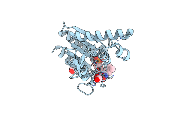 |
The Crystal Structure Of Azor (Azoreductase) From Escherichia Coli: Oxidized Azor In Tetragonal Crystals (The Resolution Has Improved From 1.8 (1V4B) To 1.4 Angstrom)
Organism: Escherichia coli
Method: X-RAY DIFFRACTION Resolution:1.40 Å Release Date: 2008-03-11 Classification: OXIDOREDUCTASE Ligands: FMN, EDO |
Organism: Escherichia coli
Method: X-RAY DIFFRACTION
Release Date: 2008-03-11
Ligands: FMN, EDO
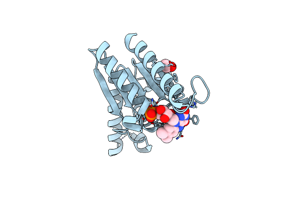 |
The Crystal Structure Of Azor (Azoreductase) From Escherichia Coli: Reduced Azor In Tetragonal Crystals
Organism: Escherichia coli
Method: X-RAY DIFFRACTION Resolution:1.70 Å Release Date: 2008-03-11 Classification: OXIDOREDUCTASE Ligands: FMN, EDO |
Organism: Escherichia coli
Method: X-RAY DIFFRACTION
Release Date: 2008-03-11
Ligands: FMN, EDO
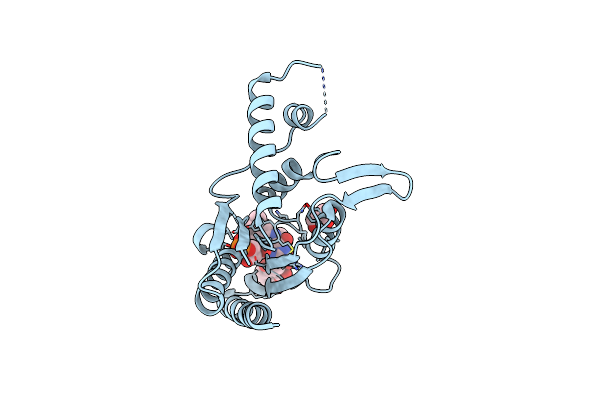 |
The Crystal Structure Of Azor (Azoreductase) From Escherichia Coli: Azor In Complex With Dicoumarol
Organism: Escherichia coli
Method: X-RAY DIFFRACTION Resolution:2.30 Å Release Date: 2008-03-11 Classification: OXIDOREDUCTASE Ligands: FMN, DTC, GOL |
Organism: Escherichia coli
Method: X-RAY DIFFRACTION
Release Date: 2008-03-11
Ligands: FMN, DTC, GOL
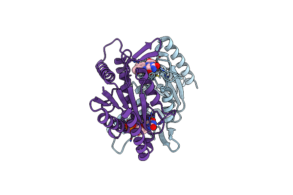 |
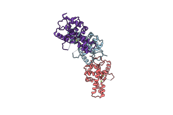
The Crystal Structure Of Azor (Azoreductase) From Escherichia Coli: Oxidized Azor In Orthorhombic Crystals
Organism: Escherichia coli
Method: X-RAY DIFFRACTION Resolution:2.10 Å Release Date: 2008-03-11 Classification: OXIDOREDUCTASE Ligands: FMN |
Organism: Escherichia coli
Method: X-RAY DIFFRACTION
Release Date: 2008-03-11
Ligands: FMN
 |
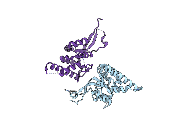
Crystal Structure Of The C-Terminal Rnase Iii Domain Of Human Dicer
Organism: Homo sapiens
Method: X-RAY DIFFRACTION Resolution:2.00 Å Release Date: 2007-11-06 Classification: HYDROLASE Ligands: MG |
Organism: Homo sapiens
Method: X-RAY DIFFRACTION
Release Date: 2007-11-06
Ligands: MG
 |
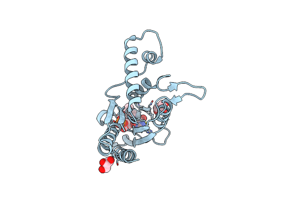
Crystal Structure Of The Pin Domain Of Human Est1A
Organism: Homo sapiens
Method: X-RAY DIFFRACTION Resolution:1.80 Å Release Date: 2007-05-01 Classification: UNKNOWN FUNCTION |
Organism: Homo sapiens
Method: X-RAY DIFFRACTION
Release Date: 2007-05-01
 |
The Crystal Structure Of Azor (Azo Reductase) From Escherichia Coli
Organism: Escherichia coli
Method: X-RAY DIFFRACTION Resolution:2.20 Å Release Date: 2006-05-16 Classification: OXIDOREDUCTASE Ligands: FMN, GOL |
Organism: Escherichia coli
Method: X-RAY DIFFRACTION
Release Date: 2006-05-16
Ligands: FMN, GOL
 |
Binding Of Coumarins To Nad(P)H:Fmn Oxidoreductase
Organism: Aliivibrio fischeri
Method: X-RAY DIFFRACTION Resolution:1.90 Å Release Date: 2005-03-08 Classification: OXIDOREDUCTASE Ligands: FMN, 4HC |
Organism: Aliivibrio fischeri
Method: X-RAY DIFFRACTION
Release Date: 2005-03-08
Ligands: FMN, 4HC
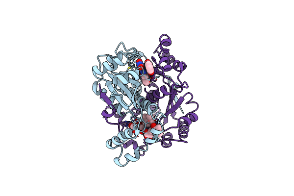 |
Binding Of Coumarins To Nad(P)H:Fmn Oxidoreductase
Organism: Aliivibrio fischeri
Method: X-RAY DIFFRACTION Resolution:2.00 Å Release Date: 2005-03-08 Classification: OXIDOREDUCTASE Ligands: FMN, 2HC |
Organism: Aliivibrio fischeri
Method: X-RAY DIFFRACTION
Release Date: 2005-03-08
Ligands: FMN, 2HC
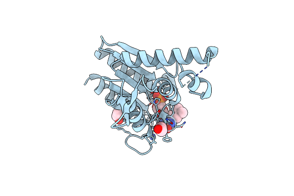 |
The Crystal Structure Of Azor (Azo Reductase) From Escherichia Coli: Oxidized Form
Organism: Escherichia coli
Method: X-RAY DIFFRACTION Resolution:1.80 Å Release Date: 2005-01-18 Classification: OXIDOREDUCTASE Ligands: FMN, EDO, IPA |
Organism: Escherichia coli
Method: X-RAY DIFFRACTION
Release Date: 2005-01-18
Ligands: FMN, EDO, IPA
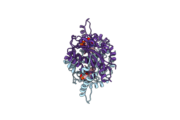 |
Structure And Site-Directed Mutagenesis Of A Flavoprotein From Escherichia Coli That Reduces Nitrocompounds. Alteration Of Pyridine Nucleotide Binding By A Single Amino Acid Substitution
Organism: Escherichia coli
Method: X-RAY DIFFRACTION Resolution:1.70 Å Release Date: 2001-02-14 Classification: OXIDOREDUCTASE Ligands: FMN |
Organism: Escherichia coli
Method: X-RAY DIFFRACTION
Release Date: 2001-02-14
Ligands: FMN
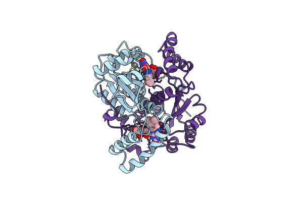 |
The Major Nad(P)H:Fmn Oxidoreductase From Vibrio Fischeri
Organism: Aliivibrio fischeri
Method: X-RAY DIFFRACTION Resolution:1.80 Å Release Date: 1999-02-16 Classification: OXIDOREDUCTASE Ligands: FMN |
Organism: Aliivibrio fischeri
Method: X-RAY DIFFRACTION
Release Date: 1999-02-16
Ligands: FMN