

Search Count: 40
 |
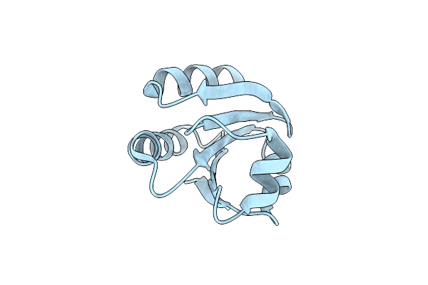
Crystallographic And Spectroscopic Characterization Of Sulfolobus Solfataricus Trxa1 Provide Insights Into The Determinants Of Thioredoxin Fold Stability
Organism: Sulfolobus solfataricus
Method: X-RAY DIFFRACTION Resolution:1.90 Å Release Date: 2011-11-30 Classification: OXIDOREDUCTASE Ligands: EDO |
Organism: Sulfolobus solfataricus
Method: X-RAY DIFFRACTION
Release Date: 2011-11-30
Ligands: EDO
 |
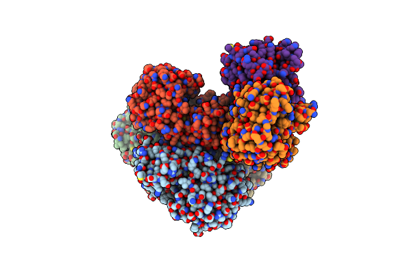
The Crystal Structure Of The Thioredoxin A2 From Sulfolobus Solfataricus
Organism: Sulfolobus solfataricus
Method: X-RAY DIFFRACTION Resolution:1.83 Å Release Date: 2009-10-27 Classification: OXIDOREDUCTASE |
Organism: Sulfolobus solfataricus
Method: X-RAY DIFFRACTION
Release Date: 2009-10-27
 |
Crystal Structure Of Sulfolobus Solfataricus Adh(Ssadh) Double Mutant (W95L,N249Y)
Organism: Sulfolobus solfataricus
Method: X-RAY DIFFRACTION Resolution:2.00 Å Release Date: 2009-07-21 Classification: OXIDOREDUCTASE Ligands: ZN |
Organism: Sulfolobus solfataricus
Method: X-RAY DIFFRACTION
Release Date: 2009-07-21
Ligands: ZN
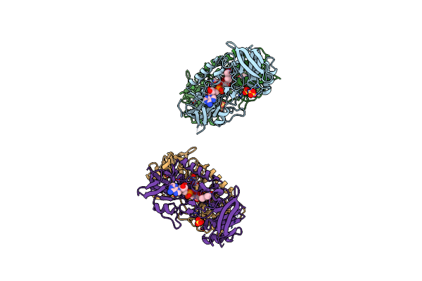 |
Structure Of Sulfolobus Solfataricus Thioredoxin Reductase Mutant C147A
Organism: Sulfolobus solfataricus
Method: X-RAY DIFFRACTION Resolution:1.40 Å Release Date: 2009-01-13 Classification: OXIDOREDUCTASE Ligands: FAD, SO4, ACY |
Organism: Sulfolobus solfataricus
Method: X-RAY DIFFRACTION
Release Date: 2009-01-13
Ligands: FAD, SO4, ACY
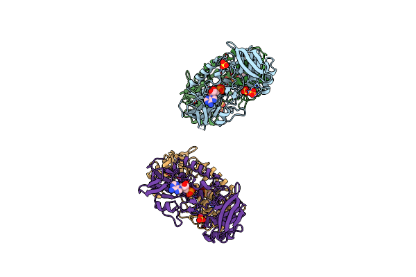 |
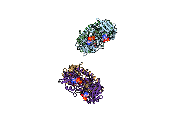
Structure Of Sulfolobus Solfataricus Trxr-B3
Organism: Sulfolobus solfataricus
Method: X-RAY DIFFRACTION Resolution:1.80 Å Release Date: 2009-01-13 Classification: OXIDOREDUCTASE Ligands: NAD, SO4, ACY |
Organism: Sulfolobus solfataricus
Method: X-RAY DIFFRACTION
Release Date: 2009-01-13
Ligands: NAD, SO4, ACY
 |
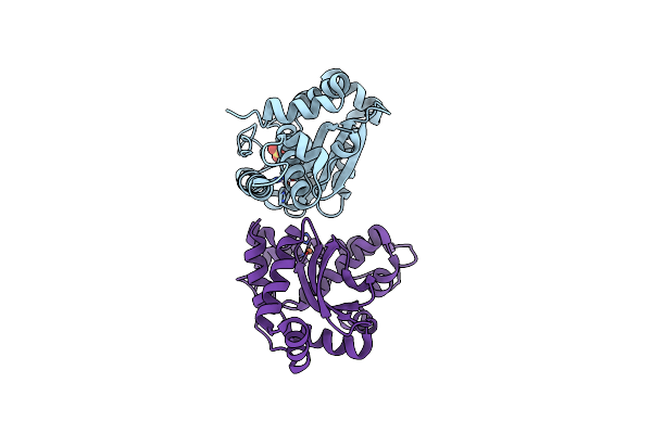
Crystal Structure Of Sulfolobus Solfataricus Thioredoxin Reductase B3 In Complex With Two Nadp Molecules
Organism: Sulfolobus solfataricus
Method: X-RAY DIFFRACTION Resolution:1.95 Å Release Date: 2009-01-13 Classification: OXIDOREDUCTASE Ligands: NAP |
Organism: Sulfolobus solfataricus
Method: X-RAY DIFFRACTION
Release Date: 2009-01-13
Ligands: NAP
 |
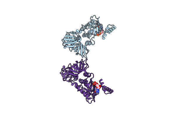
Crystal Structure Of Superoxide Dismutase From Helicobacter Pylori
Organism: Helicobacter pylori
Method: X-RAY DIFFRACTION Resolution:2.40 Å Release Date: 2008-06-10 Classification: OXIDOREDUCTASE Ligands: FE, SO4 |
Organism: Helicobacter pylori
Method: X-RAY DIFFRACTION
Release Date: 2008-06-10
Ligands: FE, SO4
 |
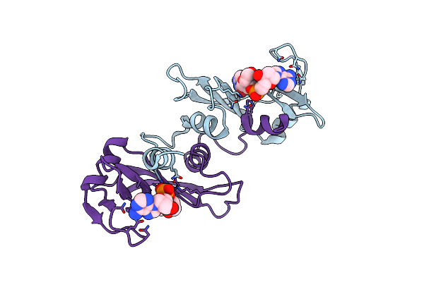
The Crystal Structure Of Sulfolobus Solfataricus Elongation Factor 1-Alpha In Complex With Magnesium And Gdp
Organism: Sulfolobus solfataricus
Method: X-RAY DIFFRACTION Resolution:1.80 Å Release Date: 2004-07-20 Classification: TRANSLATION Ligands: MG, GDP |
Organism: Sulfolobus solfataricus
Method: X-RAY DIFFRACTION
Release Date: 2004-07-20
Ligands: MG, GDP
 |
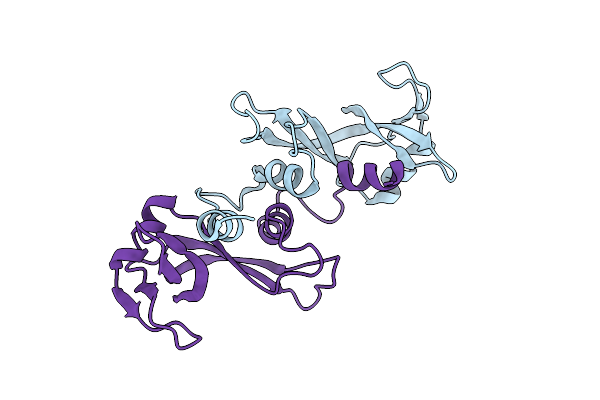
X-Ray Structure Of The Complex Of Bovine Seminal Ribonuclease Swapping Dimer With D(Cpa)
Organism: Bos taurus
Method: X-RAY DIFFRACTION Resolution:2.10 Å Release Date: 2004-04-13 Classification: HYDROLASE Ligands: CPA |
Organism: Bos taurus
Method: X-RAY DIFFRACTION
Release Date: 2004-04-13
Ligands: CPA
 |
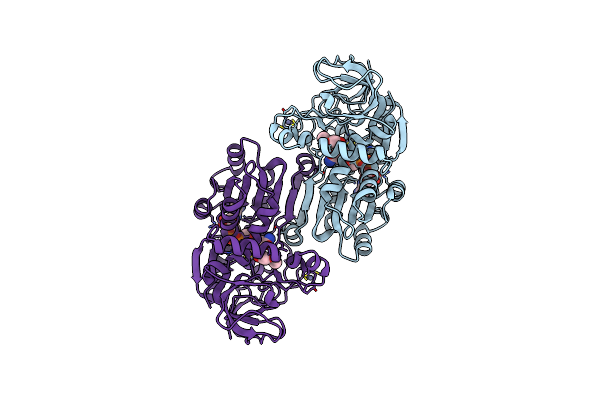
X-Ray Structure Of Bovine Seminal Ribonuclease Swapping Dimer From A New Crystal Form
Organism: Bos taurus
Method: X-RAY DIFFRACTION Resolution:2.50 Å Release Date: 2004-04-13 Classification: HYDROLASE |
Organism: Bos taurus
Method: X-RAY DIFFRACTION
Release Date: 2004-04-13
 |
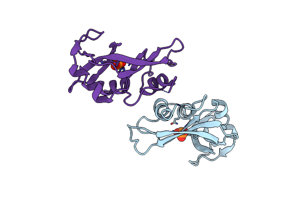
Alcohol Dehydrogenase From Sulfolobus Solfataricus Complexed With Nad(H) And 2-Ethoxyethanol
Organism: Sulfolobus solfataricus
Method: X-RAY DIFFRACTION Resolution:2.30 Å Release Date: 2004-02-10 Classification: OXIDOREDUCTASE Ligands: ZN, NAD, ETX |
Organism: Sulfolobus solfataricus
Method: X-RAY DIFFRACTION
Release Date: 2004-02-10
Ligands: ZN, NAD, ETX
 |
Crystal Structure Of The Dimeric Unswapped Form Of Bovine Seminal Ribonuclease
Organism: Bos taurus
Method: X-RAY DIFFRACTION Resolution:2.20 Å Release Date: 2003-11-18 Classification: HYDROLASE Ligands: PO4 |
Organism: Bos taurus
Method: X-RAY DIFFRACTION
Release Date: 2003-11-18
Ligands: PO4
 |
Crystal Structure Analysis Of The Monomeric [S-Carboxyamidomethyl-Cys31, S-Carboxyamidomethyl-Cys32] Bovine Seminal Ribonuclease
Organism: Bos taurus
Method: X-RAY DIFFRACTION Resolution:1.45 Å Release Date: 2003-08-26 Classification: HYDROLASE |
Organism: Bos taurus
Method: X-RAY DIFFRACTION
Release Date: 2003-08-26
 |
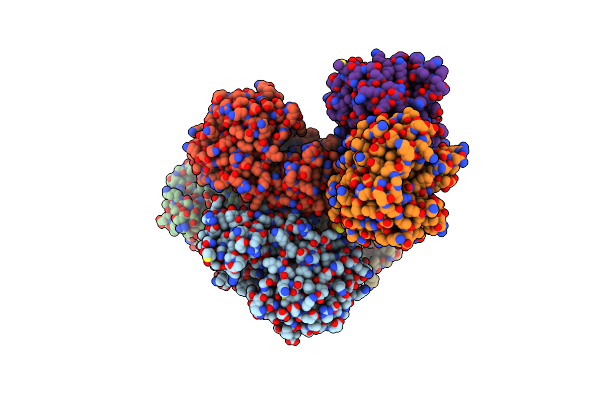
Crystal Structure Of The [S-Carboxyamidomethyl-Cys31, S-Carboxyamidomethyl-Cys32] Monomeric Derivative Of The Bovine Seminal Ribonuclease In The Liganded State
Organism: Bos taurus
Method: X-RAY DIFFRACTION Resolution:1.65 Å Release Date: 2003-08-26 Classification: HYDROLASE Ligands: U3P, ADN |
Organism: Bos taurus
Method: X-RAY DIFFRACTION
Release Date: 2003-08-26
Ligands: U3P, ADN
 |
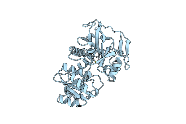
N249Y Mutant Of Alcohol Dehydrogenase From The Archaeon Sulfolobus Solfataricus-Monoclinic Crystal Form
Organism: Sulfolobus solfataricus
Method: X-RAY DIFFRACTION Resolution:1.94 Å Release Date: 2003-08-26 Classification: OXIDOREDUCTASE Ligands: ZN |
Organism: Sulfolobus solfataricus
Method: X-RAY DIFFRACTION
Release Date: 2003-08-26
Ligands: ZN
 |
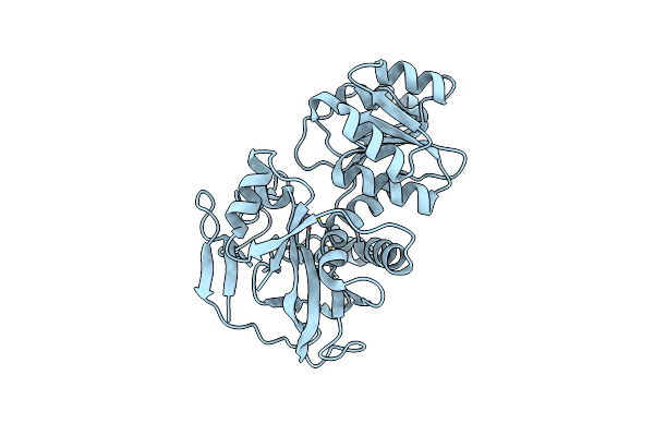
N249Y Mutant Of The Alcohol Dehydrogenase From The Archaeon Sulfolobus Solfataricus-Tetragonal Crystal Form
Organism: Sulfolobus solfataricus
Method: X-RAY DIFFRACTION Resolution:2.50 Å Release Date: 2003-08-26 Classification: OXIDOREDUCTASE Ligands: ZN |
Organism: Sulfolobus solfataricus
Method: X-RAY DIFFRACTION
Release Date: 2003-08-26
Ligands: ZN
 |
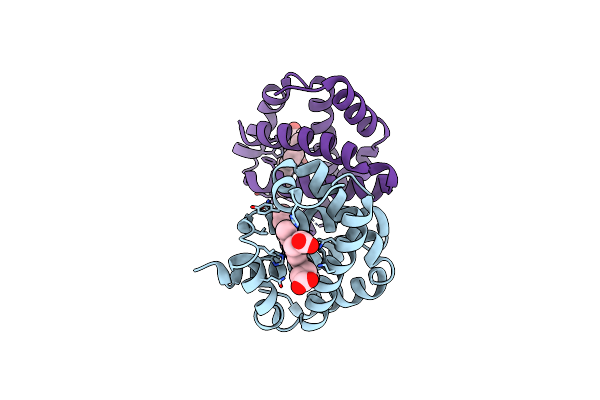
Alcohol Dehydrogenase From The Archaeon Sulfolobus Solfataricus
Organism: Sulfolobus solfataricus
Method: X-RAY DIFFRACTION Resolution:1.85 Å Release Date: 2002-08-29 Classification: OXIDOREDUCTASE Ligands: ZN |
Organism: Sulfolobus solfataricus
Method: X-RAY DIFFRACTION
Release Date: 2002-08-29
Ligands: ZN
 |
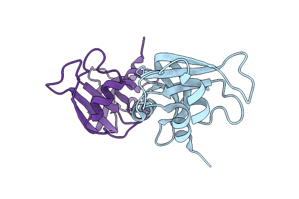
The Crystal Structure Of Trematomus Newnesi Hemoglobin In A Partial Hemichrome State
Organism: Trematomus newnesi
Method: X-RAY DIFFRACTION Resolution:2.00 Å Release Date: 2002-07-31 Classification: OXYGEN STORAGE/TRANSPORT Ligands: HEM, CMO |
Organism: Trematomus newnesi
Method: X-RAY DIFFRACTION
Release Date: 2002-07-31
Ligands: HEM, CMO
 |
Crystal Structure Of Ribonuclease A (Ligand-Free Form)
Organism: Bos taurus
Method: X-RAY DIFFRACTION Resolution:2.05 Å Release Date: 2002-06-05 Classification: HYDROLASE |
Organism: Bos taurus
Method: X-RAY DIFFRACTION
Release Date: 2002-06-05
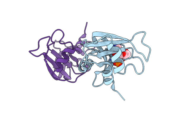 |
Crystal Structure Of Ribonuclease A (Complexed Form)
Organism: Bos taurus
Method: X-RAY DIFFRACTION Resolution:1.78 Å Release Date: 2002-06-05 Classification: HYDROLASE Ligands: C2P |
Organism: Bos taurus
Method: X-RAY DIFFRACTION
Release Date: 2002-06-05
Ligands: C2P