

Search Count: 23
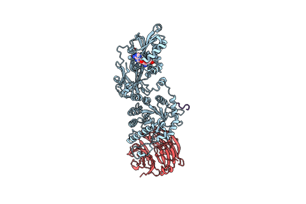 |
Prmt5 Bound To Pbm Peptide From Riok1
Organism: Homo sapiens
Method: X-RAY DIFFRACTION Resolution:2.11 Å Release Date: 2020-08-26 Classification: TRANSFERASE/SPLICING Ligands: SFG |
Organism: Homo sapiens
Method: X-RAY DIFFRACTION
Release Date: 2020-08-26
Ligands: SFG
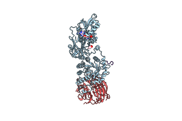 |
Prmt5 Bound To The Pbm Peptide From Picln
Organism: Homo sapiens
Method: X-RAY DIFFRACTION Resolution:2.86 Å Release Date: 2020-08-26 Classification: Transferase/Splicing Ligands: SFG, ACE |
Organism: Homo sapiens
Method: X-RAY DIFFRACTION
Release Date: 2020-08-26
Ligands: SFG, ACE
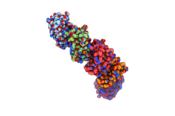 |
Structure Of The Major Structural Protein D135 Of Acidianus Tailed Spindle Virus (Atsv)
Organism: Acidianus tailed spindle virus
Method: X-RAY DIFFRACTION Resolution:1.68 Å Release Date: 2016-11-16 Classification: STRUCTURAL PROTEIN Ligands: NO3 |
Organism: Acidianus tailed spindle virus
Method: X-RAY DIFFRACTION
Release Date: 2016-11-16
Ligands: NO3
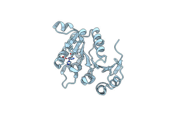 |
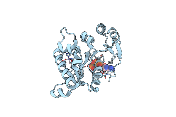
The Crystal Structure Of B204, The Dna-Packaging Atpase From Sulfolobus Turreted Icosahedral Virus
Organism: Sulfolobus turreted icosahedral virus 1
Method: X-RAY DIFFRACTION Resolution:1.96 Å Release Date: 2016-01-13 Classification: VIRAL PROTEIN Ligands: ZN |
Organism: Sulfolobus turreted icosahedral virus 1
Method: X-RAY DIFFRACTION
Release Date: 2016-01-13
Ligands: ZN
 |
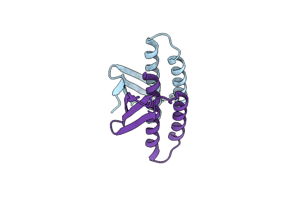
The Crystal Structure Of Stiv B204 Complexed With Amp-Pnp
Organism: Sulfolobus turreted icosahedral virus 1
Method: X-RAY DIFFRACTION Resolution:2.05 Å Release Date: 2016-01-13 Classification: VIRAL PROTEIN Ligands: ZN, ANP |
Organism: Sulfolobus turreted icosahedral virus 1
Method: X-RAY DIFFRACTION
Release Date: 2016-01-13
Ligands: ZN, ANP
 |
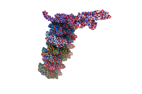
A100, A Dna Binding Scaffold From Sulfolobus Spindle-Shape Virus 1
Organism: Sulfolobus spindle-shaped virus 1
Method: X-RAY DIFFRACTION Resolution:3.00 Å Release Date: 2014-09-24 Classification: VIRAL PROTEIN |
Organism: Sulfolobus spindle-shaped virus 1
Method: X-RAY DIFFRACTION
Release Date: 2014-09-24
 |
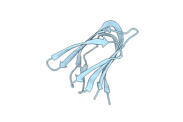
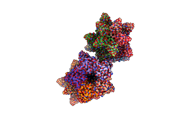
Life In The Extremes: Atomic Structure Of Sulfolobus Turreted Icosahedral Virus
Organism: Sulfolobus turreted icosahedral virus
Method: ELECTRON MICROSCOPY Release Date: 2013-05-01 Classification: VIRUS |
Organism: Sulfolobus turreted icosahedral virus
Method: ELECTRON MICROSCOPY
Release Date: 2013-05-01
 |
Crystal Structure Of A223 C-Terminal Domain, A Structural Protein From Sulfolobus Turreted Icosahedral Virus (Stiv)
Organism: Sulfolobus turreted icosahedral virus
Method: X-RAY DIFFRACTION Resolution:1.40 Å Release Date: 2013-04-03 Classification: VIRAL PROTEIN |
Organism: Sulfolobus turreted icosahedral virus
Method: X-RAY DIFFRACTION
Release Date: 2013-04-03
 |
The Triple Jelly Roll Fold And Turret Assembly In An Archaeal Virus
Organism: Sulfolobus turreted icosahedral virus
Method: X-RAY DIFFRACTION Resolution:1.80 Å Release Date: 2013-01-23 Classification: VIRAL PROTEIN Ligands: 2HP |
Organism: Sulfolobus turreted icosahedral virus
Method: X-RAY DIFFRACTION
Release Date: 2013-01-23
Ligands: 2HP
 |
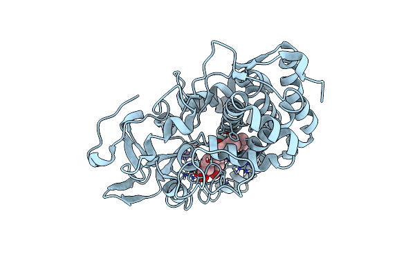
The Structure Of The Catalytic Domain Of The Sulfolobus Spindle-Shaped Viral Integrase Reveals An Evolutionarily Conserved Catalytic Core And Supports A Mechanism Of Dna Cleavage In Trans
Organism: Sulfolobus virus 1
Method: X-RAY DIFFRACTION Resolution:2.71 Å Release Date: 2012-05-16 Classification: RECOMBINATION Ligands: PO4 |
Organism: Sulfolobus virus 1
Method: X-RAY DIFFRACTION
Release Date: 2012-05-16
Ligands: PO4
 |
Reduced And Co-Bound Cytochrome P450Cam (Cyp101A1)
Organism: Pseudomonas putida
Method: SOLUTION NMR Release Date: 2012-05-09 Classification: HYDROLASE Ligands: HEM, CMO, K |
Organism: Pseudomonas putida
Method: SOLUTION NMR
Release Date: 2012-05-09
Ligands: HEM, CMO, K
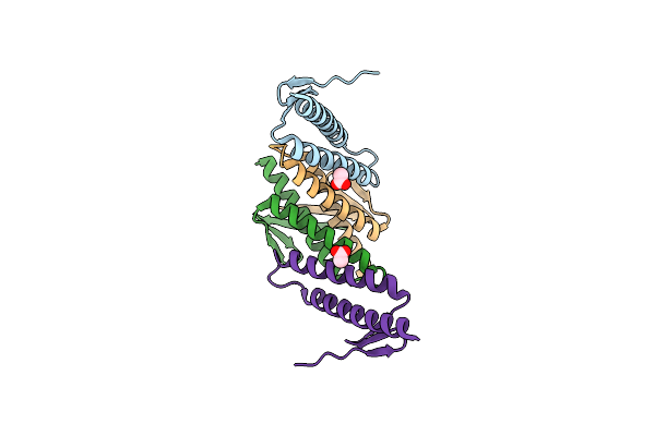 |
The Crystal Structure Of A81 From Sulfolobus Turreted Icosahedral Virus
Organism: Sulfolobus turreted icosahedral virus
Method: X-RAY DIFFRACTION Resolution:1.70 Å Release Date: 2012-05-09 Classification: VIRAL PROTEIN Ligands: ACT |
Organism: Sulfolobus turreted icosahedral virus
Method: X-RAY DIFFRACTION
Release Date: 2012-05-09
Ligands: ACT
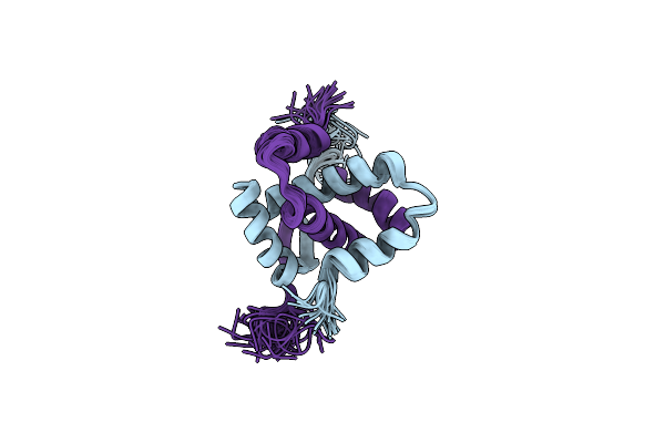 |
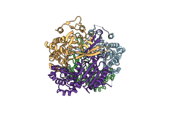
Thermostable Protein From Hyperthermophilic Virus Ssv-Rh
Organism: Sulfolobus virus ragged hills
Method: SOLUTION NMR Release Date: 2012-01-11 Classification: VIRAL PROTEIN |
Organism: Sulfolobus virus ragged hills
Method: SOLUTION NMR
Release Date: 2012-01-11
 |
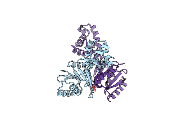
The Structure Of The Crispr-Associated Protein, Csa2, From Sulfolobus Solfataricus
Organism: Sulfolobus solfataricus
Method: X-RAY DIFFRACTION Resolution:2.00 Å Release Date: 2011-04-20 Classification: RNA BINDING PROTEIN |
Organism: Sulfolobus solfataricus
Method: X-RAY DIFFRACTION
Release Date: 2011-04-20
 |
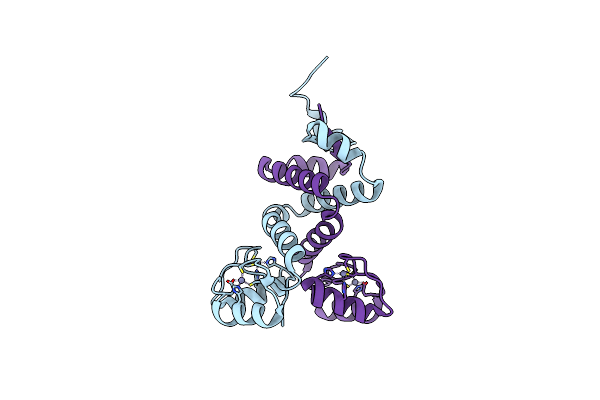
The Structure Of The Crispr-Associated Protein, Csa3, From Sulfolobus Solfataricus At 1.8 Angstrom Resolution.
Organism: Sulfolobus solfataricus
Method: X-RAY DIFFRACTION Resolution:1.80 Å Release Date: 2010-09-22 Classification: ANTIVIRAL PROTEIN Ligands: PEG |
Organism: Sulfolobus solfataricus
Method: X-RAY DIFFRACTION
Release Date: 2010-09-22
Ligands: PEG
 |
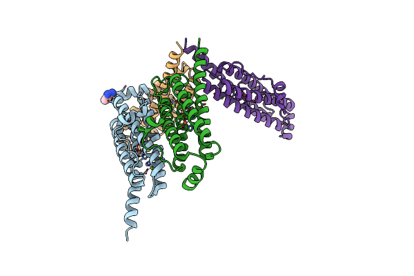
The Structure Of A Double C2H2 Zinc Finger Protein From A Hyperthermophilic Archaeal Virus In The Absence Of Dna
Organism: Sulfolobus virus 1
Method: X-RAY DIFFRACTION Resolution:2.70 Å Release Date: 2010-03-31 Classification: DNA BINDING PROTEIN Ligands: ZN |
Organism: Sulfolobus virus 1
Method: X-RAY DIFFRACTION
Release Date: 2010-03-31
Ligands: ZN
 |
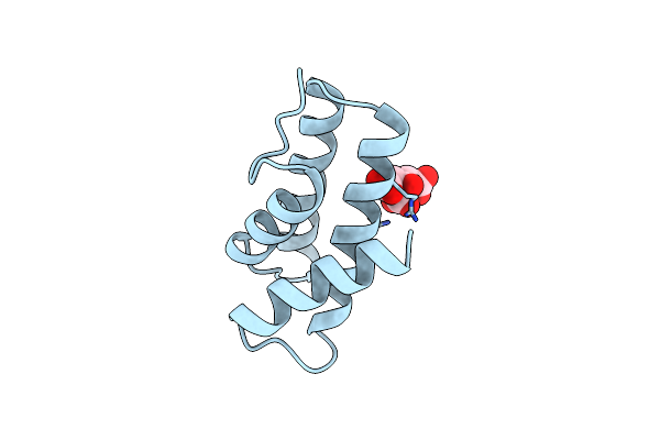
A Dodecameric Thioferritin In The Bacterial Domain, Characterization Of The Bacterioferritin-Related Protein From Bacteroides Fragilis
Organism: Bacteroides fragilis
Method: X-RAY DIFFRACTION Resolution:2.30 Å Release Date: 2009-11-17 Classification: METAL TRANSPORT Ligands: MG, BEN, FE |
Organism: Bacteroides fragilis
Method: X-RAY DIFFRACTION
Release Date: 2009-11-17
Ligands: MG, BEN, FE
 |
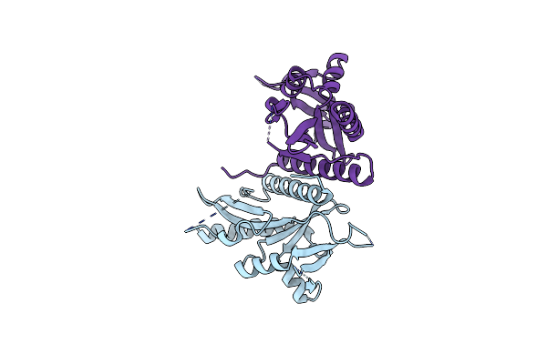
Crystal Structure Of Yellowstone Sirv Coat Protein C-Terminus
Organism: Sulfolobus islandicus rudivirus 1 variant ynp
Method: X-RAY DIFFRACTION Resolution:1.67 Å Release Date: 2009-04-21 Classification: VIRAL PROTEIN Ligands: CIT |
Organism: Sulfolobus islandicus rudivirus 1 variant ynp
Method: X-RAY DIFFRACTION
Release Date: 2009-04-21
Ligands: CIT
 |
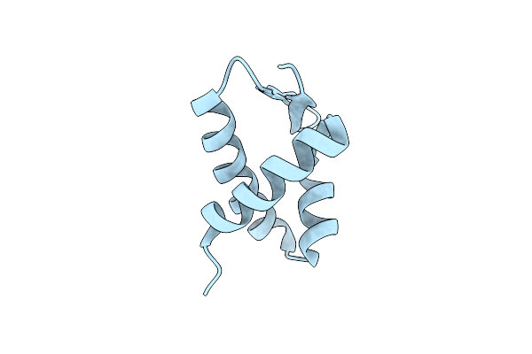
Structure Of D212, A Nuclease From A Fusselovirus.
Organism: Sulfolobus virus ragged hills
Method: X-RAY DIFFRACTION Resolution:2.40 Å Release Date: 2009-02-03 Classification: HYDROLASE |
Organism: Sulfolobus virus ragged hills
Method: X-RAY DIFFRACTION
Release Date: 2009-02-03
 |
Structure Of A Dna Binding Winged-Helix Protein, F-112, From Sulfolobus Spindle-Shaped Virus 1.
Organism: Sulfolobus virus-like particle ssv1
Method: X-RAY DIFFRACTION Resolution:2.30 Å Release Date: 2008-05-06 Classification: DNA BINDING PROTEIN |
Organism: Sulfolobus virus-like particle ssv1
Method: X-RAY DIFFRACTION
Release Date: 2008-05-06