

Search Count: 39
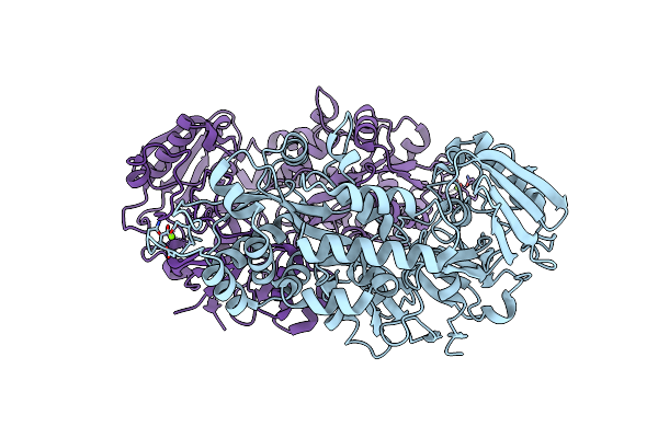 |
Crystal Structure Of Chitin Oligosaccharide Binding Protein From Vibrio Cholera.
Organism: Vibrio cholerae
Method: X-RAY DIFFRACTION Resolution:1.60 Å Release Date: 2024-01-03 Classification: SUGAR BINDING PROTEIN Ligands: MG |
Organism: Vibrio cholerae
Method: X-RAY DIFFRACTION
Release Date: 2024-01-03
Ligands: MG
 |
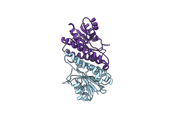
Crystal Structure Of Chitin Oligosaccharide Binding Protein From Vibrio Cholera In Complex With Chitotriose.
Organism: Vibrio cholerae
Method: X-RAY DIFFRACTION Resolution:1.22 Å Release Date: 2024-01-03 Classification: SUGAR BINDING PROTEIN Ligands: MG |
Organism: Vibrio cholerae
Method: X-RAY DIFFRACTION
Release Date: 2024-01-03
Ligands: MG
 |
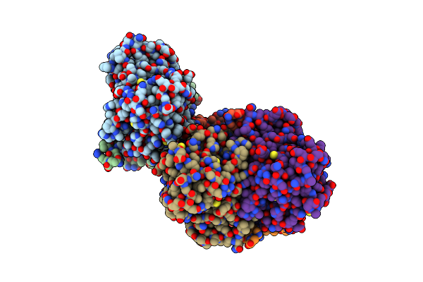
Crystal Structure Of Serine Dehydrogenase From Escherichia Coli
Organism: Escherichia coli
Method: X-RAY DIFFRACTION Resolution:1.90 Å Release Date: 2011-10-12 Classification: OXIDOREDUCTASE |
Organism: Escherichia coli
Method: X-RAY DIFFRACTION
Release Date: 2011-10-12
 |
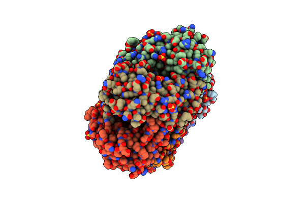
The Closed Form Of Serine Dehydrogenase Complexed With Nadp+
Organism: Escherichia coli
Method: X-RAY DIFFRACTION Resolution:2.70 Å Release Date: 2011-10-12 Classification: OXIDOREDUCTASE Ligands: NAP, PO4 |
Organism: Escherichia coli
Method: X-RAY DIFFRACTION
Release Date: 2011-10-12
Ligands: NAP, PO4
 |
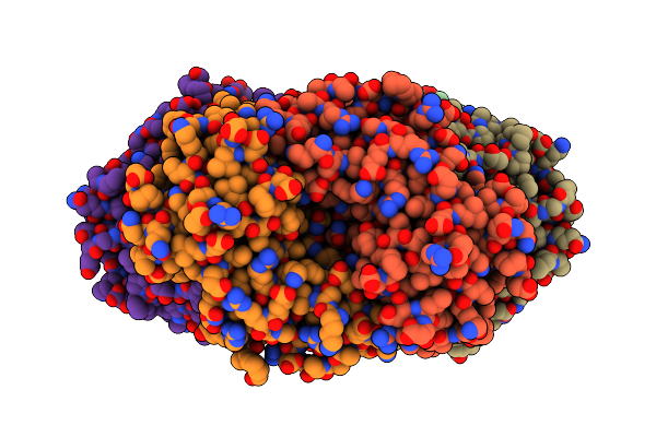
Creatininase Complexed With 1-Methylguanidine
Organism: Pseudomonas putida
Method: X-RAY DIFFRACTION Resolution:1.90 Å Release Date: 2010-02-09 Classification: HYDROLASE Ligands: MN, ZN, MGX, SO4 |
Organism: Pseudomonas putida
Method: X-RAY DIFFRACTION
Release Date: 2010-02-09
Ligands: MN, ZN, MGX, SO4
 |
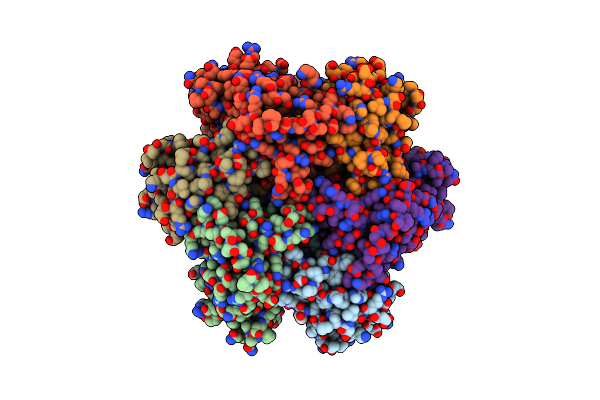
W174F Mutant Creatininase, Type I
Organism: Pseudomonas putida
Method: X-RAY DIFFRACTION Resolution:2.00 Å Release Date: 2010-02-09 Classification: HYDROLASE Ligands: MN, ZN, CAC |
Organism: Pseudomonas putida
Method: X-RAY DIFFRACTION
Release Date: 2010-02-09
Ligands: MN, ZN, CAC
 |
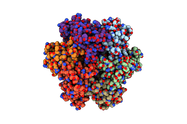
W174F Mutant Creatininase, Type Ii
Organism: Pseudomonas putida
Method: X-RAY DIFFRACTION Resolution:1.78 Å Release Date: 2010-02-09 Classification: HYDROLASE Ligands: MN, ZN, CAC |
Organism: Pseudomonas putida
Method: X-RAY DIFFRACTION
Release Date: 2010-02-09
Ligands: MN, ZN, CAC
 |
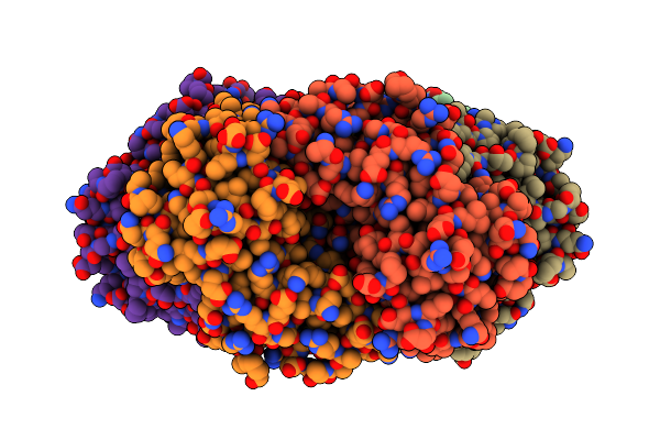
W154F Mutant Creatininase
Organism: Pseudomonas putida
Method: X-RAY DIFFRACTION Resolution:2.00 Å Release Date: 2010-02-09 Classification: HYDROLASE Ligands: MN, ZN |
Organism: Pseudomonas putida
Method: X-RAY DIFFRACTION
Release Date: 2010-02-09
Ligands: MN, ZN
 |
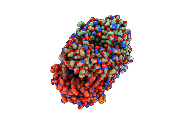
W154A Mutant Creatininase
Organism: Pseudomonas putida
Method: X-RAY DIFFRACTION Resolution:2.00 Å Release Date: 2010-02-09 Classification: HYDROLASE Ligands: MN, ZN, CL |
Organism: Pseudomonas putida
Method: X-RAY DIFFRACTION
Release Date: 2010-02-09
Ligands: MN, ZN, CL
 |
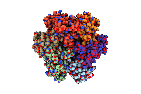
E122Q Mutant Creatininase Complexed With Creatine
Organism: Pseudomonas putida
Method: X-RAY DIFFRACTION Resolution:2.00 Å Release Date: 2010-02-09 Classification: HYDROLASE Ligands: ZN, CRN, SO4 |
Organism: Pseudomonas putida
Method: X-RAY DIFFRACTION
Release Date: 2010-02-09
Ligands: ZN, CRN, SO4
 |
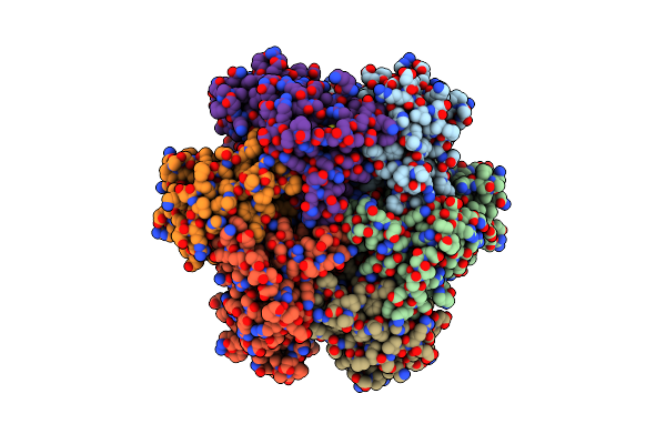
The E122Q Mutant Creatininase, Mn-Zn Type
Organism: Pseudomonas putida
Method: X-RAY DIFFRACTION Resolution:2.20 Å Release Date: 2010-02-09 Classification: HYDROLASE Ligands: MN, ZN, CL |
Organism: Pseudomonas putida
Method: X-RAY DIFFRACTION
Release Date: 2010-02-09
Ligands: MN, ZN, CL
 |
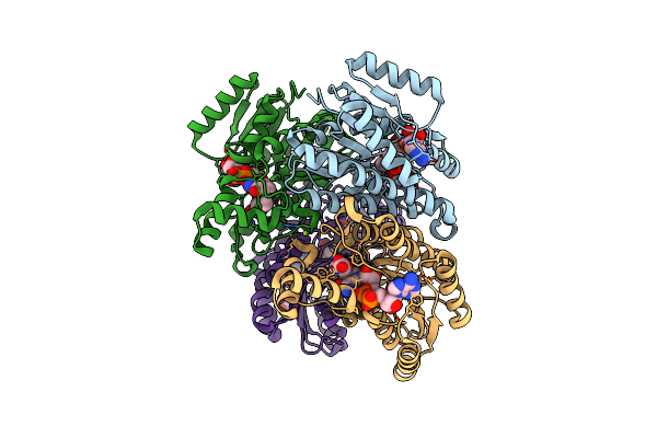
E122Q Mutant Creatininase, Zn-Zn Type
Organism: Pseudomonas putida
Method: X-RAY DIFFRACTION Resolution:2.00 Å Release Date: 2010-02-09 Classification: HYDROLASE Ligands: ZN, CL |
Organism: Pseudomonas putida
Method: X-RAY DIFFRACTION
Release Date: 2010-02-09
Ligands: ZN, CL
 |
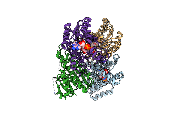
Closed Conformation Of D-3-Hydroxybutyrate Dehydrogenase Complexed With Nad+ And L-3-Hydroxybutyrate
Organism: Pseudomonas fragi
Method: X-RAY DIFFRACTION Resolution:1.80 Å Release Date: 2009-08-25 Classification: OXIDOREDUCTASE Ligands: NAD, 3HL, MG, GOL |
Organism: Pseudomonas fragi
Method: X-RAY DIFFRACTION
Release Date: 2009-08-25
Ligands: NAD, 3HL, MG, GOL
 |
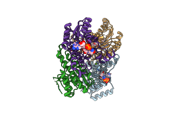
T190S Mutant Of D-3-Hydroxybutyrate Dehydrogenase
Organism: Pseudomonas fragi
Method: X-RAY DIFFRACTION Resolution:2.30 Å Release Date: 2009-08-25 Classification: OXIDOREDUCTASE Ligands: NAD, 3HL, MG |
Organism: Pseudomonas fragi
Method: X-RAY DIFFRACTION
Release Date: 2009-08-25
Ligands: NAD, 3HL, MG
 |
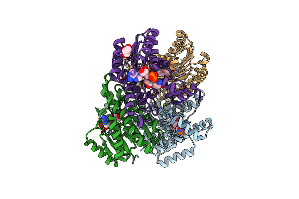
T190A Mutant Of D-3-Hydroxybutyrate Dehydrogenase Complexed With Nad+
Organism: Pseudomonas fragi
Method: X-RAY DIFFRACTION Resolution:2.00 Å Release Date: 2009-08-25 Classification: OXIDOREDUCTASE Ligands: NAD, MG |
Organism: Pseudomonas fragi
Method: X-RAY DIFFRACTION
Release Date: 2009-08-25
Ligands: NAD, MG
 |
The Binary Complex Of D-3-Hydroxybutyrate Dehydrogenase With Nad+
Organism: Pseudomonas fragi
Method: X-RAY DIFFRACTION Resolution:1.95 Å Release Date: 2009-08-25 Classification: OXIDOREDUCTASE Ligands: NAD, MG, GOL |
Organism: Pseudomonas fragi
Method: X-RAY DIFFRACTION
Release Date: 2009-08-25
Ligands: NAD, MG, GOL
 |
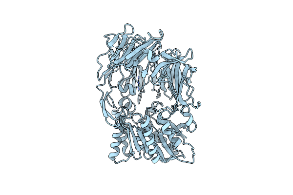
Aminopeptidase N Complexed With The Aminophosphinic Inhibitor Of Pl250, A Transition State Analogue
Organism: Escherichia coli k-12
Method: X-RAY DIFFRACTION Resolution:1.55 Å Release Date: 2009-08-25 Classification: HYDROLASE Ligands: ZN, S23, SO4, GOL |
Organism: Escherichia coli k-12
Method: X-RAY DIFFRACTION
Release Date: 2009-08-25
Ligands: ZN, S23, SO4, GOL
 |
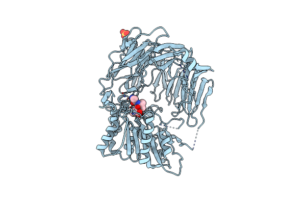
Crystal Structure Of Dipeptidyl Aminopeptidase Iv From Stenotrophomonas Maltophilia
Organism: Stenotrophomonas maltophilia
Method: X-RAY DIFFRACTION Resolution:2.80 Å Release Date: 2008-02-26 Classification: HYDROLASE |
Organism: Stenotrophomonas maltophilia
Method: X-RAY DIFFRACTION
Release Date: 2008-02-26
 |
Prolyl Tripeptidyl Aminopeptidase Complexed With An Inhibitor
Organism: Porphyromonas gingivalis
Method: X-RAY DIFFRACTION Resolution:2.20 Å Release Date: 2008-02-19 Classification: HYDROLASE Ligands: SO4, AIO |
Organism: Porphyromonas gingivalis
Method: X-RAY DIFFRACTION
Release Date: 2008-02-19
Ligands: SO4, AIO
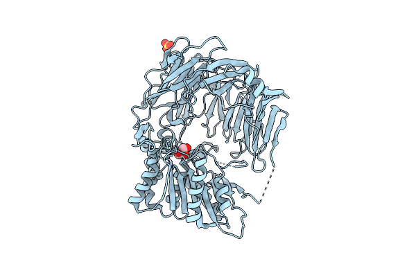 |
Prolyl Tripeptidyl Aminopeptidase Mutant E636A
Organism: Porphyromonas gingivalis
Method: X-RAY DIFFRACTION Resolution:2.00 Å Release Date: 2008-02-19 Classification: HYDROLASE Ligands: SO4, GOL |
Organism: Porphyromonas gingivalis
Method: X-RAY DIFFRACTION
Release Date: 2008-02-19
Ligands: SO4, GOL