

Search Count: 17
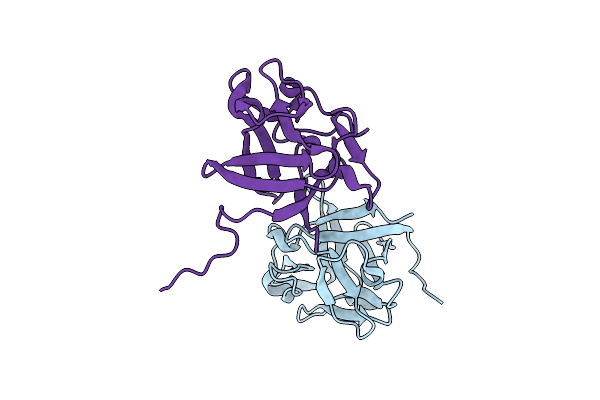 |
Linear Form Of Fgf10 From Homo Sapiens
Organism: Homo sapiens
Method: X-RAY DIFFRACTION Resolution:2.56 Å Release Date: 2025-03-26 Classification: HORMONE |
Organism: Homo sapiens
Method: X-RAY DIFFRACTION
Release Date: 2025-03-26
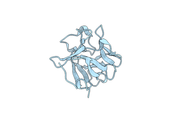 |
Crystal Structure Of Fgf2-M1 Mutant - D28E/C78L/C96I/S137P
Organism: Homo sapiens
Method: X-RAY DIFFRACTION Resolution:1.40 Å Release Date: 2024-06-26 Classification: SIGNALING PROTEIN |
Organism: Homo sapiens
Method: X-RAY DIFFRACTION
Release Date: 2024-06-26
 |
Crystal Structure Of Fgf2-M2 Mutant - D28E/C78I/C96I/S137P
Organism: Homo sapiens
Method: X-RAY DIFFRACTION Resolution:1.48 Å Release Date: 2024-06-26 Classification: SIGNALING PROTEIN |
Organism: Homo sapiens
Method: X-RAY DIFFRACTION
Release Date: 2024-06-26
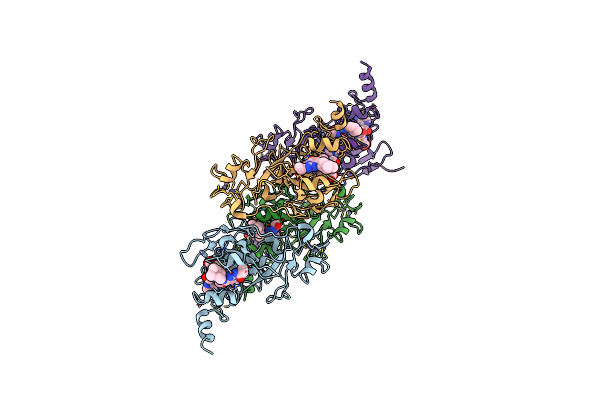 |
Structure Of Human G9A Set-Domain (Ehmt2) In Complex With Covalent Inhibitor (Compound 1)
Organism: Homo sapiens
Method: X-RAY DIFFRACTION Resolution:2.20 Å Release Date: 2022-07-06 Classification: GENE REGULATION Ligands: ZN, SAM, G5U, G4R |
Organism: Homo sapiens
Method: X-RAY DIFFRACTION
Release Date: 2022-07-06
Ligands: ZN, SAM, G5U, G4R
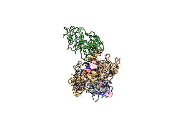 |
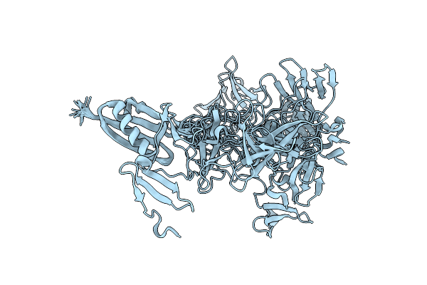
Structure Of Human Glp Set-Domain (Ehmt1) In Complex With Covalent Inhibitor (Compound 1)
Organism: Homo sapiens
Method: X-RAY DIFFRACTION Resolution:2.85 Å Release Date: 2022-07-06 Classification: TRANSFERASE/INHIBITOR Ligands: ZN, G4R, G5U |
Organism: Homo sapiens
Method: X-RAY DIFFRACTION
Release Date: 2022-07-06
Ligands: ZN, G4R, G5U
 |
Solution Structure Of The Fourth Ww Domain Of Wwp2 With Gb1-Tag
Organism: Homo sapiens
Method: SOLUTION NMR Release Date: 2019-10-09 Classification: PROTEIN BINDING |
Organism: Homo sapiens
Method: SOLUTION NMR
Release Date: 2019-10-09
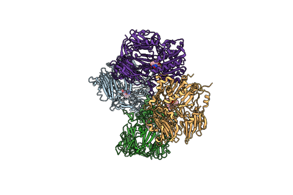 |
Complex Structure Of Dipeptidyl Peptidase Iv And A Oxadiazolyl Ketone
Organism: Homo sapiens
Method: X-RAY DIFFRACTION Resolution:3.10 Å Release Date: 2008-07-15 Classification: HYDROLASE Ligands: 13Z |
Organism: Homo sapiens
Method: X-RAY DIFFRACTION
Release Date: 2008-07-15
Ligands: 13Z
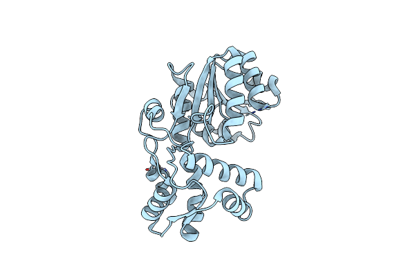 |
Crystal Structure Of A New Type Of Nadph-Dependent Quinone Oxidoreductase (Qor2) From Escherichia Coli
Organism: Escherichia coli
Method: X-RAY DIFFRACTION Resolution:1.80 Å Release Date: 2008-05-27 Classification: OXIDOREDUCTASE Ligands: CU |
Organism: Escherichia coli
Method: X-RAY DIFFRACTION
Release Date: 2008-05-27
Ligands: CU
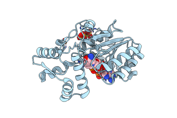 |
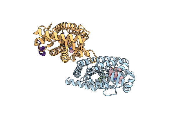
Crystal Structure Of Nadph-Dependent Quinone Oxidoreductase Qor2 Complexed With Nadph From Escherichia Coli
Organism: Escherichia coli
Method: X-RAY DIFFRACTION Resolution:2.30 Å Release Date: 2008-05-27 Classification: OXIDOREDUCTASE Ligands: CU, SO4, NDP |
Organism: Escherichia coli
Method: X-RAY DIFFRACTION
Release Date: 2008-05-27
Ligands: CU, SO4, NDP
 |
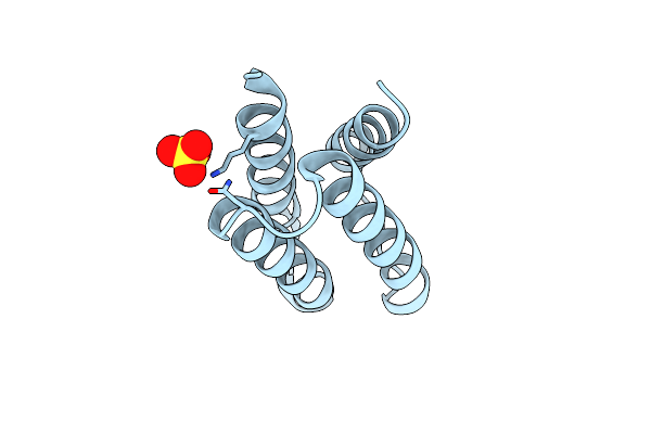
The Crystal Structure Of The Human Pparaplpha Ligand Binding Domain In Complex With A A-Hydroxyimino Phenylpropanoic Acid
Organism: Homo sapiens
Method: X-RAY DIFFRACTION Resolution:2.30 Å Release Date: 2007-10-30 Classification: TRANSCRIPTION Ligands: MMB |
Organism: Homo sapiens
Method: X-RAY DIFFRACTION
Release Date: 2007-10-30
Ligands: MMB
 |
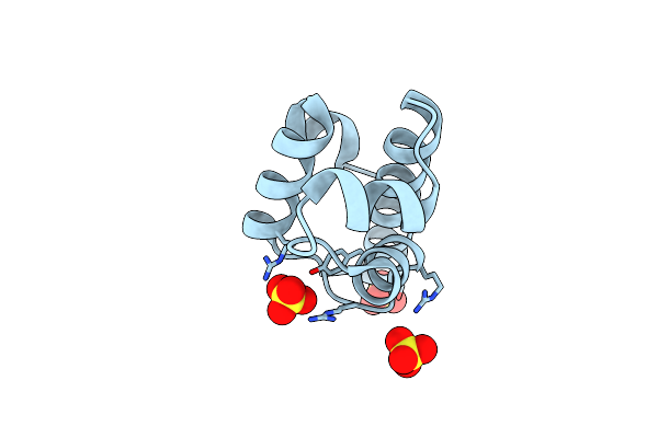
Crystal Structure Of Pedb
Organism: Pediococcus pentosaceus
Method: X-RAY DIFFRACTION Resolution:1.35 Å Release Date: 2007-10-02 Classification: ANTIMICROBIAL PROTEIN Ligands: SO4 |
Organism: Pediococcus pentosaceus
Method: X-RAY DIFFRACTION
Release Date: 2007-10-02
Ligands: SO4
 |
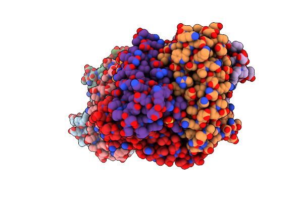
Crystal Structure Of The Dna-Binding Domain Of Bldd
Organism: Streptomyces coelicolor
Method: X-RAY DIFFRACTION Resolution:1.81 Å Release Date: 2006-06-13 Classification: DNA BINDING PROTEIN Ligands: SO4 |
Organism: Streptomyces coelicolor
Method: X-RAY DIFFRACTION
Release Date: 2006-06-13
Ligands: SO4
 |
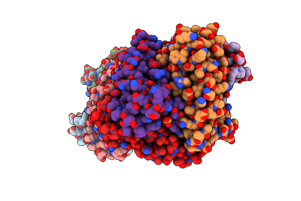
Crystal Structure Of Ni-Containing Superoxide Dismutase With Ni-Ligation Corresponding To The Oxidized State
Organism: Streptomyces seoulensis
Method: X-RAY DIFFRACTION Resolution:2.20 Å Release Date: 2004-05-18 Classification: OXIDOREDUCTASE Ligands: SO4, 3NI |
Organism: Streptomyces seoulensis
Method: X-RAY DIFFRACTION
Release Date: 2004-05-18
Ligands: SO4, 3NI
 |
Crystal Structure Of Ni-Containing Superoxide Dismutase With Ni-Ligation Corresponding To The State After Partial X-Ray-Induced Reduction
Organism: Streptomyces seoulensis
Method: X-RAY DIFFRACTION Resolution:2.20 Å Release Date: 2004-05-18 Classification: OXIDOREDUCTASE Ligands: SO4, 3NI |
Organism: Streptomyces seoulensis
Method: X-RAY DIFFRACTION
Release Date: 2004-05-18
Ligands: SO4, 3NI
 |
Crystal Structure Of Ni-Containing Superoxide Dismutase With Ni-Ligation Corresponding To The State After Full X-Ray-Induced Reduction
Organism: Streptomyces seoulensis
Method: X-RAY DIFFRACTION Resolution:1.60 Å Release Date: 2004-05-18 Classification: OXIDOREDUCTASE Ligands: NI, SO4 |
Organism: Streptomyces seoulensis
Method: X-RAY DIFFRACTION
Release Date: 2004-05-18
Ligands: NI, SO4
 |
Crystal Structure Of Ni-Containing Superoxide Dismutase With Ni-Ligation Corresponding To The Thiosulfate-Reduced State
Organism: Streptomyces seoulensis
Method: X-RAY DIFFRACTION Resolution:2.10 Å Release Date: 2004-05-18 Classification: OXIDOREDUCTASE Ligands: NI, SO4, THJ |
Organism: Streptomyces seoulensis
Method: X-RAY DIFFRACTION
Release Date: 2004-05-18
Ligands: NI, SO4, THJ
 |
Crystal Structure Of Ni-Containing Superoxide Dismutase With Ni-Ligation Corresponding To The State After Full X-Ray-Induced Reduction
Organism: Streptomyces seoulensis
Method: X-RAY DIFFRACTION Resolution:1.68 Å Release Date: 2004-05-18 Classification: OXIDOREDUCTASE Ligands: NI, SO4, ACY |
Organism: Streptomyces seoulensis
Method: X-RAY DIFFRACTION
Release Date: 2004-05-18
Ligands: NI, SO4, ACY