

Search Count: 28
 |
Crystal Structure Of A Single-Chain Monellin Mutant C41A
Organism: Dioscoreophyllum cumminsii
Method: X-RAY DIFFRACTION Resolution:1.23 Å Release Date: 2023-10-11 Classification: PLANT PROTEIN |
Organism: Dioscoreophyllum cumminsii
Method: X-RAY DIFFRACTION
Release Date: 2023-10-11
 |
Crystal Structure Of A Single-Chain Monellin Mutant C41V
Organism: Dioscoreophyllum cumminsii
Method: X-RAY DIFFRACTION Resolution:1.24 Å Release Date: 2023-10-11 Classification: PLANT PROTEIN |
Organism: Dioscoreophyllum cumminsii
Method: X-RAY DIFFRACTION
Release Date: 2023-10-11
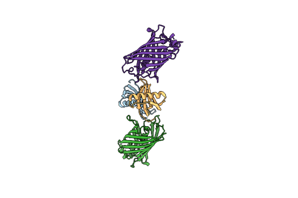 |
Gfp-40/Gfpuv Complex, Form I
Organism: Aequorea victoria, Synthetic construct
Method: X-RAY DIFFRACTION Resolution:1.70 Å Release Date: 2021-01-13 Classification: PROTEIN BINDING |
Organism: Aequorea victoria, Synthetic construct
Method: X-RAY DIFFRACTION
Release Date: 2021-01-13
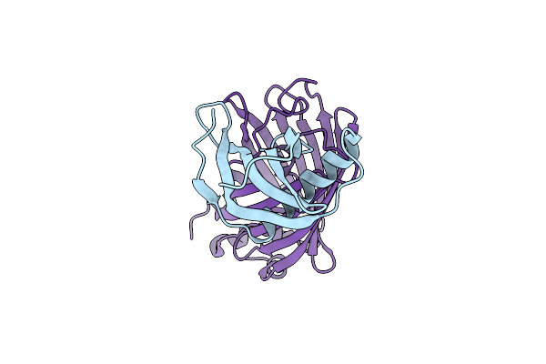 |
Gfp-40/Gfpuv Complex, Form Ii
Organism: Aequorea victoria, Synthetic construct
Method: X-RAY DIFFRACTION Resolution:2.00 Å Release Date: 2021-01-13 Classification: PROTEIN BINDING |
Organism: Aequorea victoria, Synthetic construct
Method: X-RAY DIFFRACTION
Release Date: 2021-01-13
 |
Crystal Structure Of Reelin N-Terminal Region
Organism: Mus musculus
Method: X-RAY DIFFRACTION Resolution:2.00 Å Release Date: 2019-06-19 Classification: SIGNALING PROTEIN Ligands: CA, NAG |
Organism: Mus musculus
Method: X-RAY DIFFRACTION
Release Date: 2019-06-19
Ligands: CA, NAG
 |
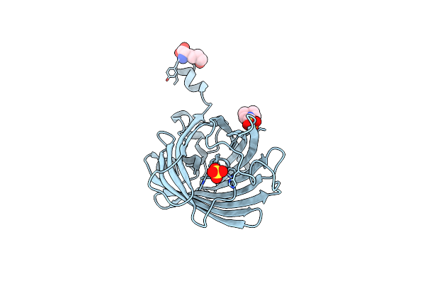
Green Fluorescent Protein Variant Gfpuv With The Native Lysine Residue At The C-Terminus
Organism: Aequorea victoria
Method: X-RAY DIFFRACTION Resolution:1.64 Å Release Date: 2019-04-03 Classification: FLUORESCENT PROTEIN Ligands: SO4 |
Organism: Aequorea victoria
Method: X-RAY DIFFRACTION
Release Date: 2019-04-03
Ligands: SO4
 |
Green Fluorescent Protein Variant Gfpuv With The Modification To 6-Hydroxynorleucine At The C-Terminus
Organism: Aequorea victoria
Method: X-RAY DIFFRACTION Resolution:1.28 Å Release Date: 2019-04-03 Classification: FLUORESCENT PROTEIN Ligands: LDO, SO4, MES |
Organism: Aequorea victoria
Method: X-RAY DIFFRACTION
Release Date: 2019-04-03
Ligands: LDO, SO4, MES
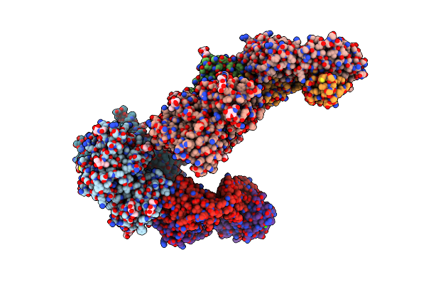 |
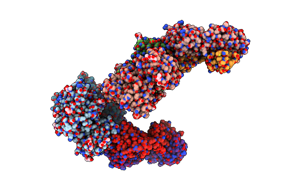
Crystal Structure Of The Medaka Fish Taste Receptor T1R2A-T1R3 Ligand Binding Domains In Complex With L-Glutamine
Organism: Oryzias latipes, Mus musculus
Method: X-RAY DIFFRACTION Resolution:2.21 Å Release Date: 2017-05-24 Classification: SIGNALING PROTEIN/IMMUNE SYSTEM Ligands: NAG, GLN, NA, CL, CA |
Organism: Oryzias latipes, Mus musculus
Method: X-RAY DIFFRACTION
Release Date: 2017-05-24
Ligands: NAG, GLN, NA, CL, CA
 |
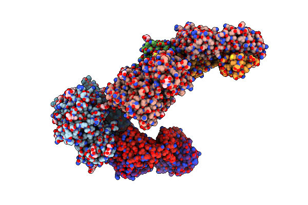
Crystal Structure Of The Medaka Fish Taste Receptor T1R2A-T1R3 Ligand Binding Domains In Complex With L-Alanine
Organism: Oryzias latipes, Mus musculus
Method: X-RAY DIFFRACTION Resolution:2.20 Å Release Date: 2017-05-24 Classification: SIGNALING PROTEIN/IMMUNE SYSTEM Ligands: NAG, ALA, NA, CL |
Organism: Oryzias latipes, Mus musculus
Method: X-RAY DIFFRACTION
Release Date: 2017-05-24
Ligands: NAG, ALA, NA, CL
 |
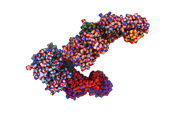
Crystal Structure Of The Medaka Fish Taste Receptor T1R2A-T1R3 Ligand Binding Domains In Complex With L-Arginine
Organism: Oryzias latipes, Mus musculus
Method: X-RAY DIFFRACTION Resolution:2.60 Å Release Date: 2017-05-24 Classification: SIGNALING PROTEIN/IMMUNE SYSTEM Ligands: NAG, ARG, NA, CL, CA |
Organism: Oryzias latipes, Mus musculus
Method: X-RAY DIFFRACTION
Release Date: 2017-05-24
Ligands: NAG, ARG, NA, CL, CA
 |
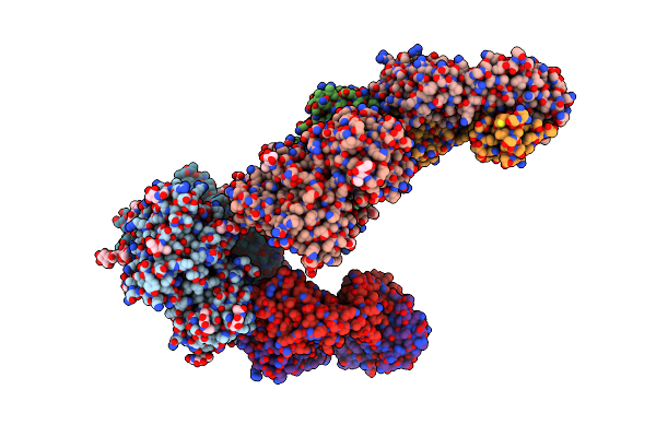
Crystal Structure Of The Medaka Fish Taste Receptor T1R2A-T1R3 Ligand Binding Domains In Complex With L-Glutamate
Organism: Oryzias latipes, Mus musculus
Method: X-RAY DIFFRACTION Resolution:2.61 Å Release Date: 2017-05-24 Classification: SIGNALING PROTEIN/IMMUNE SYSTEM Ligands: NAG, GLU, NA, CL, CA |
Organism: Oryzias latipes, Mus musculus
Method: X-RAY DIFFRACTION
Release Date: 2017-05-24
Ligands: NAG, GLU, NA, CL, CA
 |
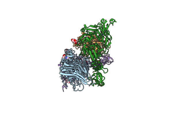
Crystal Structure Of The Medaka Fish Taste Receptor T1R2A-T1R3 Ligand Binding Domains In Complex With Glycine
Organism: Oryzias latipes, Mus musculus
Method: X-RAY DIFFRACTION Resolution:2.60 Å Release Date: 2017-05-24 Classification: SIGNALING PROTEIN/IMMUNE SYSTEM Ligands: NAG, GLY, NA, CL, CA |
Organism: Oryzias latipes, Mus musculus
Method: X-RAY DIFFRACTION
Release Date: 2017-05-24
Ligands: NAG, GLY, NA, CL, CA
 |
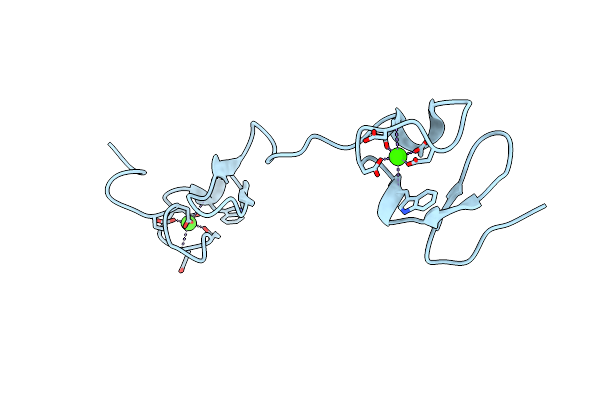
Crystal Structure Of The Apoer2 Ectodomain In Complex With The Reelin R56 Fragment
Organism: Mus musculus, Homo sapiens
Method: X-RAY DIFFRACTION Resolution:3.20 Å Release Date: 2017-04-26 Classification: SIGNALING PROTEIN/ENDOCYTOSIS Ligands: CA, NAG |
Organism: Mus musculus, Homo sapiens
Method: X-RAY DIFFRACTION
Release Date: 2017-04-26
Ligands: CA, NAG
 |
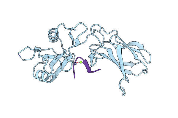
Crystal Structure Of The La12 Fragment Of Apoer2
Organism: Homo sapiens
Method: X-RAY DIFFRACTION Resolution:1.90 Å Release Date: 2017-04-26 Classification: SIGNALING PROTEIN Ligands: CA |
Organism: Homo sapiens
Method: X-RAY DIFFRACTION
Release Date: 2017-04-26
Ligands: CA
 |
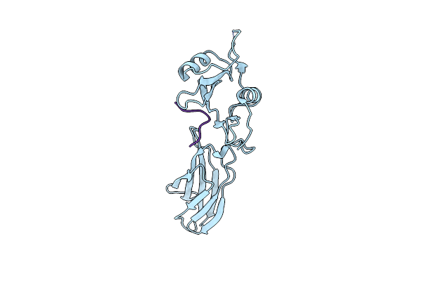
Crystal Structure Of The Synthetic Protein In Complex With Py Peptide
Organism: Synthetic construct, Homo sapiens
Method: X-RAY DIFFRACTION Resolution:1.40 Å Release Date: 2014-04-23 Classification: DE NOVO PROTEIN Ligands: MG |
Organism: Synthetic construct, Homo sapiens
Method: X-RAY DIFFRACTION
Release Date: 2014-04-23
Ligands: MG
 |
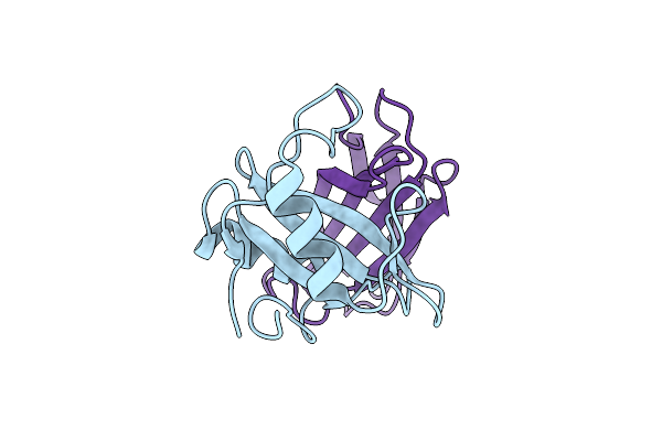
Crystal Structure Of Synthetic Protein In Complex With Double Py Peptide
Organism: Synthetic construct, Homo sapiens
Method: X-RAY DIFFRACTION Resolution:2.41 Å Release Date: 2014-04-23 Classification: DE NOVO PROTEIN |
Organism: Synthetic construct, Homo sapiens
Method: X-RAY DIFFRACTION
Release Date: 2014-04-23
 |
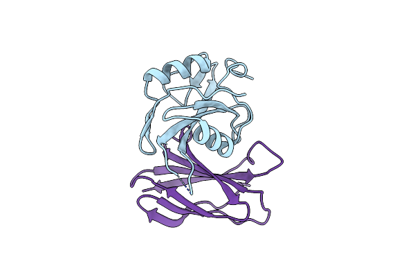
Crystal Structure Of Monobody Nsa1/Shp2 N-Sh2 Domain Complex
Organism: Homo sapiens
Method: X-RAY DIFFRACTION Resolution:2.31 Å Release Date: 2013-08-28 Classification: Signaling protein/protein binding |
Organism: Homo sapiens
Method: X-RAY DIFFRACTION
Release Date: 2013-08-28
 |
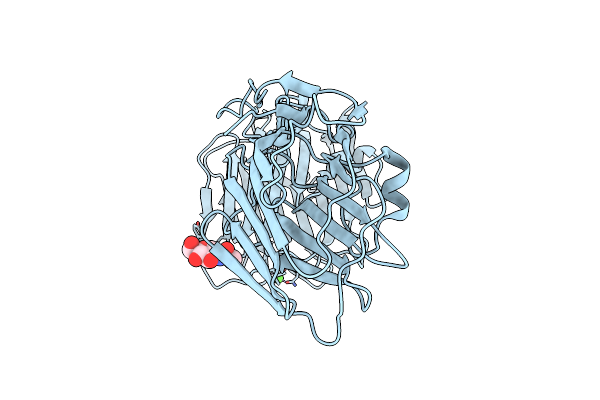
Crystal Structure Of Monobody Cs1/Shp2 C-Sh2 Domain Complex
Organism: Homo sapiens
Method: X-RAY DIFFRACTION Resolution:2.30 Å Release Date: 2013-08-28 Classification: Signaling protein/protein binding |
Organism: Homo sapiens
Method: X-RAY DIFFRACTION
Release Date: 2013-08-28
 |
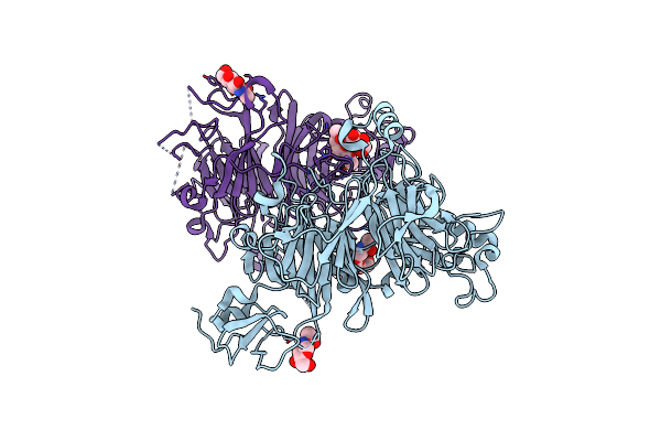
Alpha-Neurexin-1 Ectodomain Fragment; Lns5-Egf3-Lns6
Organism: Bos taurus
Method: X-RAY DIFFRACTION Resolution:2.30 Å Release Date: 2011-04-13 Classification: CELL ADHESION Ligands: CA, NAG |
Organism: Bos taurus
Method: X-RAY DIFFRACTION
Release Date: 2011-04-13
Ligands: CA, NAG
 |
Mouse Semaphorin 6A Extracellular Domain
Organism: Mus musculus
Method: X-RAY DIFFRACTION Resolution:2.50 Å Release Date: 2010-10-06 Classification: SIGNALING PROTEIN Ligands: NAG |
Organism: Mus musculus
Method: X-RAY DIFFRACTION
Release Date: 2010-10-06
Ligands: NAG