

Search Count: 24
 |
Crystal Structure Of Sod1 By Serial Synchrotron Crystallography
Organism: Homo sapiens
Method: X-RAY DIFFRACTION Resolution:2.30 Å Release Date: 2026-04-01 Classification: OXIDOREDUCTASE Ligands: CU, ZN |
Organism: Homo sapiens
Method: X-RAY DIFFRACTION
Release Date: 2026-04-01
Ligands: CU, ZN
 |
Cryogenic Structure Of Sod1, Determined As The Ssx Counterpart
Organism: Homo sapiens
Method: X-RAY DIFFRACTION Resolution:2.37 Å Release Date: 2026-04-01 Classification: OXIDOREDUCTASE Ligands: CU, ZN |
Organism: Homo sapiens
Method: X-RAY DIFFRACTION
Release Date: 2026-04-01
Ligands: CU, ZN
 |
Time-Resolved Ambient Temperature Kineto-Crystallographic Structure Of Initiation Factor In Complex With Ribosome
Organism: Thermus thermophilus hb8
Method: X-RAY DIFFRACTION Resolution:3.59 Å Release Date: 2025-04-16 Classification: RIBOSOME Ligands: MG, ZN |
Organism: Thermus thermophilus hb8
Method: X-RAY DIFFRACTION
Release Date: 2025-04-16
Ligands: MG, ZN
 |
Crystal Structure Of Hsod1 In C121 Space Group
Organism: Homo sapiens
Method: X-RAY DIFFRACTION Resolution:2.34 Å Release Date: 2024-08-14 Classification: OXIDOREDUCTASE Ligands: CU, ZN, ACT, SO4 |
Organism: Homo sapiens
Method: X-RAY DIFFRACTION
Release Date: 2024-08-14
Ligands: CU, ZN, ACT, SO4
 |
Crystal Structure Of Chicken Egg Lysozyme
Organism: Gallus gallus
Method: X-RAY DIFFRACTION Resolution:1.50 Å Release Date: 2023-04-19 Classification: HYDROLASE Ligands: NO3 |
Organism: Gallus gallus
Method: X-RAY DIFFRACTION
Release Date: 2023-04-19
Ligands: NO3
 |
Crystal Structure Of Chicken Egg Lysozyme At Ambient Temperature
Organism: Gallus gallus
Method: X-RAY DIFFRACTION Resolution:2.39 Å Release Date: 2023-03-22 Classification: HYDROLASE |
Organism: Gallus gallus
Method: X-RAY DIFFRACTION
Release Date: 2023-03-22
 |
Crystal Structure Of The R132K:R111L Mutant Of Cellular Retinoic Acid Binding Proteinii Complexed With A Synthetic Ligand (Merocyanine) At 1.73 Angstrom Resolution.
Organism: Homo sapiens
Method: X-RAY DIFFRACTION Resolution:1.73 Å Release Date: 2015-01-28 Classification: PROTEIN BINDING Ligands: LMC |
Organism: Homo sapiens
Method: X-RAY DIFFRACTION
Release Date: 2015-01-28
Ligands: LMC
 |
Crystal Structure Of The R132K:R111L:L121D Mutant Of Cellular Retinoic Acid Binding Proteinii Complexed With A Synthetic Ligand (Merocyanine) At 1.77 Angstrom Resolution
Organism: Homo sapiens
Method: X-RAY DIFFRACTION Resolution:1.77 Å Release Date: 2015-01-28 Classification: PROTEIN BINDING Ligands: LMC, B3P |
Organism: Homo sapiens
Method: X-RAY DIFFRACTION
Release Date: 2015-01-28
Ligands: LMC, B3P
 |
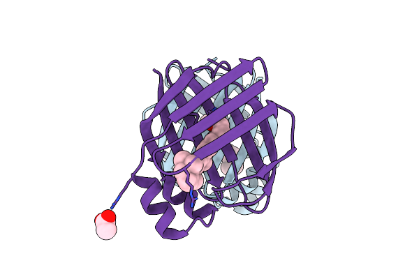
Crystal Structure Of The R132K:R111L:L121E Mutant Of Cellular Retinoic Acid Binding Proteinii Complexed With A Synthetic Ligand (Merocyanine) At 1.47 Angstrom Resolution
Organism: Homo sapiens
Method: X-RAY DIFFRACTION Resolution:1.47 Å Release Date: 2015-01-28 Classification: PROTEIN BINDING Ligands: LMC |
Organism: Homo sapiens
Method: X-RAY DIFFRACTION
Release Date: 2015-01-28
Ligands: LMC
 |
The Crystal Structures Of Holo-Wt Human Cellular Retinol Binding Protein Ii (Hcrbpii) Bound To Retinol
Organism: Homo sapiens
Method: X-RAY DIFFRACTION Resolution:1.19 Å Release Date: 2014-12-31 Classification: TRANSPORT PROTEIN Ligands: RTL, ACT |
Organism: Homo sapiens
Method: X-RAY DIFFRACTION
Release Date: 2014-12-31
Ligands: RTL, ACT
 |
The Crystal Structures Of Holo-Wt Human Cellular Retinol Binding Protein Ii (Hcrbpii) Bound To Retinal
Organism: Homo sapiens
Method: X-RAY DIFFRACTION Resolution:1.62 Å Release Date: 2014-12-10 Classification: TRANSPORT PROTEIN Ligands: RET, ACT |
Organism: Homo sapiens
Method: X-RAY DIFFRACTION
Release Date: 2014-12-10
Ligands: RET, ACT
 |
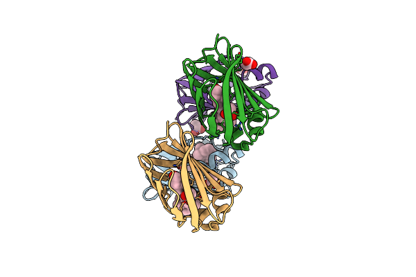
Crystal Structure Of Wild Type Human Cellular Retinol Binding Protein Ii (Hcrbpii) Bound To Retinol At 7 Kev Beam Energy
Organism: Homo sapiens
Method: X-RAY DIFFRACTION Resolution:1.90 Å Release Date: 2014-12-10 Classification: PROTEIN BINDING Ligands: RTL, ACT |
Organism: Homo sapiens
Method: X-RAY DIFFRACTION
Release Date: 2014-12-10
Ligands: RTL, ACT
 |
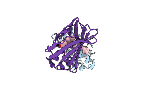
Crystal Structure Of Wild Type Human Cellular Retinol Binding Protein Ii (Hcrbpii) Bound To Retinol At 11 Kev Beam Energy
Organism: Homo sapiens
Method: X-RAY DIFFRACTION Resolution:1.50 Å Release Date: 2014-12-10 Classification: PROTEIN BINDING Ligands: RTL, ACT, GOL |
Organism: Homo sapiens
Method: X-RAY DIFFRACTION
Release Date: 2014-12-10
Ligands: RTL, ACT, GOL
 |
Crystal Structure Of The Q108K:K40L Mutant Of Human Cellular Retinol Binding Proteinii In Complex With All-Trans-Retinal After 24 Hour Incubation At 1.4 Angstrom Resolution
Organism: Homo sapiens
Method: X-RAY DIFFRACTION Resolution:1.40 Å Release Date: 2014-12-10 Classification: TRANSPORT PROTEIN Ligands: RET, ACT |
Organism: Homo sapiens
Method: X-RAY DIFFRACTION
Release Date: 2014-12-10
Ligands: RET, ACT
 |
Crystal Structure Of The R111K:R132L:Y134F:T54V:R59W:A32W Mutant Of The Cellular Retinoic Acid Binding Protein Type Ii In Complex With All-Trans Retinal At 2.6 Angstrom Resolution
Organism: Homo sapiens
Method: X-RAY DIFFRACTION Resolution:2.60 Å Release Date: 2013-10-09 Classification: TRANSPORT PROTEIN Ligands: RET |
Organism: Homo sapiens
Method: X-RAY DIFFRACTION
Release Date: 2013-10-09
Ligands: RET
 |
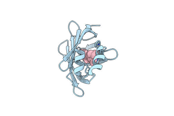
Crystal Structure Of The R111K:R132L:Y134F:T54V:R59W Mutant Of The Cellular Retinoic Acid Binding Protein Type Ii In Complex With All-Trans Retinal At 2.58 Angstrom Resolution
Organism: Homo sapiens
Method: X-RAY DIFFRACTION Resolution:2.58 Å Release Date: 2013-10-09 Classification: TRANSPORT PROTEIN Ligands: RET |
Organism: Homo sapiens
Method: X-RAY DIFFRACTION
Release Date: 2013-10-09
Ligands: RET
 |
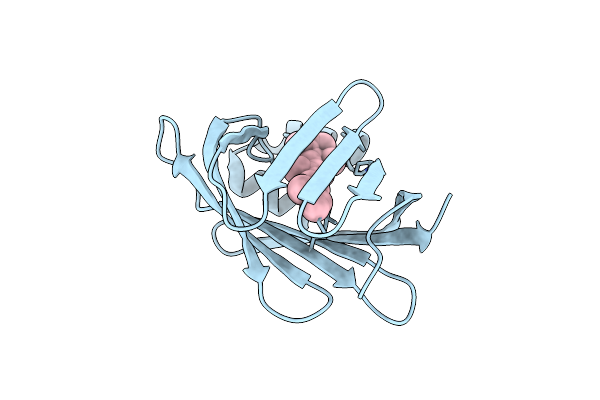
Crystal Structure Of The R111K:R132Y:Y134F:T54V:R59W:A32W Mutant Of The Cellular Retinoic Acid Binding Protein Type Ii In Complex With All-Trans Retinal At 2.38 Angstrom Resolution
Organism: Homo sapiens
Method: X-RAY DIFFRACTION Resolution:2.47 Å Release Date: 2013-10-09 Classification: TRANSPORT PROTEIN Ligands: RET |
Organism: Homo sapiens
Method: X-RAY DIFFRACTION
Release Date: 2013-10-09
Ligands: RET
 |
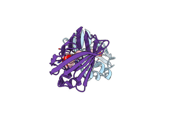
Crystal Structure Of The R111K:R132Q:Y134F:T54V:R59W:A32W Mutant Of The Cellular Retinoic Acid Binding Protein Type Ii In Complex With All-Trans Retinal At 2.57 Angstrom Resolution
Organism: Homo sapiens
Method: X-RAY DIFFRACTION Resolution:2.57 Å Release Date: 2013-10-09 Classification: TRANSPORT PROTEIN Ligands: RET |
Organism: Homo sapiens
Method: X-RAY DIFFRACTION
Release Date: 2013-10-09
Ligands: RET
 |
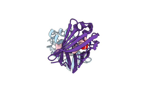
Crystal Structure Of The Q108K:K40L:T51V:T53C:Y19W:R58W:T29L:A33W Mutant Of Cellular Retinol Binding Protein Type Ii In Complex With All-Trans-Retinal At 1.4 Angstrom Resolution
Organism: Homo sapiens
Method: X-RAY DIFFRACTION Resolution:1.40 Å Release Date: 2012-12-26 Classification: TRANSPORT PROTEIN Ligands: ACT, RET |
Organism: Homo sapiens
Method: X-RAY DIFFRACTION
Release Date: 2012-12-26
Ligands: ACT, RET
 |
Crystal Structure Of The Q108K:K40L:T51V:T53C:Y19W:R58W:T29L:Q4R Mutant Of Cellular Retinol Binding Protein Type Ii In Complex With All-Trans-Retinal At 1.5 Angstrom Resolution
Organism: Homo sapiens
Method: X-RAY DIFFRACTION Resolution:1.50 Å Release Date: 2012-12-26 Classification: TRANSPORT PROTEIN Ligands: RET, ACT |
Organism: Homo sapiens
Method: X-RAY DIFFRACTION
Release Date: 2012-12-26
Ligands: RET, ACT