

Search Count: 144
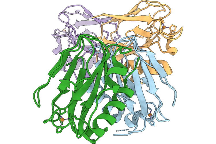 |
X-Ray Crystal Structure Of Pseudoazurin Met16Gly Variant (Tris-Hcl Ph 7.6)
Organism: Achromobacter cycloclastes
Method: X-RAY DIFFRACTION Resolution:1.50 Å Release Date: 2026-08-05 Classification: ELECTRON TRANSPORT Ligands: CU, CL |
Organism: Achromobacter cycloclastes
Method: X-RAY DIFFRACTION
Release Date: 2026-08-05
Ligands: CU, CL
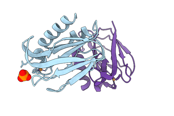 |
X-Ray Crystal Structure Of Pseudoazurin Met16Arg Variant, Oxidized Form At Ph 7.0
Organism: Achromobacter cycloclastes
Method: X-RAY DIFFRACTION Resolution:1.22 Å Release Date: 2026-03-11 Classification: ELECTRON TRANSPORT Ligands: CU, PO4 |
Organism: Achromobacter cycloclastes
Method: X-RAY DIFFRACTION
Release Date: 2026-03-11
Ligands: CU, PO4
 |
P Ring On Polyrod-P Ring Complex From Salmonella Th26292 Strain
Organism: Salmonella enterica subsp. enterica serovar typhimurium
Method: ELECTRON MICROSCOPY Resolution:2.33 Å Release Date: 2026-03-04 Classification: MOTOR PROTEIN |
Organism: Salmonella enterica subsp. enterica serovar typhimurium
Method: ELECTRON MICROSCOPY
Release Date: 2026-03-04
 |
Polyrod Formed By Flgg (G65V) From The Salmonella Th26292 Strain
Organism: Salmonella enterica subsp. enterica serovar typhimurium
Method: ELECTRON MICROSCOPY Release Date: 2026-02-04 Classification: MOTOR PROTEIN |
Organism: Salmonella enterica subsp. enterica serovar typhimurium
Method: ELECTRON MICROSCOPY
Release Date: 2026-02-04
 |
Cryo-Em Structure Of The Lgi1 Lrr-Lgi1 Eptp-Adam22 Ecd Complex
Organism: Homo sapiens
Method: ELECTRON MICROSCOPY Release Date: 2025-06-25 Classification: CELL ADHESION Ligands: CA |
Organism: Homo sapiens
Method: ELECTRON MICROSCOPY
Release Date: 2025-06-25
Ligands: CA
 |
Cryo-Em Structure Of The 3:3 Lgi1-Adam22 Complex
Organism: Homo sapiens
Method: ELECTRON MICROSCOPY Release Date: 2025-06-25 Classification: CELL ADHESION |
Organism: Homo sapiens
Method: ELECTRON MICROSCOPY
Release Date: 2025-06-25
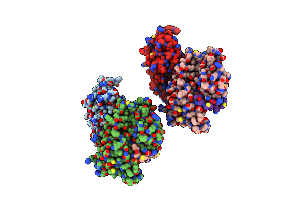 |
Crystal Structure Of The Mutant Human Ror Gamma Ligand Binding Domain With Jte-151
Organism: Homo sapiens
Method: X-RAY DIFFRACTION Resolution:2.30 Å Release Date: 2024-11-27 Classification: NUCLEAR PROTEIN/INHIBITOR Ligands: YAL |
Organism: Homo sapiens
Method: X-RAY DIFFRACTION
Release Date: 2024-11-27
Ligands: YAL
 |
X-Ray Crystal Structure Of Pseudoazurin Met16Gly Variant
Organism: Achromobacter cycloclastes
Method: X-RAY DIFFRACTION Resolution:1.20 Å Release Date: 2024-10-16 Classification: ELECTRON TRANSPORT Ligands: CU, PEG |
Organism: Achromobacter cycloclastes
Method: X-RAY DIFFRACTION
Release Date: 2024-10-16
Ligands: CU, PEG
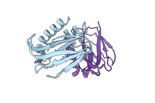 |
X-Ray Crystal Structure Of Pseudoazurin Met16His Variant At Ph 4.0
Organism: Achromobacter cycloclastes
Method: X-RAY DIFFRACTION Resolution:1.36 Å Release Date: 2024-01-31 Classification: ELECTRON TRANSPORT Ligands: CU |
Organism: Achromobacter cycloclastes
Method: X-RAY DIFFRACTION
Release Date: 2024-01-31
Ligands: CU
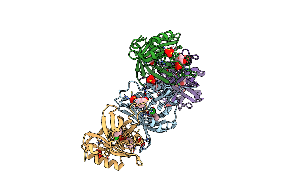 |
Hydroxynitrile Lyase From The Millipede
Organism: Oxidus gracilis
Method: X-RAY DIFFRACTION Resolution:2.01 Å Release Date: 2024-01-24 Classification: LYASE Ligands: HBX, CL, SO4, GOL |
Organism: Oxidus gracilis
Method: X-RAY DIFFRACTION
Release Date: 2024-01-24
Ligands: HBX, CL, SO4, GOL
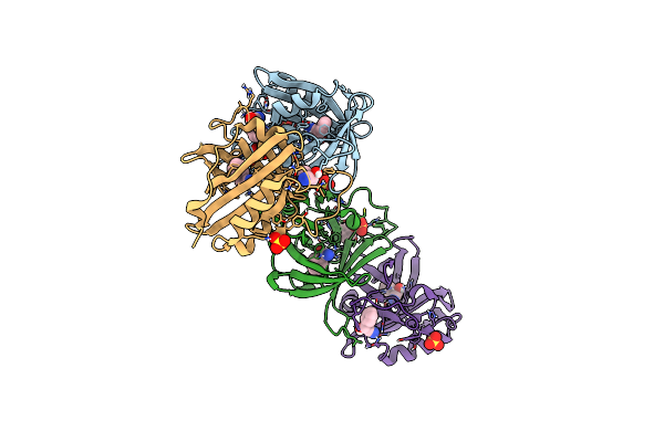 |
Hydroxynitrile Lyase From The Millipede, Oxidus Gracilis Bound With (R)-(+)-Alpha-Hydroxybenzene-Acetonitrile
Organism: Oxidus gracilis
Method: X-RAY DIFFRACTION Resolution:2.01 Å Release Date: 2024-01-24 Classification: LYASE Ligands: MXN, SO4 |
Organism: Oxidus gracilis
Method: X-RAY DIFFRACTION
Release Date: 2024-01-24
Ligands: MXN, SO4
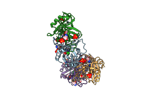 |
Hydroxynitrile Lyase From The Millipede, Oxidus Gracilis In Acetonitrile
Organism: Oxidus gracilis
Method: X-RAY DIFFRACTION Resolution:2.01 Å Release Date: 2024-01-24 Classification: LYASE Ligands: CNH, CL, SO4 |
Organism: Oxidus gracilis
Method: X-RAY DIFFRACTION
Release Date: 2024-01-24
Ligands: CNH, CL, SO4
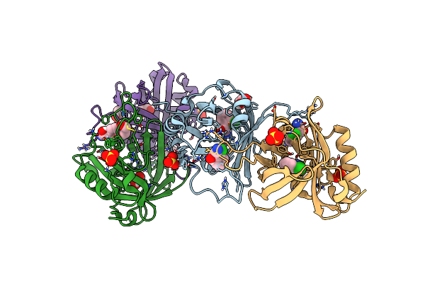 |
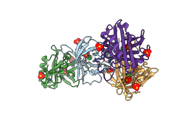
Hydroxynitrile Lyase From The Millipede, Oxidus Gracilis Complexed With (R)-2-Chloromandelonitrile
Organism: Oxidus gracilis
Method: X-RAY DIFFRACTION Resolution:2.01 Å Release Date: 2024-01-24 Classification: LYASE Ligands: IJ5, GOL, SO4 |
Organism: Oxidus gracilis
Method: X-RAY DIFFRACTION
Release Date: 2024-01-24
Ligands: IJ5, GOL, SO4
 |
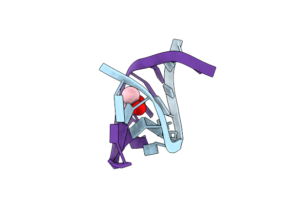
Hydroxynitrile Lyase From The Millipede,
Organism: Oxidus gracilis
Method: X-RAY DIFFRACTION Resolution:2.01 Å Release Date: 2024-01-17 Classification: LYASE Ligands: SO4, CL |
Organism: Oxidus gracilis
Method: X-RAY DIFFRACTION
Release Date: 2024-01-17
Ligands: SO4, CL
 |
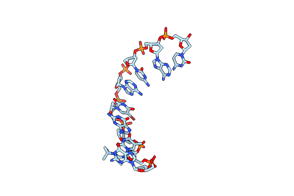
Crystal Structure Of Dna Decamer Containing Guna[Me,Tbu]
Organism: Synthetic construct
Method: X-RAY DIFFRACTION Resolution:2.01 Å Release Date: 2023-08-09 Classification: DNA Ligands: CAD |
Organism: Synthetic construct
Method: X-RAY DIFFRACTION
Release Date: 2023-08-09
Ligands: CAD
 |
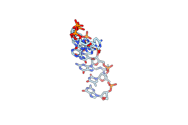
Crystal Structure Of Dna Octamer Containing Guna[Me,Tbu]
Organism: Synthetic construct
Method: X-RAY DIFFRACTION Resolution:0.93 Å Release Date: 2023-08-09 Classification: DNA |
Organism: Synthetic construct
Method: X-RAY DIFFRACTION
Release Date: 2023-08-09
 |
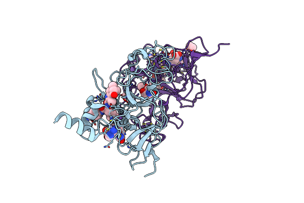
Crystal Structure Of Dna Octamer Containing Guna[Me,Me]
Organism: Synthetic construct
Method: X-RAY DIFFRACTION Resolution:0.95 Å Release Date: 2023-08-09 Classification: DNA |
Organism: Synthetic construct
Method: X-RAY DIFFRACTION
Release Date: 2023-08-09
 |
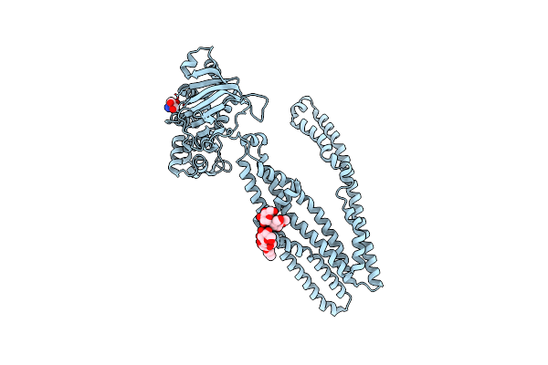
Structure Of G9A In Complex With Rk-701
Organism: Homo sapiens
Method: X-RAY DIFFRACTION Resolution:1.49 Å Release Date: 2022-12-21 Classification: TRANSFERASE Ligands: ZN, SFG, J26, EDO, TRS |
Organism: Homo sapiens
Method: X-RAY DIFFRACTION
Release Date: 2022-12-21
Ligands: ZN, SFG, J26, EDO, TRS
 |
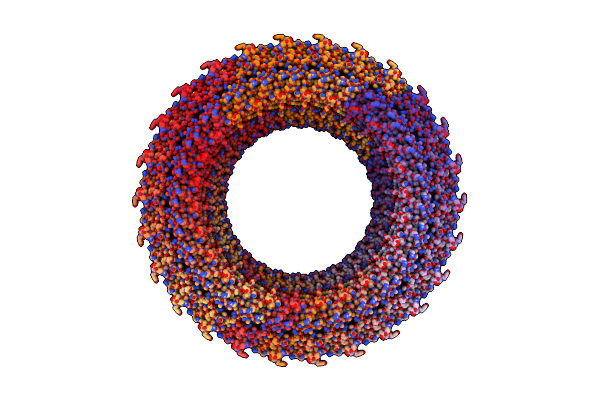
Crystal Structure Of Cmabcb1 W114Y/W161Y/W363Y/W364Y/M391W (4Wy/M391W) Mutant
Organism: Cyanidioschyzon merolae (strain 10d)
Method: X-RAY DIFFRACTION Resolution:3.00 Å Release Date: 2022-09-07 Classification: TRANSPORT PROTEIN Ligands: DMU, TRS |
Organism: Cyanidioschyzon merolae (strain 10d)
Method: X-RAY DIFFRACTION
Release Date: 2022-09-07
Ligands: DMU, TRS
 |
Cryoem Structure Of S.Typhimurium Flagellar Lp Ring
Organism: Salmonella enterica subsp. enterica serovar typhimurium
Method: ELECTRON MICROSCOPY Release Date: 2021-06-02 Classification: MOTOR PROTEIN |
Organism: Salmonella enterica subsp. enterica serovar typhimurium
Method: ELECTRON MICROSCOPY
Release Date: 2021-06-02