

Search Count: 11
 |
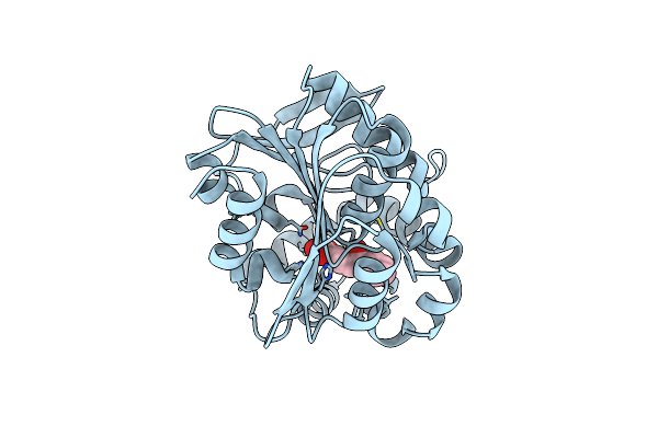
Crystal Structure Of The S105A Mutant Of A Carbon-Carbon Bond Hydrolase, Dxnb2 From Sphingomonas Wittichii Rw1, In Complex With 5,8-Dif Hopda
Organism: Sphingomonas wittichii rw1
Method: X-RAY DIFFRACTION Resolution:2.17 Å Release Date: 2013-10-09 Classification: HYDROLASE Ligands: 22J, NA |
Organism: Sphingomonas wittichii rw1
Method: X-RAY DIFFRACTION
Release Date: 2013-10-09
Ligands: 22J, NA
 |
Crystal Structure Of The S105A Mutant Of A C-C Hydrolase, Dxnb2 From Sphingomonas Wittichii Rw1, In Complex With Substrate Hopda
Organism: Sphingomonas wittichii
Method: X-RAY DIFFRACTION Resolution:2.33 Å Release Date: 2013-10-09 Classification: HYDROLASE Ligands: HPK |
Organism: Sphingomonas wittichii
Method: X-RAY DIFFRACTION
Release Date: 2013-10-09
Ligands: HPK
 |
Crystal Structure Of Dxnb2, A Carbon - Carbon Bond Hydrolase From Sphingomonas Wittichii Rw1
Organism: Sphingomonas wittichii rw1
Method: X-RAY DIFFRACTION Resolution:2.22 Å Release Date: 2013-09-18 Classification: HYDROLASE Ligands: SO4 |
Organism: Sphingomonas wittichii rw1
Method: X-RAY DIFFRACTION
Release Date: 2013-09-18
Ligands: SO4
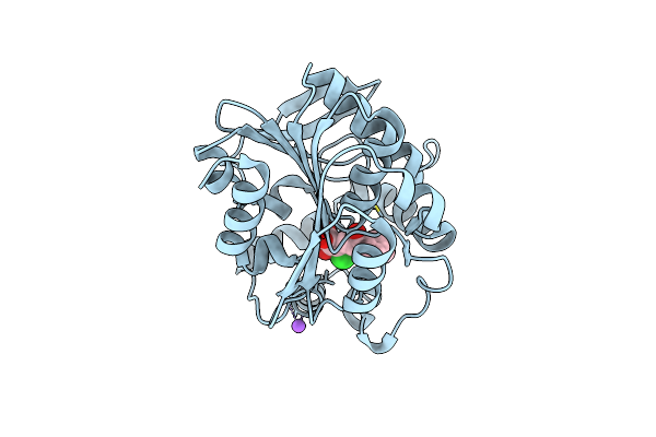 |
Crystal Structure Of The S105A Mutant Of A Carbon-Carbon Bond Hydrolase, Dxnb2 From Sphingomonas Wittichii Rw1, In Complex With 3-Cl Hopda
Organism: Sphingomonas wittichii rw1
Method: X-RAY DIFFRACTION Resolution:2.02 Å Release Date: 2013-09-18 Classification: HYDROLASE Ligands: C1E, NA |
Organism: Sphingomonas wittichii rw1
Method: X-RAY DIFFRACTION
Release Date: 2013-09-18
Ligands: C1E, NA
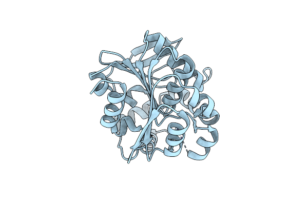 |
Crystal Structure Of The S105A Mutant Of A C-C Hydrolase, Dxnb2 From Sphingomonas Wittichii Rw1
Organism: Sphingomonas wittichii
Method: X-RAY DIFFRACTION Resolution:2.26 Å Release Date: 2013-09-18 Classification: HYDROLASE |
Organism: Sphingomonas wittichii
Method: X-RAY DIFFRACTION
Release Date: 2013-09-18
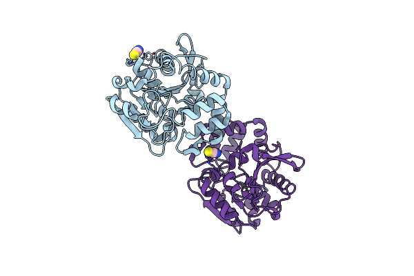 |
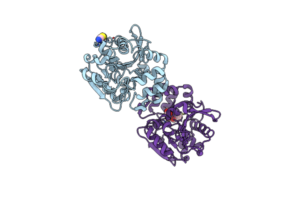
Crystal Structure Of S114A Mutant Of Hsad From Mycobacterium Tuberculosis
Organism: Mycobacterium tuberculosis
Method: X-RAY DIFFRACTION Resolution:2.10 Å Release Date: 2009-10-20 Classification: HYDROLASE Ligands: SCN |
Organism: Mycobacterium tuberculosis
Method: X-RAY DIFFRACTION
Release Date: 2009-10-20
Ligands: SCN
 |
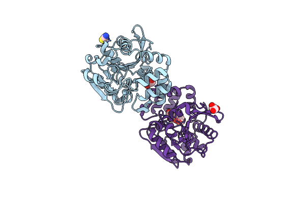
Crystal Structure Of S114A Mutant Of Hsad From Mycobacterium Tuberculosis In Complex With Hopoda
Organism: Mycobacterium tuberculosis
Method: X-RAY DIFFRACTION Resolution:1.80 Å Release Date: 2009-10-20 Classification: HYDROLASE Ligands: SCN, KEK |
Organism: Mycobacterium tuberculosis
Method: X-RAY DIFFRACTION
Release Date: 2009-10-20
Ligands: SCN, KEK
 |
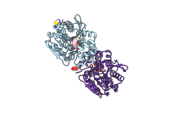
Crystal Structure Of S114A Mutant Of Hsad From Mycobacterium Tuberculosis In Complex With 4,9Dsha
Organism: Mycobacterium tuberculosis
Method: X-RAY DIFFRACTION Resolution:1.90 Å Release Date: 2009-10-20 Classification: HYDROLASE Ligands: GOL, SCN, KEM |
Organism: Mycobacterium tuberculosis
Method: X-RAY DIFFRACTION
Release Date: 2009-10-20
Ligands: GOL, SCN, KEM
 |
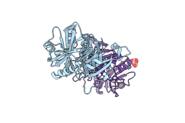
Crystal Structure Of S114A Mutant Of Hsad From Mycobacterium Tuberculosis In Complex With Hopda
Organism: Mycobacterium tuberculosis
Method: X-RAY DIFFRACTION Resolution:1.80 Å Release Date: 2009-10-20 Classification: HYDROLASE Ligands: HPK, SCN, GOL |
Organism: Mycobacterium tuberculosis
Method: X-RAY DIFFRACTION
Release Date: 2009-10-20
Ligands: HPK, SCN, GOL
 |
Crystal Structure Of The Hsac Extradiol Dioxygenase From M. Tuberculosis
Organism: Mycobacterium tuberculosis
Method: X-RAY DIFFRACTION Resolution:2.00 Å Release Date: 2009-03-10 Classification: OXIDOREDUCTASE Ligands: FE2, TAR |
Organism: Mycobacterium tuberculosis
Method: X-RAY DIFFRACTION
Release Date: 2009-03-10
Ligands: FE2, TAR
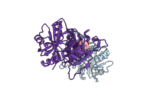 |
Crystal Structure Of The Hsac Extradiol Dioxygenase From M. Tuberculosis In Complex With 3,4-Dihydroxy-9,10-Seconandrost-1,3,5(10)-Triene-9,17-Dione (Dhsa)
Organism: Mycobacterium tuberculosis
Method: X-RAY DIFFRACTION Resolution:2.20 Å Release Date: 2009-02-24 Classification: OXIDOREDUCTASE Ligands: FE2, SDT |
Organism: Mycobacterium tuberculosis
Method: X-RAY DIFFRACTION
Release Date: 2009-02-24
Ligands: FE2, SDT