

Search Count: 14
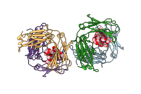 |
An Engineered Disulfide Bond Reversibly Traps The Ige-Fc3-4 In A Closed, Non-Receptor Binding Conformation
Organism: Homo sapiens
Method: X-RAY DIFFRACTION Resolution:2.61 Å Release Date: 2012-09-12 Classification: IMMUNE SYSTEM |
Organism: Homo sapiens
Method: X-RAY DIFFRACTION
Release Date: 2012-09-12
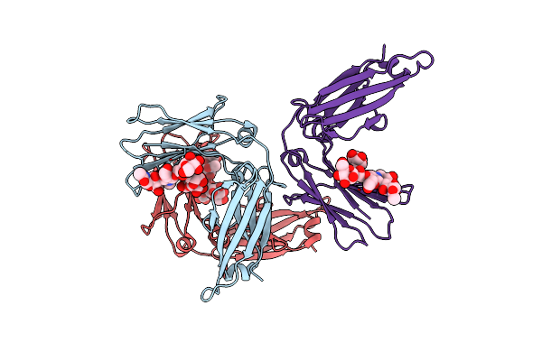 |
Crystal Structure Of The Ige-Fc3-4 Domains
Organism: Homo sapiens
Method: X-RAY DIFFRACTION Resolution:2.23 Å Release Date: 2009-09-08 Classification: IMMUNE SYSTEM Ligands: NH4 |
Organism: Homo sapiens
Method: X-RAY DIFFRACTION
Release Date: 2009-09-08
Ligands: NH4
 |
Crystal Structure Of The Ige-Fc3-4 Domains
Organism: Homo sapiens
Method: X-RAY DIFFRACTION Resolution:2.45 Å Release Date: 2009-09-08 Classification: IMMUNE SYSTEM Ligands: NH4 |
Organism: Homo sapiens
Method: X-RAY DIFFRACTION
Release Date: 2009-09-08
Ligands: NH4
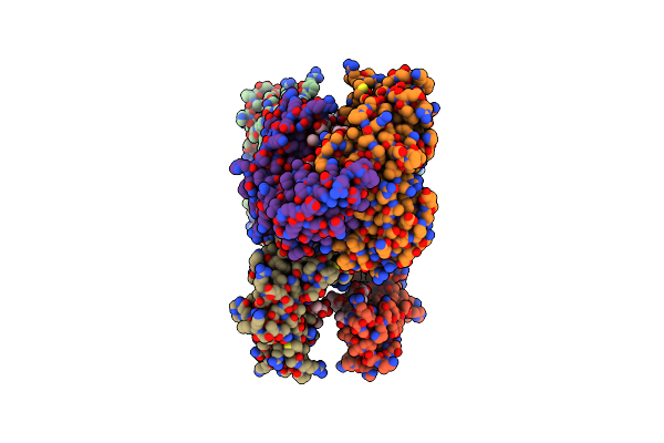 |
Crystal Structure Of The Ige-Fc3-4 Domains
Organism: Homo sapiens
Method: X-RAY DIFFRACTION Resolution:2.80 Å Release Date: 2009-09-08 Classification: IMMUNE SYSTEM |
Organism: Homo sapiens
Method: X-RAY DIFFRACTION
Release Date: 2009-09-08
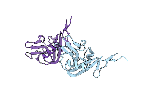 |
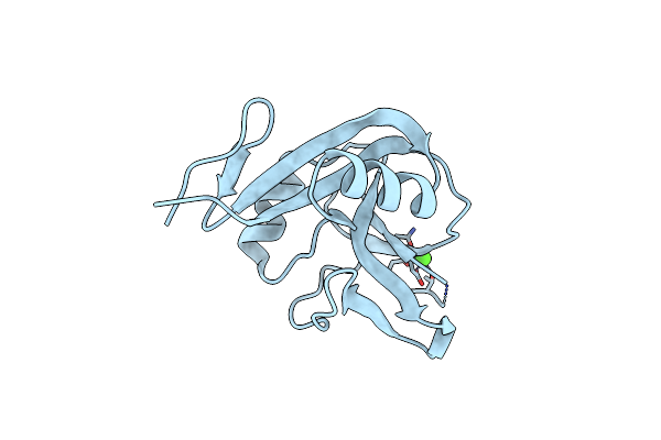
Crystal Structure Of The Human Cd23 Lectin Domain, Apo Form
Organism: Homo sapiens
Method: X-RAY DIFFRACTION Resolution:1.50 Å Release Date: 2006-06-20 Classification: IMMUNE SYSTEM |
Organism: Homo sapiens
Method: X-RAY DIFFRACTION
Release Date: 2006-06-20
 |
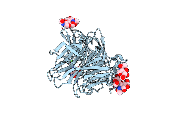
Cd23 Lectin Domain, Calcium 2+-Bound
Organism: Homo sapiens
Method: X-RAY DIFFRACTION Resolution:1.30 Å Release Date: 2006-06-20 Classification: IMMUNE SYSTEM Ligands: CA |
Organism: Homo sapiens
Method: X-RAY DIFFRACTION
Release Date: 2006-06-20
Ligands: CA
 |
Parainfluenza Virus 5 (Sv5) Hemagglutinin-Neuraminidase (Hn) With Ligand Dana (Soaked With Dana, Ph 7.0)
Organism: Simian virus 5
Method: X-RAY DIFFRACTION Resolution:2.30 Å Release Date: 2005-05-24 Classification: HYDROLASE Ligands: NAG, CA, DAN |
Organism: Simian virus 5
Method: X-RAY DIFFRACTION
Release Date: 2005-05-24
Ligands: NAG, CA, DAN
 |
Parainfluenza Virus 5 (Sv5) Hemagglutinin-Neuraminidase (Hn) With Ligand Dana (Soaked With Dana, Ph8.0)
Organism: Simian virus 5
Method: X-RAY DIFFRACTION Resolution:2.70 Å Release Date: 2005-05-24 Classification: HYDROLASE Ligands: NAG, CA, DAN |
Organism: Simian virus 5
Method: X-RAY DIFFRACTION
Release Date: 2005-05-24
Ligands: NAG, CA, DAN
 |
Parainfluenza Virus 5 (Sv5) Hemagglutinin-Neuraminidase (Hn) With Ligand Sialyllactose (Soaked With Sialyllactose, Ph8.0)
Organism: Simian virus 5
Method: X-RAY DIFFRACTION Resolution:2.50 Å Release Date: 2005-05-24 Classification: HYDROLASE Ligands: NAG, CA |
Organism: Simian virus 5
Method: X-RAY DIFFRACTION
Release Date: 2005-05-24
Ligands: NAG, CA
 |
Parainfluenza Virus 5 (Sv5) Hemagglutinin-Neuraminidase (Hn) (Ph 8.0)
Organism: Simian virus 5
Method: X-RAY DIFFRACTION Resolution:2.60 Å Release Date: 2005-05-24 Classification: HYDROLASE Ligands: NAG, CA |
Organism: Simian virus 5
Method: X-RAY DIFFRACTION
Release Date: 2005-05-24
Ligands: NAG, CA
 |
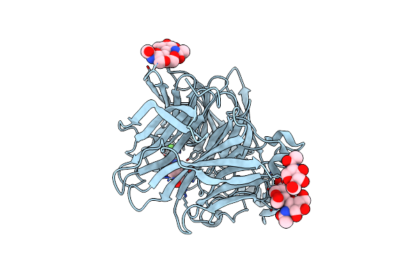
Parainfluenza Virus 5 (Sv5) Hemagglutinin-Neuraminidase (Hn) With Ligand Dana(Soaked With Sialic Acid, Ph7.0))
Organism: Simian virus 5
Method: X-RAY DIFFRACTION Resolution:2.50 Å Release Date: 2005-05-24 Classification: HYDROLASE Ligands: NAG, CA, DAN |
Organism: Simian virus 5
Method: X-RAY DIFFRACTION
Release Date: 2005-05-24
Ligands: NAG, CA, DAN
 |
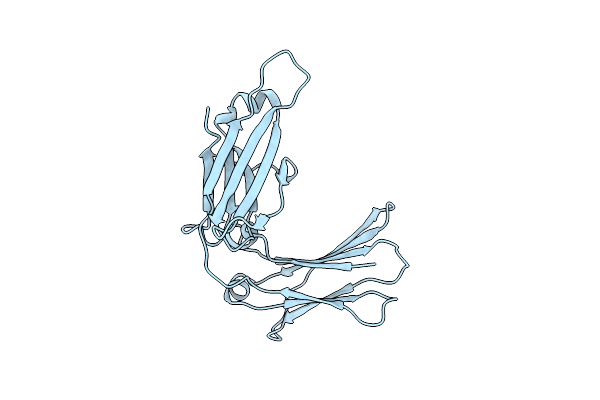
Parainfluenza Virus 5 (Sv5) Hemagglutinin-Neuraminidase (Hn) With Ligand Dana (Soaked With Sialic Acid, Ph 8.0)
Organism: Simian virus 5
Method: X-RAY DIFFRACTION Resolution:2.80 Å Release Date: 2005-05-24 Classification: HYDROLASE Ligands: NAG, CA, DAN |
Organism: Simian virus 5
Method: X-RAY DIFFRACTION
Release Date: 2005-05-24
Ligands: NAG, CA, DAN
 |
Crystal Structure Analysis Of The Human Ige-Fc Cepsilon3-Cepsilon4 Fragment.
Organism: Homo sapiens
Method: X-RAY DIFFRACTION Resolution:2.30 Å Release Date: 2000-09-27 Classification: IMMUNE SYSTEM |
Organism: Homo sapiens
Method: X-RAY DIFFRACTION
Release Date: 2000-09-27
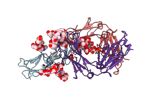 |
Structure Of The Human Ige-Fc Bound To Its High Affinity Receptor Fc(Epsilon)Ri(Alpha)
Organism: Homo sapiens
Method: X-RAY DIFFRACTION Resolution:3.50 Å Release Date: 2000-07-20 Classification: IMMUNE SYSTEM Ligands: SO4, CPS |
Organism: Homo sapiens
Method: X-RAY DIFFRACTION
Release Date: 2000-07-20
Ligands: SO4, CPS