

Search Count: 33
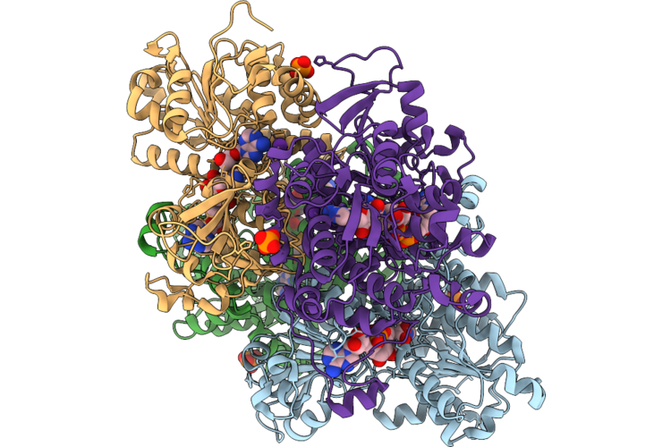 |
Crystal Structure Of S-Adenosyl-L-Homocysteine Hydrolase From P. Aeruginosa: F324A Mutant Complexed With Adenosine And Rubidium Ions Replacing Native Potassium
Organism: Pseudomonas aeruginosa pao1
Method: X-RAY DIFFRACTION Resolution:2.06 Å Release Date: 2026-07-08 Classification: HYDROLASE Ligands: NAD, ADN, PO4, RB |
Organism: Pseudomonas aeruginosa pao1
Method: X-RAY DIFFRACTION
Release Date: 2026-07-08
Ligands: NAD, ADN, PO4, RB
 |
Crystal Structure Of The S-Adenosyl-L-Homocysteine Hydrolase From P. Aeruginosa, Q65N Mutant Probed With Rubidium To Confirm Disruption Of A Potassium Binding Site.
Organism: Pseudomonas aeruginosa pao1
Method: X-RAY DIFFRACTION Resolution:1.91 Å Release Date: 2026-04-08 Classification: HYDROLASE Ligands: NAD, GOL, PO4 |
Organism: Pseudomonas aeruginosa pao1
Method: X-RAY DIFFRACTION
Release Date: 2026-04-08
Ligands: NAD, GOL, PO4
 |
Crystal Structure Of S-Adenosyl-L-Homocysteine Hydrolase From P. Aeruginosa: Q65A Mutant Soaked With Adenosine And Probed With Rubidium To Confirm Disruption Of A Potassium Binding Site.
Organism: Pseudomonas aeruginosa pao1
Method: X-RAY DIFFRACTION Resolution:2.40 Å Release Date: 2026-04-08 Classification: HYDROLASE Ligands: NAD, ADN, PO4 |
Organism: Pseudomonas aeruginosa pao1
Method: X-RAY DIFFRACTION
Release Date: 2026-04-08
Ligands: NAD, ADN, PO4
 |
Crystal Structure Of S-Adenosyl-L-Homocysteine Hydrolase From P. Aeruginosa, Q65N Mutant Soaked With Adenosine And Probed With Rubidium To Confirm Disruption Of A Potassium Binding Site.
Organism: Pseudomonas aeruginosa pao1
Method: X-RAY DIFFRACTION Resolution:2.34 Å Release Date: 2026-04-08 Classification: HYDROLASE Ligands: NAD, ADN, PO4 |
Organism: Pseudomonas aeruginosa pao1
Method: X-RAY DIFFRACTION
Release Date: 2026-04-08
Ligands: NAD, ADN, PO4
 |
Cryo-Em Structure Of Pseudomonas Aeruginosa Tetrameric S-Adenosyl-L-Homocysteine Hydrolase In The Fully Open State
Organism: Pseudomonas aeruginosa pao1
Method: ELECTRON MICROSCOPY Release Date: 2025-02-19 Classification: HYDROLASE Ligands: NAD |
Organism: Pseudomonas aeruginosa pao1
Method: ELECTRON MICROSCOPY
Release Date: 2025-02-19
Ligands: NAD
 |
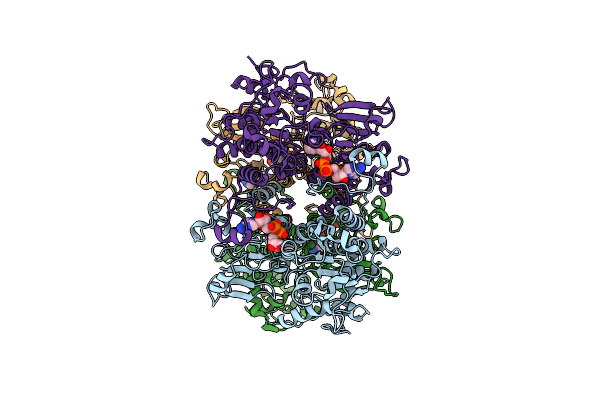
Cryo-Em Structure Of Pseudomonas Aeruginosa Tetrameric S-Adenosyl-L-Homocysteine Hydrolase With 3 Open And 1 Closed Subunits
Organism: Pseudomonas aeruginosa pao1
Method: ELECTRON MICROSCOPY Release Date: 2025-02-19 Classification: HYDROLASE Ligands: NAD, ADN, K |
Organism: Pseudomonas aeruginosa pao1
Method: ELECTRON MICROSCOPY
Release Date: 2025-02-19
Ligands: NAD, ADN, K
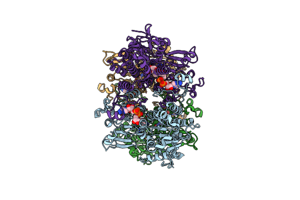 |
Cryo-Em Structure Of Pseudomonas Aeruginosa Tetrameric S-Adenosyl-L-Homocysteine Hydrolase With 1 Open And 3 Closed Subunits
Organism: Pseudomonas aeruginosa pao1
Method: ELECTRON MICROSCOPY Release Date: 2025-02-19 Classification: HYDROLASE Ligands: NAD, ADN, K |
Organism: Pseudomonas aeruginosa pao1
Method: ELECTRON MICROSCOPY
Release Date: 2025-02-19
Ligands: NAD, ADN, K
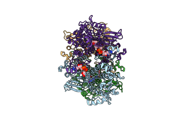 |
Cryo-Em Structure Of Pseudomonas Aeruginosa Tetrameric S-Adenosyl-L-Homocysteine Hydrolase With 2 Open And 2 Closed Subunits
Organism: Pseudomonas aeruginosa pao1
Method: ELECTRON MICROSCOPY Release Date: 2025-02-19 Classification: HYDROLASE Ligands: NAD, ADN, K |
Organism: Pseudomonas aeruginosa pao1
Method: ELECTRON MICROSCOPY
Release Date: 2025-02-19
Ligands: NAD, ADN, K
 |
Cryo-Em Structure Of Pseudomonas Aeruginosa Tetrameric S-Adenosyl-L-Homocysteine Hydrolase With 2 Wide Open, 1 Open And 1 Closed Subunits
Organism: Pseudomonas aeruginosa
Method: ELECTRON MICROSCOPY Release Date: 2025-02-19 Classification: HYDROLASE Ligands: NAD, ADN |
Organism: Pseudomonas aeruginosa
Method: ELECTRON MICROSCOPY
Release Date: 2025-02-19
Ligands: NAD, ADN
 |
Crystal Structure Of K449E Variant Of S-Adenosyl-L-Homocysteine Hydrolase From Pseudomonas Aeruginosa In Complex With Adenosine
Organism: Pseudomonas aeruginosa pao1
Method: X-RAY DIFFRACTION Resolution:1.41 Å Release Date: 2023-09-13 Classification: HYDROLASE Ligands: NAD, ADN, K, PO4 |
Organism: Pseudomonas aeruginosa pao1
Method: X-RAY DIFFRACTION
Release Date: 2023-09-13
Ligands: NAD, ADN, K, PO4
 |
Crystal Structure Of The F324A Mutant Of S-Adenosyl-L-Homocysteine Hydrolase From Pseudomonas Aeruginosa Cocrystallized With Adenosine In The Presence Of K+ Cations
Organism: Pseudomonas aeruginosa pao1
Method: X-RAY DIFFRACTION Resolution:1.68 Å Release Date: 2023-08-16 Classification: HYDROLASE Ligands: NAD, ADN, K, CL, PEG |
Organism: Pseudomonas aeruginosa pao1
Method: X-RAY DIFFRACTION
Release Date: 2023-08-16
Ligands: NAD, ADN, K, CL, PEG
 |
Crystal Structure Of The H323A Mutant Of S-Adenosyl-L-Homocysteine Hydrolase From Pseudomonas Aeruginosa Cocrystallized With Adenosine In The Presence Of K+ Cations
Organism: Pseudomonas aeruginosa pao1
Method: X-RAY DIFFRACTION Resolution:1.50 Å Release Date: 2023-08-16 Classification: HYDROLASE Ligands: ADN, NAD, TRS, PEG, K, PO4, CL |
Organism: Pseudomonas aeruginosa pao1
Method: X-RAY DIFFRACTION
Release Date: 2023-08-16
Ligands: ADN, NAD, TRS, PEG, K, PO4, CL
 |
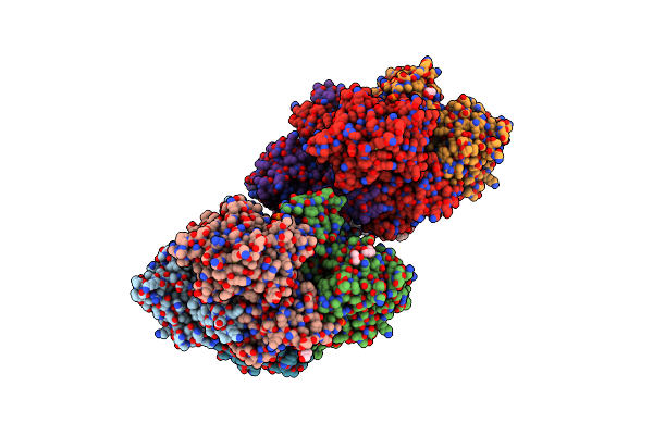
Crystal Structure Of The Q65A Mutant Of S-Adenosyl-L-Homocysteine Hydrolase From Pseudomonas Aeruginosa Cocrystallized With Adenosine In The Presence Of K+ Cations
Organism: Pseudomonas aeruginosa pao1
Method: X-RAY DIFFRACTION Resolution:1.65 Å Release Date: 2023-08-16 Classification: HYDROLASE Ligands: PO4, NAD, ADN, GOL |
Organism: Pseudomonas aeruginosa pao1
Method: X-RAY DIFFRACTION
Release Date: 2023-08-16
Ligands: PO4, NAD, ADN, GOL
 |
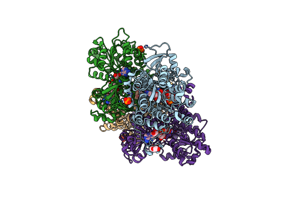
Crystal Structure Of The Q65N Mutant Of S-Adenosyl-L-Homocysteine Hydrolase From Pseudomonas Aeruginosa Crystallized In The Presence Of K+ Cations
Organism: Pseudomonas aeruginosa pao1
Method: X-RAY DIFFRACTION Resolution:1.90 Å Release Date: 2023-08-16 Classification: HYDROLASE Ligands: NAD, GOL, PO4, K, PEG |
Organism: Pseudomonas aeruginosa pao1
Method: X-RAY DIFFRACTION
Release Date: 2023-08-16
Ligands: NAD, GOL, PO4, K, PEG
 |
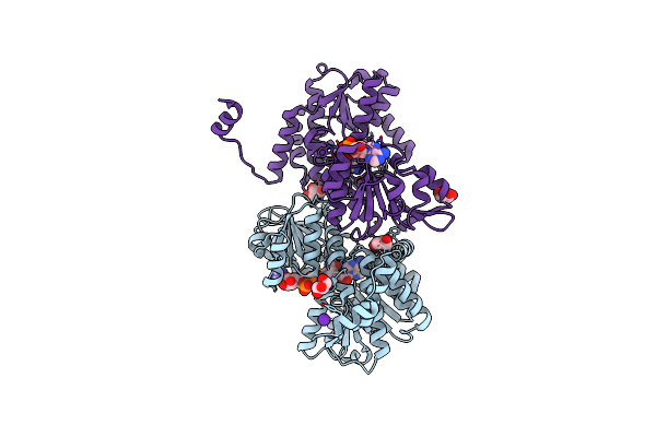
Crystal Structure Of The Q65N Mutant Of S-Adenosyl-L-Homocysteine Hydrolase From Pseudomonas Aeruginosa Cocrystallized With Adenosine In The Presence Of K+ Cations
Organism: Pseudomonas aeruginosa pao1
Method: X-RAY DIFFRACTION Resolution:1.82 Å Release Date: 2023-08-16 Classification: HYDROLASE Ligands: NAD, ADN, PEG, PO4 |
Organism: Pseudomonas aeruginosa pao1
Method: X-RAY DIFFRACTION
Release Date: 2023-08-16
Ligands: NAD, ADN, PEG, PO4
 |
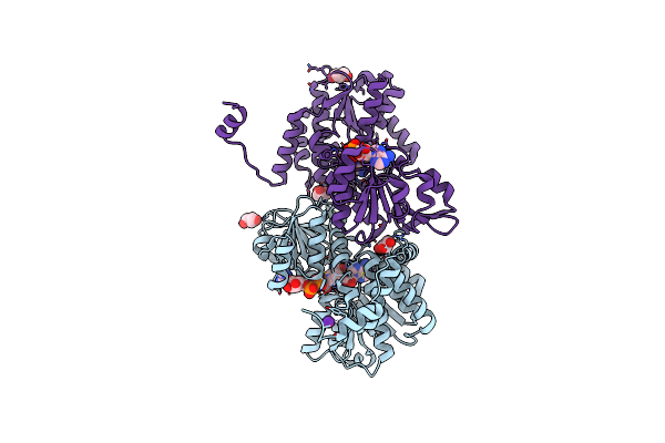
Crystal Structure Of The R24E/E352T Double Mutant Of S-Adenosyl-L-Homocysteine Hydrolase From Synechocystis Sp. Pcc 6803 Cocrystallized With Adenosine In The Presence Of Rb+ Cations
Organism: Synechocystis sp. pcc 6803
Method: X-RAY DIFFRACTION Resolution:1.90 Å Release Date: 2022-07-13 Classification: HYDROLASE Ligands: GOL, NAD, ADN, RB, CL |
Organism: Synechocystis sp. pcc 6803
Method: X-RAY DIFFRACTION
Release Date: 2022-07-13
Ligands: GOL, NAD, ADN, RB, CL
 |
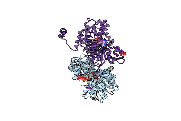
Crystal Structure Of The R24E Mutant Of S-Adenosyl-L-Homocysteine Hydrolase From Synechocystis Sp. Pcc 6803 Cocrystallized With Adenosine In The Presence Of Rb+ Cations
Organism: Synechocystis sp. pcc 6803
Method: X-RAY DIFFRACTION Resolution:2.03 Å Release Date: 2022-07-13 Classification: HYDROLASE Ligands: GOL, NAD, ADN, CL, RB |
Organism: Synechocystis sp. pcc 6803
Method: X-RAY DIFFRACTION
Release Date: 2022-07-13
Ligands: GOL, NAD, ADN, CL, RB
 |
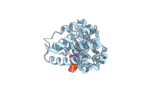
Crystal Structure Of The E352T Mutant Of S-Adenosyl-L-Homocysteine Hydrolase From Synechocystis Sp. Pcc 6803 Cocrystallized With Adenosine In The Presence Of Rb+ Cations
Organism: Synechocystis sp. pcc 6803
Method: X-RAY DIFFRACTION Resolution:1.89 Å Release Date: 2022-07-13 Classification: HYDROLASE Ligands: NAD, ADN, CL, RB, GOL |
Organism: Synechocystis sp. pcc 6803
Method: X-RAY DIFFRACTION
Release Date: 2022-07-13
Ligands: NAD, ADN, CL, RB, GOL
 |
Structure Of Bacillus Subtilis Ccrz (Previously Called Ytmp)
Organism: Bacillus subtilis py79
Method: X-RAY DIFFRACTION Resolution:2.60 Å Release Date: 2022-05-11 Classification: CELL CYCLE Ligands: ANP |
Organism: Bacillus subtilis py79
Method: X-RAY DIFFRACTION
Release Date: 2022-05-11
Ligands: ANP
 |
Crystal Structure Of S-Adenosyl-L-Homocysteine Hydrolase From Synechocystis Sp. Pcc 6803 Cocrystallized With Adenosine In The Presence Of Rb+ Cations
Organism: Synechocystis sp. (strain pcc 6803 / kazusa)
Method: X-RAY DIFFRACTION Resolution:1.74 Å Release Date: 2022-04-20 Classification: HYDROLASE Ligands: NAD, RB, ADN, CL |
Organism: Synechocystis sp. (strain pcc 6803 / kazusa)
Method: X-RAY DIFFRACTION
Release Date: 2022-04-20
Ligands: NAD, RB, ADN, CL