

Search Count: 32
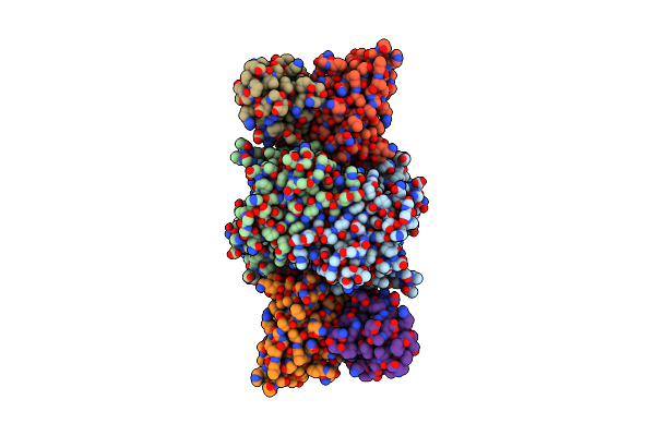 |
16E10 Fab Bound To Norovirus Gi.1 P Domain
Organism: Norovirus, Homo sapiens
Method: ELECTRON MICROSCOPY Release Date: 2025-04-09 Classification: VIRAL PROTEIN/IMMUNE SYSTEM |
Organism: Norovirus, Homo sapiens
Method: ELECTRON MICROSCOPY
Release Date: 2025-04-09
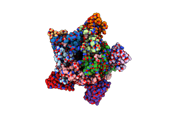 |
X-Ray Structure Of A Pentameric Ligand Gated Ion Channel From Erwinia Chrysanthemi (Elic) 7'C Pore Mutant (L238C) In Complex With Nanobody 72
Organism: Dickeya chrysanthemi, Lama glama
Method: X-RAY DIFFRACTION Resolution:2.50 Å Release Date: 2019-10-09 Classification: MEMBRANE PROTEIN Ligands: P6G, PTY, LMT, MES, NA |
Organism: Dickeya chrysanthemi, Lama glama
Method: X-RAY DIFFRACTION
Release Date: 2019-10-09
Ligands: P6G, PTY, LMT, MES, NA
 |
X-Ray Structure Of A Pentameric Ligand Gated Ion Channel From Erwinia Chrysanthemi (Elic) Delta8 Truncation Mutant In Complex With Nanobody 72
Organism: Dickeya chrysanthemi, Lama glama
Method: X-RAY DIFFRACTION Resolution:2.78 Å Release Date: 2019-10-09 Classification: MEMBRANE PROTEIN |
Organism: Dickeya chrysanthemi, Lama glama
Method: X-RAY DIFFRACTION
Release Date: 2019-10-09
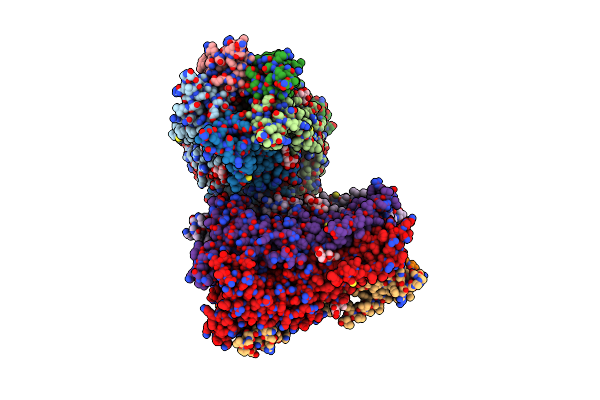 |
X-Ray Structure Of A Pentameric Ligand Gated Ion Channel From Erwinia Chrysanthemi (Elic) F16'S Pore Mutant (F247S) With Alternate M4 Conformation.
Organism: Dickeya chrysanthemi
Method: X-RAY DIFFRACTION Resolution:3.45 Å Release Date: 2019-10-09 Classification: MEMBRANE PROTEIN Ligands: LMT |
Organism: Dickeya chrysanthemi
Method: X-RAY DIFFRACTION
Release Date: 2019-10-09
Ligands: LMT
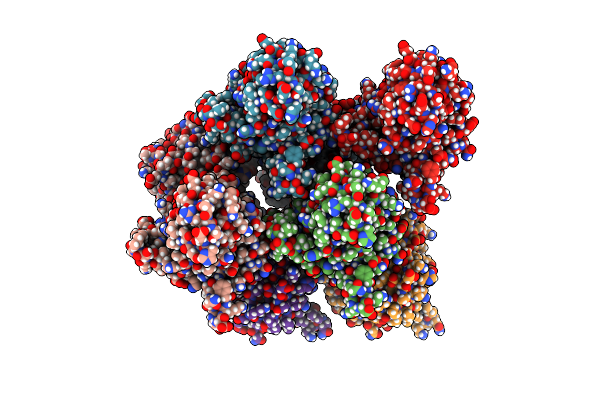 |
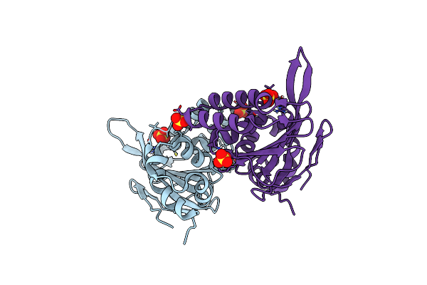
Crystal Structure Of Chalcone Isomerase From Medicago Truncatula Complexed With (2S) Naringenin
Organism: Medicago truncatula
Method: X-RAY DIFFRACTION Resolution:1.90 Å Release Date: 2019-08-21 Classification: plant protein, isomerase Ligands: NAR |
Organism: Medicago truncatula
Method: X-RAY DIFFRACTION
Release Date: 2019-08-21
Ligands: NAR
 |
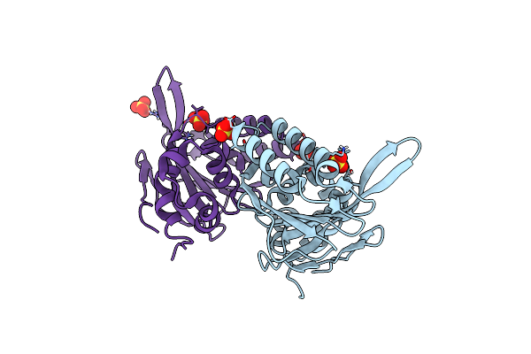
Crystal Structure Of Chalcone Isomerase From Medicago Sativa With The G95T Mutation
Organism: Medicago sativa
Method: X-RAY DIFFRACTION Resolution:2.40 Å Release Date: 2019-03-13 Classification: plant protein, isomerase Ligands: SO4 |
Organism: Medicago sativa
Method: X-RAY DIFFRACTION
Release Date: 2019-03-13
Ligands: SO4
 |
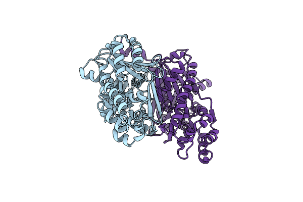
Crystal Structure Of Chalcone Isomerase From Medicago Sativa With The G95S Mutation.
Organism: Medicago sativa
Method: X-RAY DIFFRACTION Resolution:2.40 Å Release Date: 2019-03-13 Classification: plant protein, isomerase Ligands: SO4 |
Organism: Medicago sativa
Method: X-RAY DIFFRACTION
Release Date: 2019-03-13
Ligands: SO4
 |
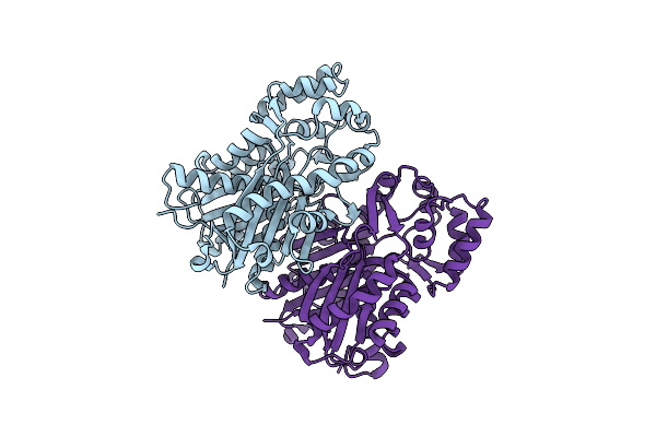
Chalcone Synthase From Malus Domestica
Organism: Malus domestica
Method: X-RAY DIFFRACTION Resolution:2.10 Å Release Date: 2017-12-13 Classification: TRANSFERASE |
Organism: Malus domestica
Method: X-RAY DIFFRACTION
Release Date: 2017-12-13
 |
Benzophenone Synthase From Hypericum Androsaemum
Organism: Hypericum androsaemum
Method: X-RAY DIFFRACTION Resolution:2.85 Å Release Date: 2017-12-13 Classification: TRANSFERASE |
Organism: Hypericum androsaemum
Method: X-RAY DIFFRACTION
Release Date: 2017-12-13
 |
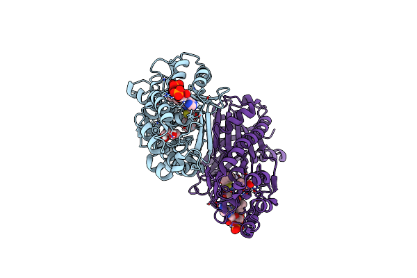
Crystal Structure Of Biphenyl Synthase From Malus Domestica
Organism: Malus domestica
Method: X-RAY DIFFRACTION Resolution:1.17 Å Release Date: 2017-12-13 Classification: TRANSFERASE Ligands: BU4 |
Organism: Malus domestica
Method: X-RAY DIFFRACTION
Release Date: 2017-12-13
Ligands: BU4
 |
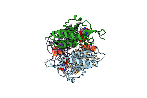
Crystal Structure Of Biphenyl Synthase From Malus Domestic Complexed With Benzoyl-Coa
Organism: Malus domestica
Method: X-RAY DIFFRACTION Resolution:1.20 Å Release Date: 2017-12-13 Classification: TRANSFERASE Ligands: BU4, BYC |
Organism: Malus domestica
Method: X-RAY DIFFRACTION
Release Date: 2017-12-13
Ligands: BU4, BYC
 |
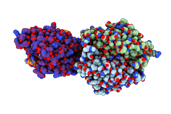
Crystal Structures Of Mycobacterium Tuberculosis 6-Oxopurine Phosphoribosyltransferase Which Is A Potential Target For Drug Development Against This Disease
Organism: Mycobacterium tuberculosis
Method: X-RAY DIFFRACTION Resolution:2.76 Å Release Date: 2015-05-20 Classification: TRANSFERASE Ligands: POP, 5GP, MG |
Organism: Mycobacterium tuberculosis
Method: X-RAY DIFFRACTION
Release Date: 2015-05-20
Ligands: POP, 5GP, MG
 |
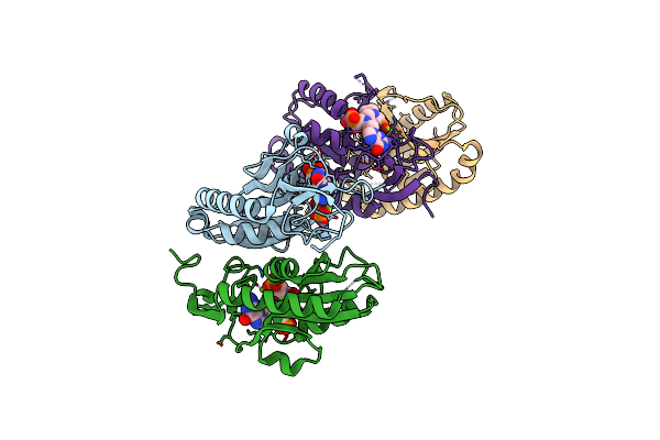
Crystal Structures Of Mycobacterium Tuberculosis 6-Oxopurine Phosphoribosyltransferase Which Is A Potential Target For Drug Development Against This Disease
Organism: Mycobacterium tuberculosis
Method: X-RAY DIFFRACTION Resolution:2.57 Å Release Date: 2015-05-20 Classification: transferase/transferase inhibitor Ligands: 3QE, MG, 45T |
Organism: Mycobacterium tuberculosis
Method: X-RAY DIFFRACTION
Release Date: 2015-05-20
Ligands: 3QE, MG, 45T
 |
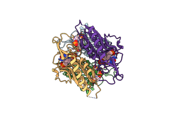
Structures Of Mycobacterium Tuberculosis 6-Oxopurine Phosphoribosyltransferase Which Is A Potential Target For Drug Development Against This Disease
Organism: Mycobacterium tuberculosis
Method: X-RAY DIFFRACTION Resolution:2.03 Å Release Date: 2015-05-20 Classification: transferase/transferase inhibitor Ligands: 3QF, MG |
Organism: Mycobacterium tuberculosis
Method: X-RAY DIFFRACTION
Release Date: 2015-05-20
Ligands: 3QF, MG
 |
Crystal Structures Of Mycobacterium Tuberculosis 6-Oxopurine Phosphoribosyltransferase Which Is A Potential Target For Drug Development Against This Disease
Organism: Mycobacterium tuberculosis
Method: X-RAY DIFFRACTION Resolution:2.32 Å Release Date: 2015-05-20 Classification: transferase/transferase inhibitor Ligands: MG, 3QG |
Organism: Mycobacterium tuberculosis
Method: X-RAY DIFFRACTION
Release Date: 2015-05-20
Ligands: MG, 3QG
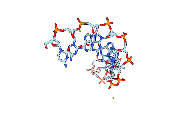 |
Crystal Structure Of One Turn Of G/C Rich B-Dna Revisited
Method: X-RAY DIFFRACTION
Resolution:0.96 Å Release Date: 2011-11-09 Classification: DNA Ligands: MG |
Method: X-RAY DIFFRACTION
Release Date: 2011-11-09
Ligands: MG
 |
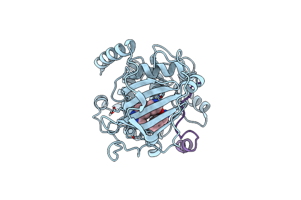
Parp Complexed With A968427
Organism: Homo sapiens
Method: X-RAY DIFFRACTION Resolution:2.30 Å Release Date: 2010-03-09 Classification: TRANSFERASE Ligands: GJW |
Organism: Homo sapiens
Method: X-RAY DIFFRACTION
Release Date: 2010-03-09
Ligands: GJW
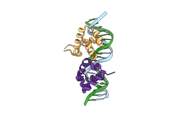 |
Crystal Structure Of The P22 C2 Repressor Protein In Complex With The Synthetic Operator 9T
Organism: Enterobacteria phage p22
Method: X-RAY DIFFRACTION Resolution:1.53 Å Release Date: 2008-04-29 Classification: TRANSCRIPTION/DNA |
Organism: Enterobacteria phage p22
Method: X-RAY DIFFRACTION
Release Date: 2008-04-29
 |
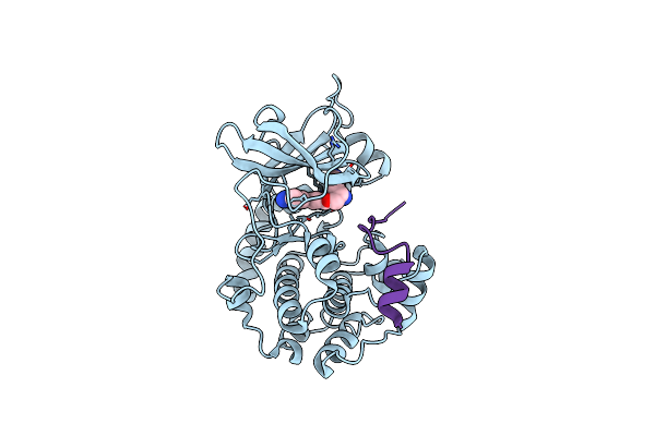
Pka Structures Of Akt, Indazole-Pyridine Inhibitors
Organism: Bos taurus
Method: X-RAY DIFFRACTION Resolution:2.10 Å Release Date: 2007-06-05 Classification: TRANSFERASE Ligands: SS3 |
Organism: Bos taurus
Method: X-RAY DIFFRACTION
Release Date: 2007-06-05
Ligands: SS3
 |
Pka Structures Of Indazole-Pyridine Series Of Akt Inhibitors
Organism: Bos taurus
Method: X-RAY DIFFRACTION Resolution:2.40 Å Release Date: 2007-06-05 Classification: TRANSFERASE Ligands: L20 |
Organism: Bos taurus
Method: X-RAY DIFFRACTION
Release Date: 2007-06-05
Ligands: L20