

Search Count: 15
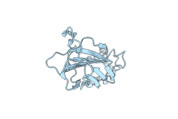 |
The Structure Of Vaccinia Virus A41
Organism: Vaccinia virus
Method: X-RAY DIFFRACTION Resolution:1.90 Å Release Date: 2008-02-26 Classification: VIRAL PROTEIN |
Organism: Vaccinia virus
Method: X-RAY DIFFRACTION
Release Date: 2008-02-26
 |
Crystal Structure Of 3-Keto-L-Gulonate 6-Phosphate Decarboxylase With Bound D-Ribulose 5-Phosphate
Organism: Escherichia coli
Method: X-RAY DIFFRACTION Resolution:1.66 Å Release Date: 2005-04-26 Classification: UNKNOWN FUNCTION Ligands: 5RP, MG |
Organism: Escherichia coli
Method: X-RAY DIFFRACTION
Release Date: 2005-04-26
Ligands: 5RP, MG
 |
Structure Of 3-Keto-L-Gulonate 6-Phosphate Decarboxylase E112D/R139V/T169A Mutant With Bound D-Ribulose 5-Phosphate
Organism: Escherichia coli
Method: X-RAY DIFFRACTION Resolution:1.81 Å Release Date: 2005-04-26 Classification: UNKNOWN FUNCTION Ligands: HMS, MG, 5RP |
Organism: Escherichia coli
Method: X-RAY DIFFRACTION
Release Date: 2005-04-26
Ligands: HMS, MG, 5RP
 |
Structure Of 3-Keto-L-Gulonate 6-Phosphate Decarboxylase E112D/T169A Mutant With Bound D-Ribulose 5-Phosphate
Organism: Escherichia coli
Method: X-RAY DIFFRACTION Resolution:1.58 Å Release Date: 2005-04-26 Classification: UNKNOWN FUNCTION Ligands: 5RP, MG |
Organism: Escherichia coli
Method: X-RAY DIFFRACTION
Release Date: 2005-04-26
Ligands: 5RP, MG
 |
Crystal Structure Of 3-Keto-L-Gulonate 6-Phosphate Decarboxylase E112D/R139V/T169A Mutant With Bound L-Xylulose 5-Phosphate
Organism: Escherichia coli
Method: X-RAY DIFFRACTION Resolution:1.80 Å Release Date: 2005-04-26 Classification: UNKNOWN FUNCTION Ligands: MG, LX1 |
Organism: Escherichia coli
Method: X-RAY DIFFRACTION
Release Date: 2005-04-26
Ligands: MG, LX1
 |
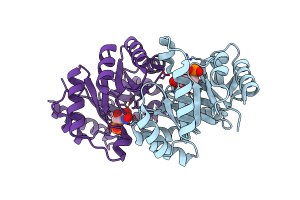
Crystal Structure Of D-Ribulose 5-Phosphate 3-Epimerase From Synechocystis To 1.6 Angstrom Resolution
Organism: Synechocystis sp.
Method: X-RAY DIFFRACTION Resolution:1.60 Å Release Date: 2004-08-31 Classification: ISOMERASE |
Organism: Synechocystis sp.
Method: X-RAY DIFFRACTION
Release Date: 2004-08-31
 |
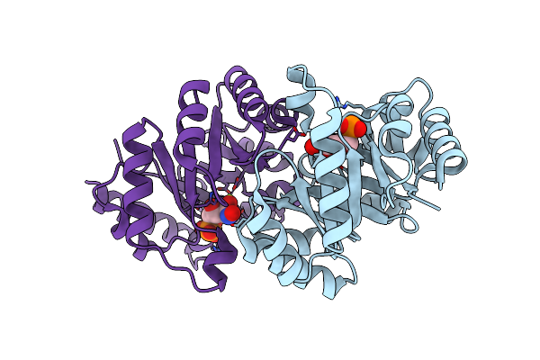
Crystal Structure Of H136A Mutant Of 3-Keto-L-Gulonate 6-Phosphate Decarboxylase With Bound L-Threonohydroxamate 4-Phosphate
Organism: Escherichia coli
Method: X-RAY DIFFRACTION Resolution:1.90 Å Release Date: 2004-06-08 Classification: LYASE Ligands: MG, TX4 |
Organism: Escherichia coli
Method: X-RAY DIFFRACTION
Release Date: 2004-06-08
Ligands: MG, TX4
 |
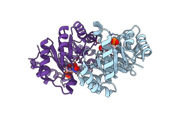
Crystal Structure Of K64A Mutant Of 3-Keto-L-Gulonate 6-Phosphate Decarboxylase With Bound L-Threonohydroxamate 4-Phosphate
Organism: Escherichia coli
Method: X-RAY DIFFRACTION Resolution:1.70 Å Release Date: 2004-06-08 Classification: LYASE Ligands: MG, TX4 |
Organism: Escherichia coli
Method: X-RAY DIFFRACTION
Release Date: 2004-06-08
Ligands: MG, TX4
 |
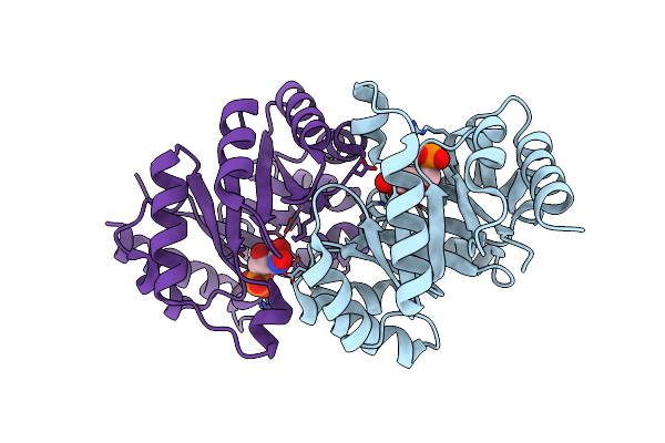
Crystal Structure Of E112Q Mutant Of 3-Keto-L-Gulonate 6-Phosphate Decarboxylase With Bound L-Threonohydroxamate 4-Phosphate
Organism: Escherichia coli
Method: X-RAY DIFFRACTION Resolution:1.80 Å Release Date: 2004-06-08 Classification: LYASE Ligands: MG, TX4 |
Organism: Escherichia coli
Method: X-RAY DIFFRACTION
Release Date: 2004-06-08
Ligands: MG, TX4
 |
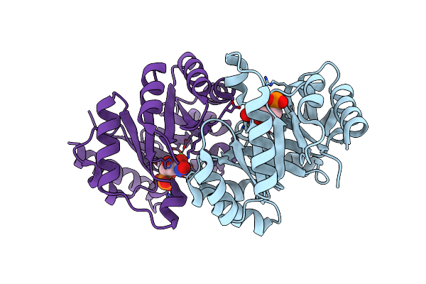
Crystal Structure Of E112Q/H136A Double Mutant Of 3-Keto-L-Gulonate 6-Phosphate Decarboxylase With Bound L-Threonohydroxamate 4-Phosphate
Organism: Escherichia coli
Method: X-RAY DIFFRACTION Resolution:1.90 Å Release Date: 2004-06-08 Classification: LYASE Ligands: MG, TX4 |
Organism: Escherichia coli
Method: X-RAY DIFFRACTION
Release Date: 2004-06-08
Ligands: MG, TX4
 |
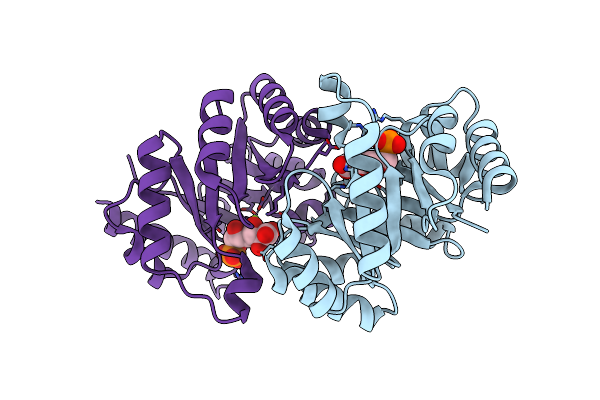
Crystal Structure Of Methanococcus Jannaschii Phosphosulfolactate Synthase
Organism: Methanocaldococcus jannaschii
Method: X-RAY DIFFRACTION Resolution:1.60 Å Release Date: 2003-12-09 Classification: LYASE Ligands: SO4 |
Organism: Methanocaldococcus jannaschii
Method: X-RAY DIFFRACTION
Release Date: 2003-12-09
Ligands: SO4
 |
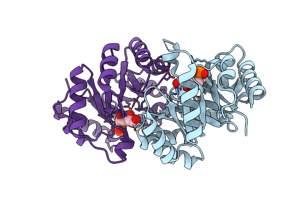
Structure Of 3-Keto-L-Gulonate 6-Phosphate Decarboxylase With Bound L-Threonohydroxamate 4-Phosphate
Organism: Escherichia coli
Method: X-RAY DIFFRACTION Resolution:1.80 Å Release Date: 2003-10-28 Classification: LYASE Ligands: MG, TX4 |
Organism: Escherichia coli
Method: X-RAY DIFFRACTION
Release Date: 2003-10-28
Ligands: MG, TX4
 |
Structure Of 3-Keto-L-Gulonate 6-Phosphate Decarboxylase With Bound L-Gulonaet 6-Phosphate
Organism: Escherichia coli
Method: X-RAY DIFFRACTION Resolution:1.20 Å Release Date: 2003-10-28 Classification: LYASE Ligands: MG, LG6 |
Organism: Escherichia coli
Method: X-RAY DIFFRACTION
Release Date: 2003-10-28
Ligands: MG, LG6
 |
Structure Of 3-Keto-L-Gulonate 6-Phosphate Decarboxylase With Bound Xylitol 5-Phosphate
Organism: Escherichia coli
Method: X-RAY DIFFRACTION Resolution:1.70 Å Release Date: 2003-10-28 Classification: LYASE Ligands: MG, LXP |
Organism: Escherichia coli
Method: X-RAY DIFFRACTION
Release Date: 2003-10-28
Ligands: MG, LXP
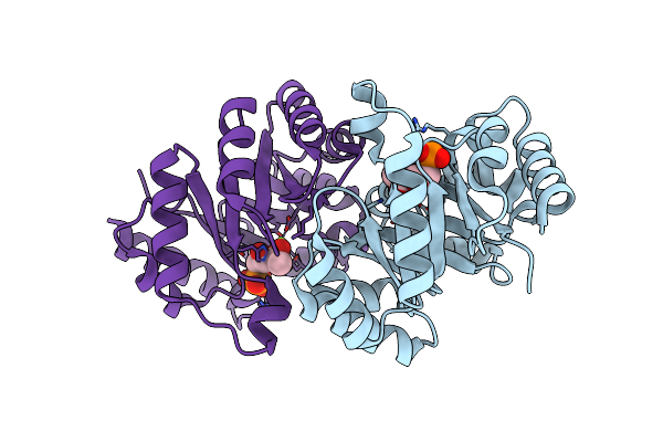 |
Structure Of 3-Keto-L-Gulonate 6-Phosphate Decarboxylase With Bound L-Xylulose 5-Phosphate
Organism: Escherichia coli
Method: X-RAY DIFFRACTION Resolution:1.76 Å Release Date: 2003-10-28 Classification: LYASE Ligands: MG, LX1 |
Organism: Escherichia coli
Method: X-RAY DIFFRACTION
Release Date: 2003-10-28
Ligands: MG, LX1