

Search Count: 119
 |
Crystal Structure Of Sh3-Like_Bac-Type Domain (79-145) Of Conserved Domain Protein Gbaa_2967 From Bacillus Anthracis Ames Ancestor
Organism: Bacillus anthracis str. ames
Method: X-RAY DIFFRACTION Resolution:1.50 Å Release Date: 2025-06-11 Classification: UNKNOWN FUNCTION Ligands: MG, FMT |
Organism: Bacillus anthracis str. ames
Method: X-RAY DIFFRACTION
Release Date: 2025-06-11
Ligands: MG, FMT
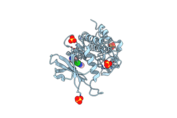 |
Crystal Structure Of Human Dapk1 Catalytic Subunit Complexed With Compound Mw01-9-039Srm.
Organism: Homo sapiens
Method: X-RAY DIFFRACTION Resolution:1.90 Å Release Date: 2024-12-25 Classification: TRANSFERASE Ligands: A1BLJ, SO4 |
Organism: Homo sapiens
Method: X-RAY DIFFRACTION
Release Date: 2024-12-25
Ligands: A1BLJ, SO4
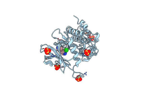 |
Crystal Structure Of Human Dapk1 Catalytic Subunit Complexed With Compound Srm-25-071
Organism: Homo sapiens
Method: X-RAY DIFFRACTION Resolution:1.50 Å Release Date: 2024-10-09 Classification: TRANSFERASE Ligands: A1BB7, CL, SO4 |
Organism: Homo sapiens
Method: X-RAY DIFFRACTION
Release Date: 2024-10-09
Ligands: A1BB7, CL, SO4
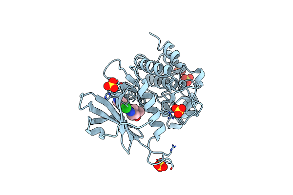 |
Crystal Structure Of Human Dapk1 Catalytic Subunit Complexed With Compound Srm-07-081A
Organism: Homo sapiens
Method: X-RAY DIFFRACTION Resolution:1.65 Å Release Date: 2024-10-09 Classification: TRANSFERASE Ligands: A1BB6, BO3, SO4 |
Organism: Homo sapiens
Method: X-RAY DIFFRACTION
Release Date: 2024-10-09
Ligands: A1BB6, BO3, SO4
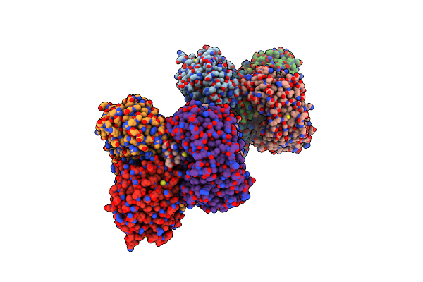 |
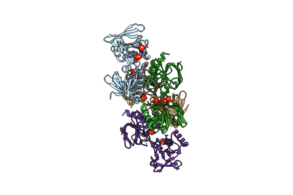
2.9 Angstrom Resolution Crystal Structure Of Gamma-Aminobutyraldehyde Dehydrogenase From Salmonella Typhimurium.
Organism: Salmonella choleraesuis
Method: X-RAY DIFFRACTION Resolution:2.90 Å Release Date: 2018-01-24 Classification: OXIDOREDUCTASE |
Organism: Salmonella choleraesuis
Method: X-RAY DIFFRACTION
Release Date: 2018-01-24
 |
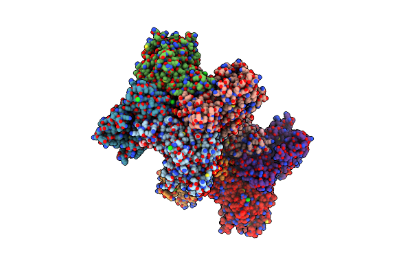
2.45 Angstrom Resolution Crystal Structure Thioredoxin Reductase From Francisella Tularensis.
Organism: Francisella tularensis subsp. tularensis (strain schu s4 / schu 4)
Method: X-RAY DIFFRACTION Resolution:2.45 Å Release Date: 2017-12-27 Classification: OXIDOREDUCTASE Ligands: CL, SO4 |
Organism: Francisella tularensis subsp. tularensis (strain schu s4 / schu 4)
Method: X-RAY DIFFRACTION
Release Date: 2017-12-27
Ligands: CL, SO4
 |
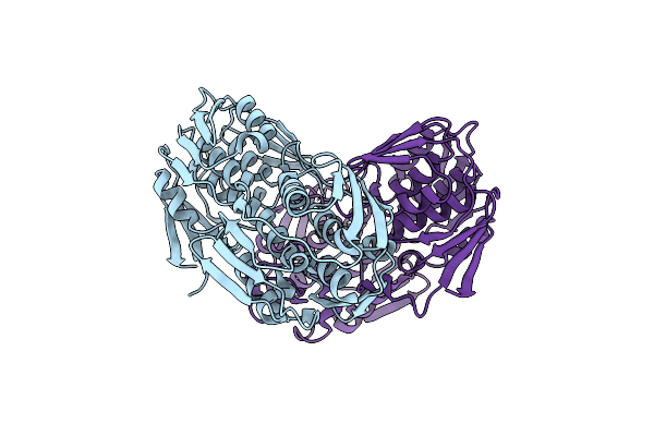
2.65 Angstrom Resolution Crystal Structure Of S-Adenosylhomocysteinase From Cryptosporidium Parvum In Complex With Sah And Nad
Organism: Cryptosporidium parvum (strain iowa ii)
Method: X-RAY DIFFRACTION Resolution:2.65 Å Release Date: 2017-03-01 Classification: HYDROLASE,OXIDOREDUCTASE Ligands: NAD, SAH, CL, SO4, P33, P6G, ADN, PEG |
Organism: Cryptosporidium parvum (strain iowa ii)
Method: X-RAY DIFFRACTION
Release Date: 2017-03-01
Ligands: NAD, SAH, CL, SO4, P33, P6G, ADN, PEG
 |
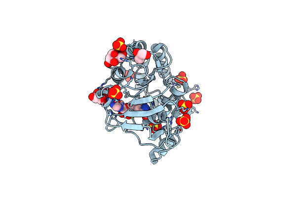
2.45 Angstrom Resolution Crystal Structure Of Udp-N-Acetylglucosamine 1-Carboxyvinyltransferase From Campylobacter Jejuni.
Organism: Campylobacter jejuni subsp. jejuni serotype o:2 (strain atcc 700819 / nctc 11168)
Method: X-RAY DIFFRACTION Resolution:2.46 Å Release Date: 2017-02-01 Classification: HYDROLASE,OXIDOREDUCTASE Ligands: CL |
Organism: Campylobacter jejuni subsp. jejuni serotype o:2 (strain atcc 700819 / nctc 11168)
Method: X-RAY DIFFRACTION
Release Date: 2017-02-01
Ligands: CL
 |
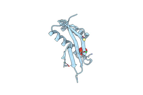
1.9 Angstrom Crystal Structure Of Ns5 Methyl Transferase From Dengue Virus 1 In Complex With S-Adenosylmethionine And Beta-D-Fructopyranose.
Organism: Dengue virus 1
Method: X-RAY DIFFRACTION Resolution:1.90 Å Release Date: 2016-03-16 Classification: TRANSFERASE Ligands: CL, SO4, SAM, BDF, MLT, GOL |
Organism: Dengue virus 1
Method: X-RAY DIFFRACTION
Release Date: 2016-03-16
Ligands: CL, SO4, SAM, BDF, MLT, GOL
 |
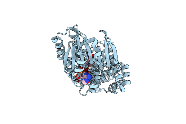
Crystal Structure Of Predicted Acyltransferase Yjdj With Acyl-Coa N-Acyltransferase Domain From Escherichia Coli Str. K-12
Organism: Escherichia coli (strain k12)
Method: X-RAY DIFFRACTION Resolution:1.92 Å Release Date: 2016-02-24 Classification: HYDROLASE Ligands: NI, CD, PO4 |
Organism: Escherichia coli (strain k12)
Method: X-RAY DIFFRACTION
Release Date: 2016-02-24
Ligands: NI, CD, PO4
 |
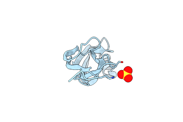
3.0 Angstrom Crystal Structure Of 3-Dehydroquinate Synthase (Arob) From Francisella Tularensis In Complex With Nad.
Organism: Francisella tularensis subsp. tularensis (strain schu s4 / schu 4)
Method: X-RAY DIFFRACTION Resolution:3.00 Å Release Date: 2016-02-10 Classification: LYASE Ligands: NAD, CL |
Organism: Francisella tularensis subsp. tularensis (strain schu s4 / schu 4)
Method: X-RAY DIFFRACTION
Release Date: 2016-02-10
Ligands: NAD, CL
 |
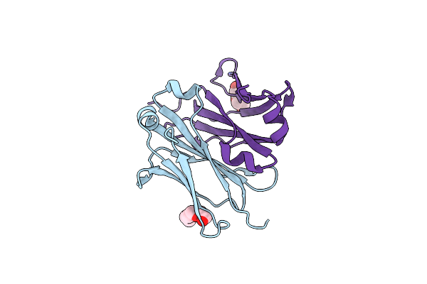
1.32 Angstrom Crystal Structure Of Ybbr Like Domain Of Lmo2119 Protein From Listeria Monocytogenes.
Organism: Listeria monocytogenes serovar 1/2a
Method: X-RAY DIFFRACTION Resolution:1.32 Å Release Date: 2016-02-03 Classification: UNKNOWN FUNCTION Ligands: CL, SO4 |
Organism: Listeria monocytogenes serovar 1/2a
Method: X-RAY DIFFRACTION
Release Date: 2016-02-03
Ligands: CL, SO4
 |
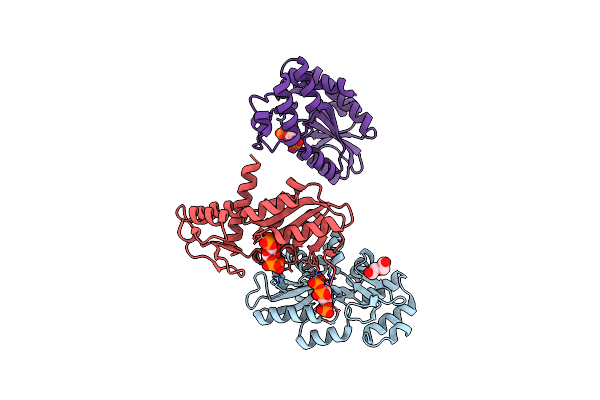
1.52 Angstrom Crystal Structure Of Fc Fragment Of Human Igg1.
Organism: Homo sapiens
Method: X-RAY DIFFRACTION Resolution:1.52 Å Release Date: 2016-02-03 Classification: IMMUNE SYSTEM Ligands: PGE |
Organism: Homo sapiens
Method: X-RAY DIFFRACTION
Release Date: 2016-02-03
Ligands: PGE
 |
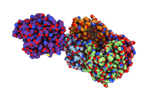
2.65 Angstrom Resolution Crystal Structure Of An Orotate Phosphoribosyltransferase From Bacillus Anthracis Str. 'Ames Ancestor' In Complex With 5-Phospho-Alpha-D-Ribosyl Diphosphate (Prpp)
Organism: Bacillus anthracis
Method: X-RAY DIFFRACTION Resolution:2.65 Å Release Date: 2014-12-17 Classification: TRANSFERASE Ligands: PRP, PEG |
Organism: Bacillus anthracis
Method: X-RAY DIFFRACTION
Release Date: 2014-12-17
Ligands: PRP, PEG
 |
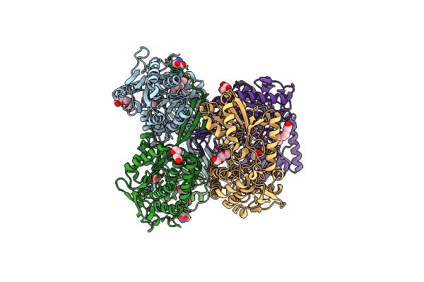
1.95 Angstrom Resolution Crystal Structure Of A Hypoxanthine-Guanine Phosphoribosyltransferase (Hpt-2) From Bacillus Anthracis Str. 'Ames Ancestor' With Hepes Molecule In The Active Site
Organism: Bacillus anthracis
Method: X-RAY DIFFRACTION Resolution:1.95 Å Release Date: 2014-08-27 Classification: TRANSFERASE Ligands: MG, PO4, EPE, GOL |
Organism: Bacillus anthracis
Method: X-RAY DIFFRACTION
Release Date: 2014-08-27
Ligands: MG, PO4, EPE, GOL
 |
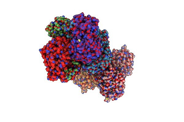
1.65 Angstrom Resolution Crystal Structure Of Betaine Aldehyde Dehydrogenase (Betb) From Staphylococcus Aureus With Bme-Modified Cys289 And Peg Molecule In Active Site
Organism: Staphylococcus aureus subsp. aureus col
Method: X-RAY DIFFRACTION Resolution:1.65 Å Release Date: 2014-07-16 Classification: OXIDOREDUCTASE |
Organism: Staphylococcus aureus subsp. aureus col
Method: X-RAY DIFFRACTION
Release Date: 2014-07-16
 |
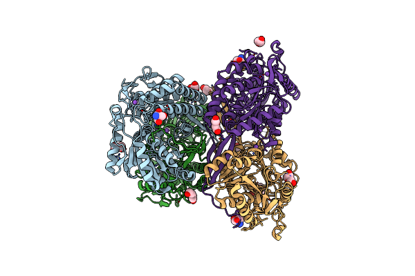
2.6 Angstrom Resolution Crystal Structure Of Betaine Aldehyde Dehydrogenase (Betb) G234S Mutant From Staphylococcus Aureus (Idp00699) In Complex With Nad+ And Bme-Free Cys289
Organism: Staphylococcus aureus subsp. aureus
Method: X-RAY DIFFRACTION Resolution:2.60 Å Release Date: 2014-07-02 Classification: OXIDOREDUCTASE Ligands: NAD, ACT |
Organism: Staphylococcus aureus subsp. aureus
Method: X-RAY DIFFRACTION
Release Date: 2014-07-02
Ligands: NAD, ACT
 |
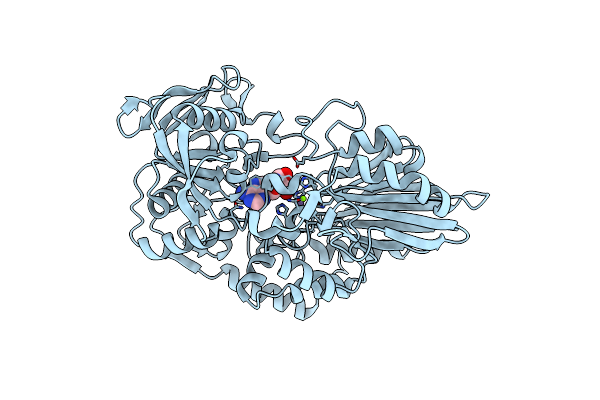
1.85 Angstrom Resolution Crystal Structure Of Apo Betaine Aldehyde Dehydrogenase (Betb) G234S Mutant From Staphylococcus Aureus (Idp00699) With Bme-Free Sulfinic Acid Form Of Cys289
Organism: Staphylococcus aureus subsp. aureus
Method: X-RAY DIFFRACTION Resolution:1.85 Å Release Date: 2014-06-18 Classification: OXIDOREDUCTASE |
Organism: Staphylococcus aureus subsp. aureus
Method: X-RAY DIFFRACTION
Release Date: 2014-06-18
 |
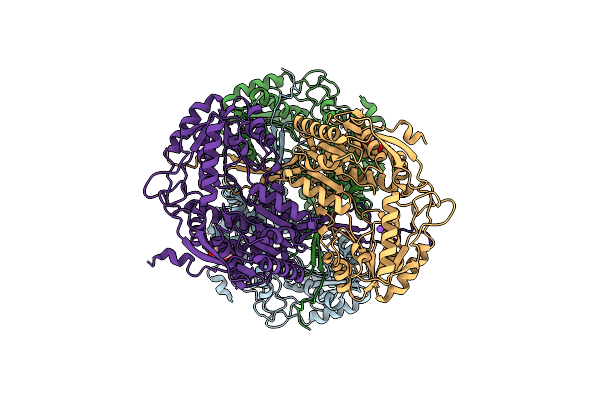
1.98 Angstrom Crystal Structure Of Putative 5'-Nucleotidase From Staphylococcus Aureus In Complex With Adenosine.
Organism: Staphylococcus aureus subsp. aureus
Method: X-RAY DIFFRACTION Resolution:1.98 Å Release Date: 2014-05-07 Classification: METAL BINDING PROTEIN Ligands: MG, MN, 3D1 |
Organism: Staphylococcus aureus subsp. aureus
Method: X-RAY DIFFRACTION
Release Date: 2014-05-07
Ligands: MG, MN, 3D1
 |
1.90 Angstrom Resolution Crystal Structure Of Apo Betaine Aldehyde Dehydrogenase (Betb) G234S Mutant From Staphylococcus Aureus (Idp00699) With Bme-Modified Cys289
Organism: Staphylococcus aureus subsp. aureus
Method: X-RAY DIFFRACTION Resolution:1.90 Å Release Date: 2014-05-07 Classification: OXIDOREDUCTASE |
Organism: Staphylococcus aureus subsp. aureus
Method: X-RAY DIFFRACTION
Release Date: 2014-05-07