
Search Count: 401
All
Selected
 |
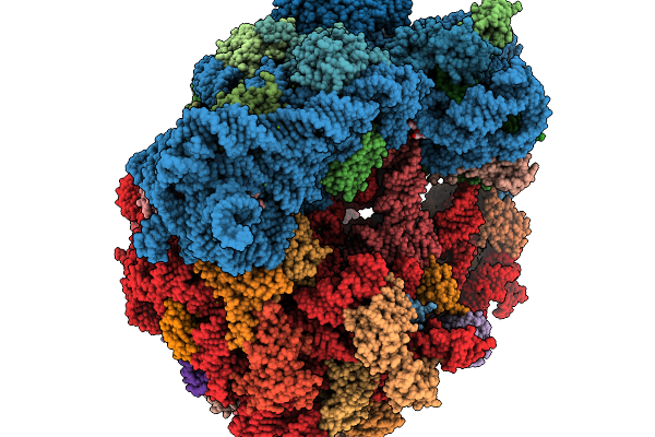
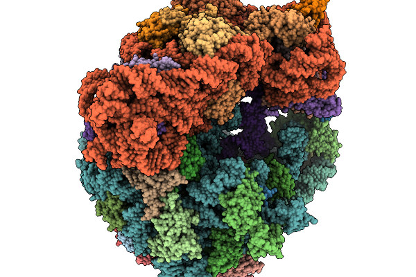
Cryo-Em Structure Of Skm-70S Ribosomal Stalled Complex In The Major State (Vacant A-Site, Canon)
Organism: Streptococcus sanguinis, Escherichia coli b
Method: ELECTRON MICROSCOPY Resolution:2.26 Å Release Date: 2026-02-18 Classification: RIBOSOME Ligands: ZN, A1JA7, MG |
 |
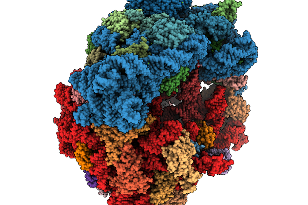
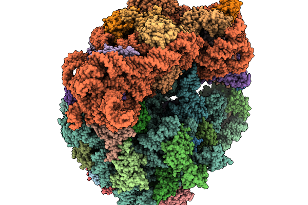
Cryo-Em Structure Of Skm-70S Ribosomal Stalled Complex In The A-Trna Positioned (Body Open) State.
Organism: Streptococcus sanguinis, Escherichia coli b
Method: ELECTRON MICROSCOPY Resolution:2.62 Å Release Date: 2026-02-18 Classification: RIBOSOME Ligands: ZN, A1JAI, MG |
 |
Cryo-Em Structure Of Skm-70S Ribosomal Stalled Complex In The Rotated State With Hybrid Trnas
Organism: Streptococcus sanguinis, Escherichia coli
Method: ELECTRON MICROSCOPY Resolution:2.60 Å Release Date: 2026-02-18 Classification: RIBOSOME Ligands: ZN, MG, A1JAI |
 |
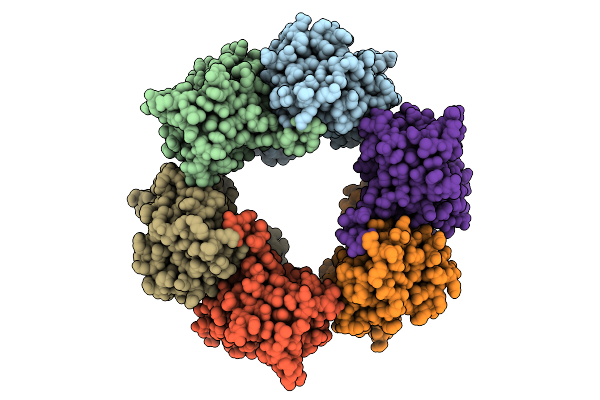
Organism: Plasmodium vivax, Mus musculus
Method: X-RAY DIFFRACTION Resolution:2.08 Å Release Date: 2026-02-04 Classification: CELL INVASION Ligands: GOL, SO4 |
 |
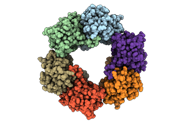
Organism: Plasmodium vivax, Vicugna pacos
Method: X-RAY DIFFRACTION Resolution:3.14 Å Release Date: 2026-01-21 Classification: CELL INVASION |
 |
Structure Of Clim-Stalled Bacillus Subtilis 70S Ribosome With Release Factor Bound In The A-Site
Organism: Clostridioides difficile 630, Bacillus subtilis subsp. subtilis str. 168
Method: ELECTRON MICROSCOPY Resolution:2.30 Å Release Date: 2026-01-14 Classification: RIBOSOME Ligands: ZN |
 |
Organism: Clostridioides difficile 630, Bacillus subtilis subsp. subtilis str. 168
Method: ELECTRON MICROSCOPY Resolution:2.30 Å Release Date: 2026-01-14 Classification: RIBOSOME |
 |
Structure Of Clim-Stalled Bacillus Subtilis 70S Ribosome With Trna-Tyr In The A-Site
Organism: Clostridioides difficile 630, Bacillus subtilis subsp. subtilis str. 168
Method: ELECTRON MICROSCOPY Resolution:2.80 Å Release Date: 2026-01-14 Classification: RIBOSOME Ligands: ZN |
 |
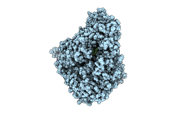
Cryo-Em Structure Of Candida Albicans Ph Regulated Antigen 1 (Pra1) Protein In The Absence Of Zn2+
Organism: Candida albicans
Method: ELECTRON MICROSCOPY Release Date: 2025-12-10 Classification: METAL BINDING PROTEIN |
 |
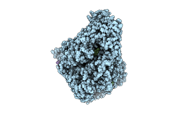
Cryo-Em Structure Of Candida Albicans Ph Regulated Antigen 1 (Pra1) Protein In Complex With Zn2+
Organism: Candida albicans
Method: ELECTRON MICROSCOPY Release Date: 2025-12-10 Classification: METAL BINDING PROTEIN Ligands: ZN |
 |
Organism: Homo sapiens
Method: X-RAY DIFFRACTION Resolution:2.95 Å Release Date: 2025-11-26 Classification: TRANSCRIPTION/INHIBITOR Ligands: BME, 9N6, NA, GOL, SO4 |
 |
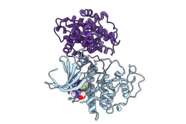
Organism: Escherichia coli, Homo sapiens
Method: X-RAY DIFFRACTION Resolution:1.28 Å Release Date: 2025-10-08 Classification: SIGNALING PROTEIN Ligands: A1CMI |
 |
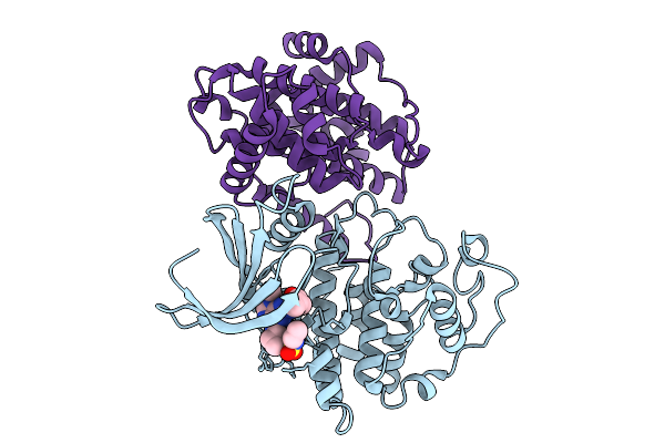
Organism: Escherichia coli, Homo sapiens
Method: X-RAY DIFFRACTION Resolution:1.53 Å Release Date: 2025-10-08 Classification: SIGNALING PROTEIN Ligands: A1CMH, EDO, FMT |
 |
Organism: Escherichia coli, Homo sapiens
Method: X-RAY DIFFRACTION Resolution:1.24 Å Release Date: 2025-10-08 Classification: SIGNALING PROTEIN Ligands: A1CMG, EDO, FMT |
 |
Organism: Homo sapiens
Method: X-RAY DIFFRACTION Resolution:1.16 Å Release Date: 2025-09-10 Classification: TRANSFERASE Ligands: A1INI |
 |
Organism: Homo sapiens
Method: X-RAY DIFFRACTION Resolution:2.16 Å Release Date: 2025-09-10 Classification: TRANSFERASE Ligands: A1IOI |
 |
Organism: Homo sapiens
Method: X-RAY DIFFRACTION Resolution:2.45 Å Release Date: 2025-09-10 Classification: TRANSFERASE Ligands: A1IOP |
 |
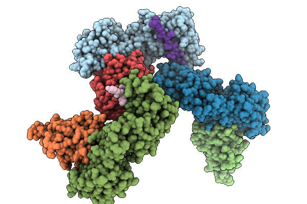
Organism: Homo sapiens, Synthetic construct
Method: ELECTRON MICROSCOPY Release Date: 2025-08-20 Classification: PEPTIDE BINDING PROTEIN |
 |
Organism: Homo sapiens, Synthetic construct
Method: ELECTRON MICROSCOPY Release Date: 2025-08-20 Classification: PEPTIDE BINDING PROTEIN |
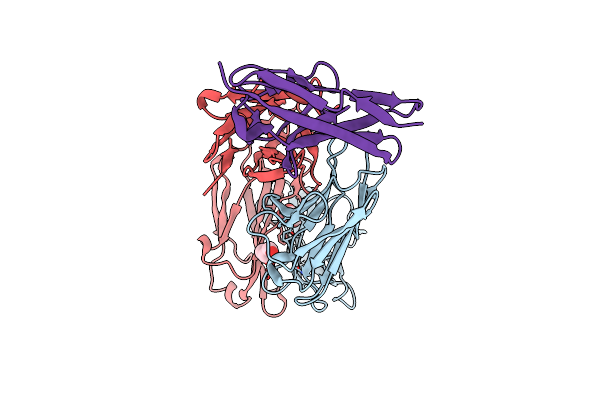 |
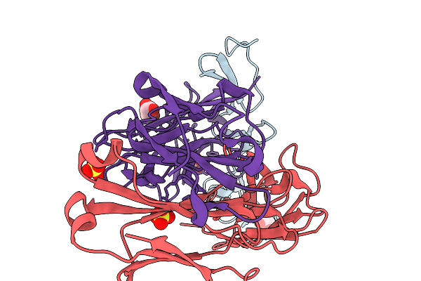
Co-Structure Of The Fab Of The Anti-Tigit Vibostolimab Antibody With Its Antigen
Organism: Mus musculus, Homo sapiens
Method: X-RAY DIFFRACTION Resolution:1.23 Å Release Date: 2025-07-02 Classification: IMMUNE SYSTEM Ligands: GOL |