

Search Count: 21
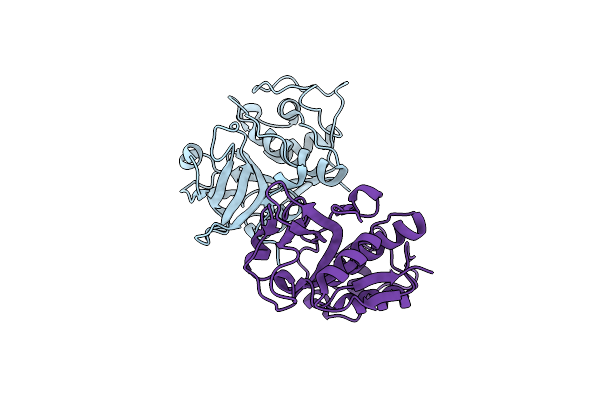 |
Native Ananain From Ananas Comosus
Organism: Ananas comosus
Method: X-RAY DIFFRACTION Resolution:1.73 Å Release Date: 2019-04-24 Classification: PLANT PROTEIN |
Organism: Ananas comosus
Method: X-RAY DIFFRACTION
Release Date: 2019-04-24
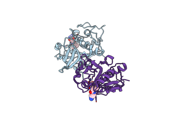 |
Native Ananain In Complex With E-64
Organism: Ananas comosus
Method: X-RAY DIFFRACTION Resolution:1.98 Å Release Date: 2018-10-03 Classification: HYDROLASE/HYDROLASE INHIBITOR Ligands: E64 |
Organism: Ananas comosus
Method: X-RAY DIFFRACTION
Release Date: 2018-10-03
Ligands: E64
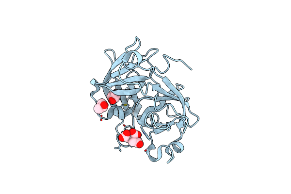 |
Coagulation Factor Xii Protease Domain Crystal Structure
Organism: Homo sapiens
Method: X-RAY DIFFRACTION Resolution:2.14 Å Release Date: 2015-02-04 Classification: BLOOD CLOTTING Ligands: IPA, FLC |
Organism: Homo sapiens
Method: X-RAY DIFFRACTION
Release Date: 2015-02-04
Ligands: IPA, FLC
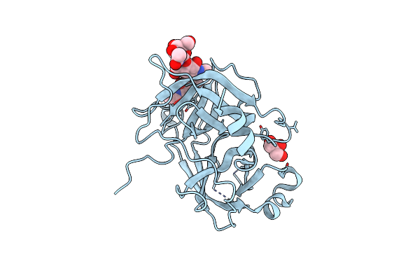 |
Coagulation Factor Xii Protease Domain Crystal Structure
Organism: Homo sapiens
Method: X-RAY DIFFRACTION Resolution:2.40 Å Release Date: 2015-02-04 Classification: BLOOD CLOTTING Ligands: GOL |
Organism: Homo sapiens
Method: X-RAY DIFFRACTION
Release Date: 2015-02-04
Ligands: GOL
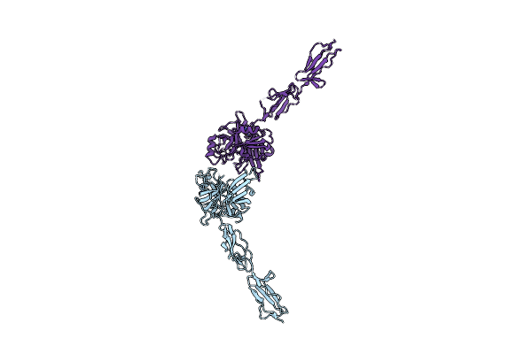 |
The X-Ray Crystal Structure Of Mannose-Binding Lectin-Associated Serine Proteinase-3 Reveals The Structural Basis For Enzyme Inactivity Associated With The 3Mc Syndrome
Organism: Homo sapiens
Method: X-RAY DIFFRACTION Resolution:2.60 Å Release Date: 2013-07-03 Classification: HYDROLASE Ligands: IMD |
Organism: Homo sapiens
Method: X-RAY DIFFRACTION
Release Date: 2013-07-03
Ligands: IMD
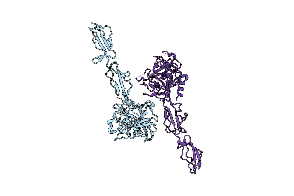 |
The X-Ray Crystal Structure Of Human Complement Protease C1S Zymogen
Organism: Homo sapiens
Method: X-RAY DIFFRACTION Resolution:2.66 Å Release Date: 2013-04-24 Classification: HYDROLASE |
Organism: Homo sapiens
Method: X-RAY DIFFRACTION
Release Date: 2013-04-24
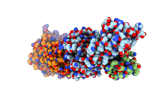 |
Crystal Structure Of C12C Tcr-Ha B2705-Kk10
Organism: Homo sapiens, Human immunodeficiency virus 1
Method: X-RAY DIFFRACTION Resolution:2.40 Å Release Date: 2013-03-20 Classification: IMMUNE SYSTEM |
Organism: Homo sapiens, Human immunodeficiency virus 1
Method: X-RAY DIFFRACTION
Release Date: 2013-03-20
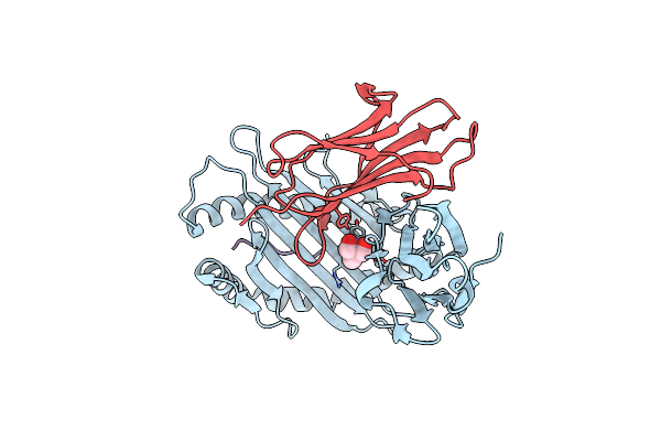 |
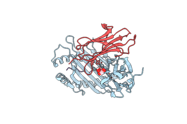
Crystal Structure Of Hla B2705-Kk10-L6M
Organism: Homo sapiens, Human immunodeficiency virus 1
Method: X-RAY DIFFRACTION Resolution:1.60 Å Release Date: 2013-03-20 Classification: IMMUNE SYSTEM Ligands: TRS |
Organism: Homo sapiens, Human immunodeficiency virus 1
Method: X-RAY DIFFRACTION
Release Date: 2013-03-20
Ligands: TRS
 |
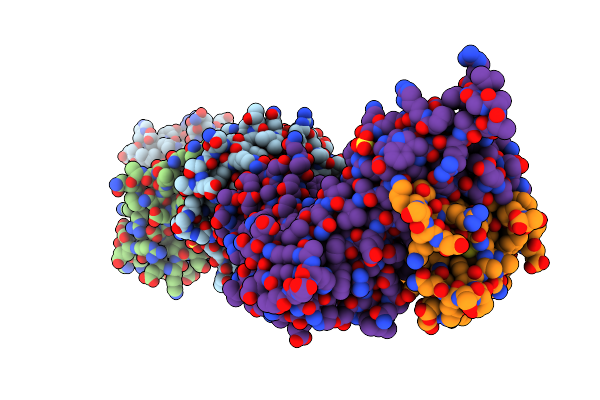
Crystal Structure Of Hla B2705-Kk10
Organism: Homo sapiens, Human immunodeficiency virus 1
Method: X-RAY DIFFRACTION Resolution:1.60 Å Release Date: 2013-03-20 Classification: IMMUNE SYSTEM Ligands: GOL |
Organism: Homo sapiens, Human immunodeficiency virus 1
Method: X-RAY DIFFRACTION
Release Date: 2013-03-20
Ligands: GOL
 |
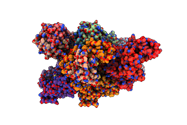
Crystal Structure Of C12C Tcr-Hlab2705-Kk10-L6M
Organism: Homo sapiens, Human immunodeficiency virus 1
Method: X-RAY DIFFRACTION Resolution:1.90 Å Release Date: 2013-03-20 Classification: IMMUNE SYSTEM Ligands: SO4 |
Organism: Homo sapiens, Human immunodeficiency virus 1
Method: X-RAY DIFFRACTION
Release Date: 2013-03-20
Ligands: SO4
 |
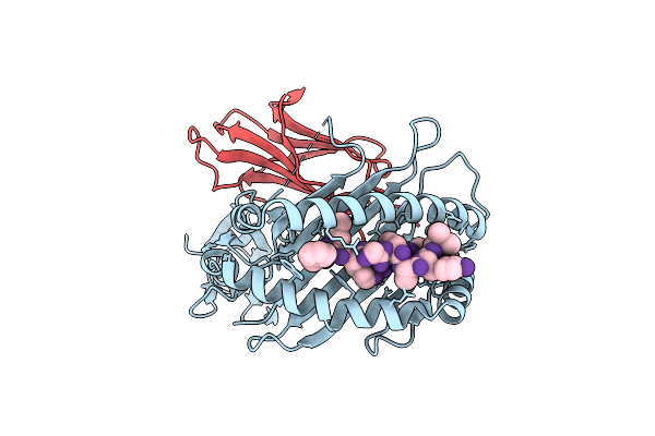
Crystal Structure Of The Rl42 Tcr In Complex With Hla-B8-Flr
Organism: Homo sapiens, Human herpesvirus 4
Method: X-RAY DIFFRACTION Resolution:3.10 Å Release Date: 2012-02-29 Classification: IMMUNE SYSTEM |
Organism: Homo sapiens, Human herpesvirus 4
Method: X-RAY DIFFRACTION
Release Date: 2012-02-29
 |
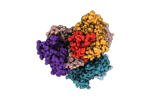
Crystal Structure Of The Hla-B8Flrgrayvl, Mutant G8V Of The Flr Peptide
Organism: Homo sapiens, Human herpesvirus 4
Method: X-RAY DIFFRACTION Resolution:1.80 Å Release Date: 2012-02-29 Classification: IMMUNE SYSTEM |
Organism: Homo sapiens, Human herpesvirus 4
Method: X-RAY DIFFRACTION
Release Date: 2012-02-29
 |
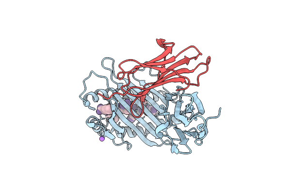
Crystal Structure Of The Rl42 Tcr Unliganded
Organism: Homo sapiens
Method: X-RAY DIFFRACTION Resolution:2.90 Å Release Date: 2012-02-29 Classification: IMMUNE SYSTEM |
Organism: Homo sapiens
Method: X-RAY DIFFRACTION
Release Date: 2012-02-29
 |
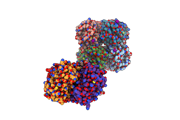
Crystal Structure Of The Hla-B8-A66-Flr, Mutant A66 Of The Hla B8
Organism: Homo sapiens, Human herpesvirus 4
Method: X-RAY DIFFRACTION Resolution:1.60 Å Release Date: 2012-02-29 Classification: IMMUNE SYSTEM |
Organism: Homo sapiens, Human herpesvirus 4
Method: X-RAY DIFFRACTION
Release Date: 2012-02-29
 |
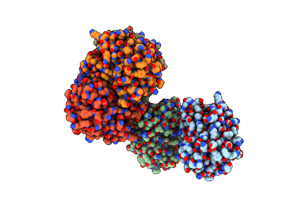
High Ph Structure Of Rtms5 H146S Variant
Organism: Montipora efflorescens
Method: X-RAY DIFFRACTION Resolution:1.80 Å Release Date: 2007-04-03 Classification: LUMINESCENT PROTEIN Ligands: IOD |
Organism: Montipora efflorescens
Method: X-RAY DIFFRACTION
Release Date: 2007-04-03
Ligands: IOD
 |
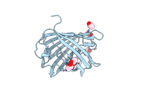
The 1.7 A Crystal Structure Of Dronpa: A Photoswitchable Green Fluorescent Protein
Organism: Echinophyllia sp. sc22
Method: X-RAY DIFFRACTION Resolution:1.70 Å Release Date: 2006-11-14 Classification: LUMINESCENT PROTEIN |
Organism: Echinophyllia sp. sc22
Method: X-RAY DIFFRACTION
Release Date: 2006-11-14
 |
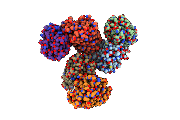
The 2.0 Angstroms Crystal Structure Of A Pocilloporin At Ph 3.5: The Structural Basis For The Linkage Between Color Transition And Halide Binding
Organism: Montipora efflorescens
Method: X-RAY DIFFRACTION Resolution:2.00 Å Release Date: 2006-09-05 Classification: LUMINESCENT PROTEIN Ligands: IOD, CL, ACY |
Organism: Montipora efflorescens
Method: X-RAY DIFFRACTION
Release Date: 2006-09-05
Ligands: IOD, CL, ACY
 |
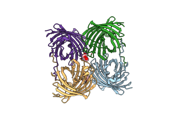
The 2.1A Crystal Structure Of Copgfp
Organism: Pontellina plumata
Method: X-RAY DIFFRACTION Resolution:2.10 Å Release Date: 2006-08-15 Classification: LUMINESCENT PROTEIN |
Organism: Pontellina plumata
Method: X-RAY DIFFRACTION
Release Date: 2006-08-15
 |
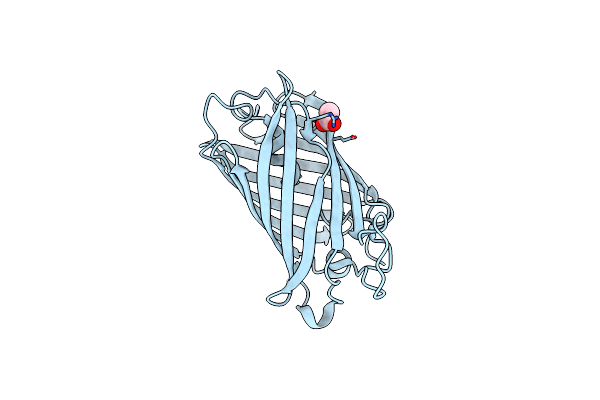
The 2.1A Crystal Structure Of The Far-Red Fluorescent Protein Hcred: Inherent Conformational Flexibility Of The Chromophore
Organism: Heteractis crispa
Method: X-RAY DIFFRACTION Resolution:2.10 Å Release Date: 2005-05-17 Classification: LUMINESCENT PROTEIN Ligands: PEG |
Organism: Heteractis crispa
Method: X-RAY DIFFRACTION
Release Date: 2005-05-17
Ligands: PEG
 |
Variations On The Gfp Chromophore Scaffold: A Fragmented 5-Membered Heterocycle Revealed In The 2.1A Crystal Structure Of A Non-Fluorescent Chromoprotein
Method: X-RAY DIFFRACTION
Resolution:2.10 Å Release Date: 2004-11-16 Classification: LUMINESCENT PROTEIN Ligands: ACY |
Method: X-RAY DIFFRACTION
Release Date: 2004-11-16
Ligands: ACY