

Search Count: 26
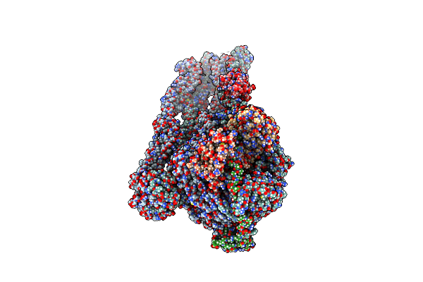 |
Mukbef In A Nucleotide-Bound State With Open Neck Gate (Heads Core)
Organism: Photorhabdus thracensis, Escherichia coli
Method: ELECTRON MICROSCOPY Release Date: 2025-03-26 Classification: DNA BINDING PROTEIN Ligands: MG, ATP |
Organism: Photorhabdus thracensis, Escherichia coli
Method: ELECTRON MICROSCOPY
Release Date: 2025-03-26
Ligands: MG, ATP
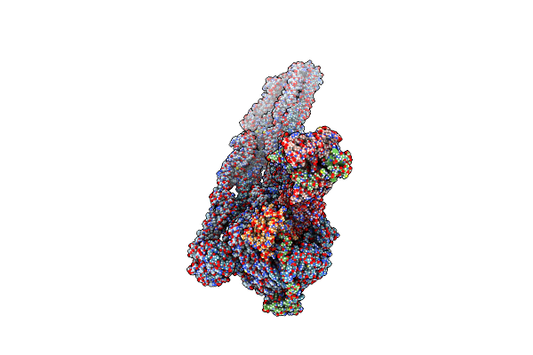 |
Mukbef In A Nucleotide-Bound State With Open Neck Gate (Monomer)
Organism: Photorhabdus thracensis, Escherichia coli
Method: ELECTRON MICROSCOPY Release Date: 2025-03-26 Classification: DNA BINDING PROTEIN Ligands: MG, ATP |
Organism: Photorhabdus thracensis, Escherichia coli
Method: ELECTRON MICROSCOPY
Release Date: 2025-03-26
Ligands: MG, ATP
 |
Mukbef In A Nucleotide-Bound State With Open Neck Gate
Organism: Photorhabdus thracensis, Escherichia coli
Method: ELECTRON MICROSCOPY Release Date: 2025-03-26 Classification: DNA BINDING PROTEIN Ligands: MG, ATP |
Organism: Photorhabdus thracensis, Escherichia coli
Method: ELECTRON MICROSCOPY
Release Date: 2025-03-26
Ligands: MG, ATP
 |
Mukbef In A Dna Capture State
Organism: Photorhabdus thracensis, Escherichia coli
Method: ELECTRON MICROSCOPY Release Date: 2025-03-26 Classification: DNA BINDING PROTEIN Ligands: MG, ATP |
Organism: Photorhabdus thracensis, Escherichia coli
Method: ELECTRON MICROSCOPY
Release Date: 2025-03-26
Ligands: MG, ATP
 |
Mukbef In A Dna Capture State (Dimer)
Organism: Photorhabdus thracensis, Escherichia coli
Method: ELECTRON MICROSCOPY Release Date: 2025-03-26 Classification: DNA BINDING PROTEIN Ligands: MG, ATP |
Organism: Photorhabdus thracensis, Escherichia coli
Method: ELECTRON MICROSCOPY
Release Date: 2025-03-26
Ligands: MG, ATP
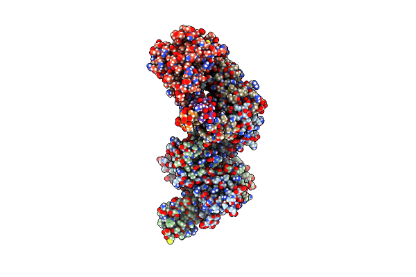 |
Mukef In Complex With The Phage Protein Gp5.9
Organism: Escherichia coli, Escherichia phage t7
Method: ELECTRON MICROSCOPY Release Date: 2025-03-26 Classification: DNA BINDING PROTEIN |
Organism: Escherichia coli, Escherichia phage t7
Method: ELECTRON MICROSCOPY
Release Date: 2025-03-26
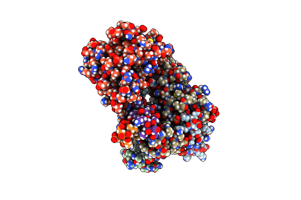 |
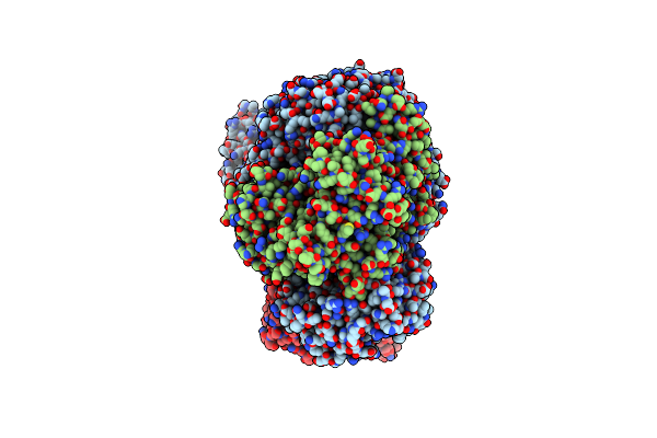
Mukef In Complex With The Phage Protein Gp5.9 (Focus)
Organism: Escherichia coli, Escherichia phage t7
Method: ELECTRON MICROSCOPY Release Date: 2025-03-26 Classification: DNA BINDING PROTEIN |
Organism: Escherichia coli, Escherichia phage t7
Method: ELECTRON MICROSCOPY
Release Date: 2025-03-26
 |
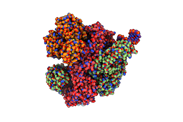
Recbcd In Complex With The Phage Protein Gp5.9
Organism: Escherichia coli, Escherichia phage t7
Method: ELECTRON MICROSCOPY Release Date: 2022-12-28 Classification: DNA BINDING PROTEIN Ligands: MG |
Organism: Escherichia coli, Escherichia phage t7
Method: ELECTRON MICROSCOPY
Release Date: 2022-12-28
Ligands: MG
 |
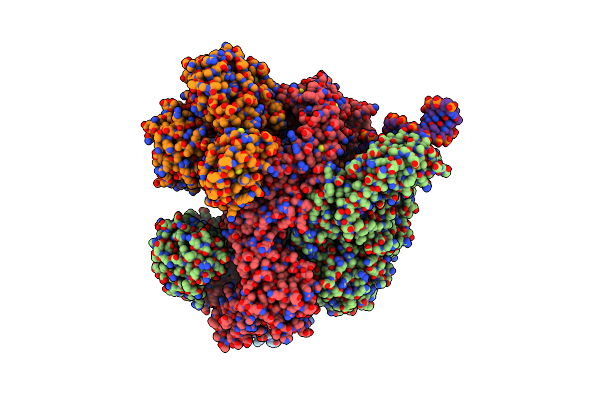
Recbcd-Dna In Complex With The Phage Protein Abc2
Organism: Escherichia coli, Salmonella phage p22, Synthetic construct
Method: ELECTRON MICROSCOPY Release Date: 2022-12-28 Classification: DNA BINDING PROTEIN Ligands: ANP, MG |
Organism: Escherichia coli, Salmonella phage p22, Synthetic construct
Method: ELECTRON MICROSCOPY
Release Date: 2022-12-28
Ligands: ANP, MG
 |
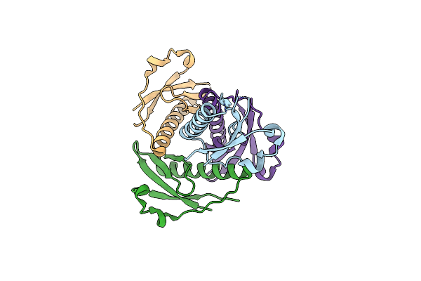
Recbcd-Dna In Complex With The Phage Protein Abc2 And Host Ppib
Organism: Escherichia coli, Salmonella phage p22, Synthetic construct
Method: ELECTRON MICROSCOPY Release Date: 2022-12-28 Classification: DNA BINDING PROTEIN Ligands: ANP, MG |
Organism: Escherichia coli, Salmonella phage p22, Synthetic construct
Method: ELECTRON MICROSCOPY
Release Date: 2022-12-28
Ligands: ANP, MG
 |
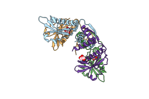
Crystal Structure Of The N-Terminal Domain Of Burkholderia Pseudomallei Antitoxin Hicb
Organism: Burkholderia pseudomallei k96243
Method: X-RAY DIFFRACTION Resolution:1.56 Å Release Date: 2018-10-31 Classification: ANTITOXIN |
Organism: Burkholderia pseudomallei k96243
Method: X-RAY DIFFRACTION
Release Date: 2018-10-31
 |
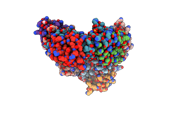
Crystal Structure Of The Burkholderia Pseudomallei Antitoxin Hicb
Organism: Burkholderia pseudomallei
Method: X-RAY DIFFRACTION Resolution:1.85 Å Release Date: 2018-10-31 Classification: ANTITOXIN Ligands: CL, GOL |
Organism: Burkholderia pseudomallei
Method: X-RAY DIFFRACTION
Release Date: 2018-10-31
Ligands: CL, GOL
 |
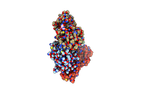
The Crystal Structure Of The Burkholderia Pseudomallei Hicab Complex
Organism: Burkholderia pseudomallei k96243
Method: X-RAY DIFFRACTION Resolution:2.49 Å Release Date: 2018-10-31 Classification: ANTITOXIN Ligands: SO4, EDO, PGE |
Organism: Burkholderia pseudomallei k96243
Method: X-RAY DIFFRACTION
Release Date: 2018-10-31
Ligands: SO4, EDO, PGE
 |
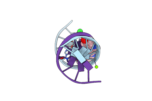
Crystal Structure Of The Brct Domains Of 53Bp1 In Complex With P53 And H2Ax-Pser139 (Gammah2Ax)
Organism: Homo sapiens
Method: X-RAY DIFFRACTION Resolution:3.00 Å Release Date: 2015-12-16 Classification: ANTITUMOR PROTEIN Ligands: ZN |
Organism: Homo sapiens
Method: X-RAY DIFFRACTION
Release Date: 2015-12-16
Ligands: ZN
 |
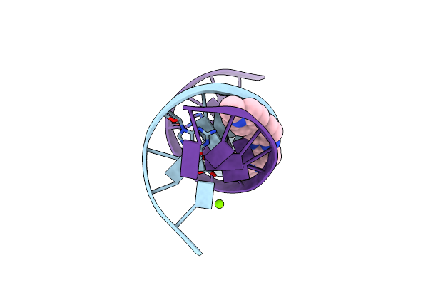
O6-Carboxymethylguanine In Dna Forms A Sequence Context Dependent Wobble Base Pair Structure With Thymine
Method: X-RAY DIFFRACTION
Resolution:1.60 Å Release Date: 2014-07-02 Classification: DNA Ligands: HT, MG, SR |
Method: X-RAY DIFFRACTION
Release Date: 2014-07-02
Ligands: HT, MG, SR
 |
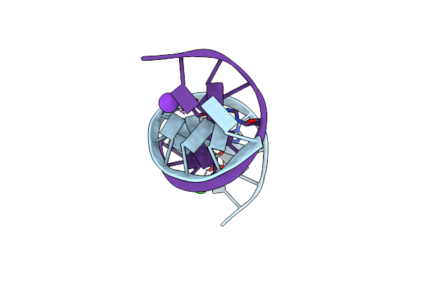
O6-Carboxymethylguanine In Dna Forms A Sequence Context Dependent Wobble Base Pair Structure With Thymine.
Method: X-RAY DIFFRACTION
Resolution:1.60 Å Release Date: 2014-07-02 Classification: DNA Ligands: HT, MG |
Method: X-RAY DIFFRACTION
Release Date: 2014-07-02
Ligands: HT, MG
 |
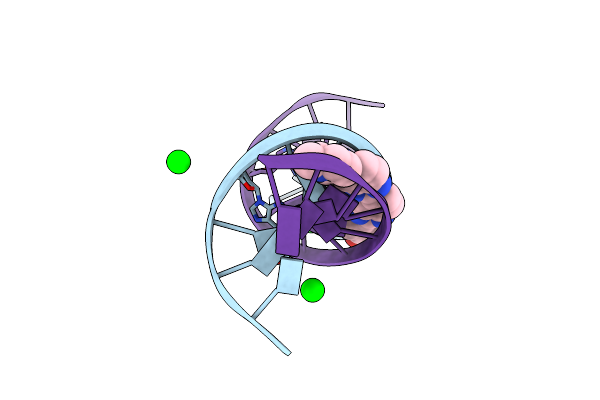
O6-Carboxymethylguanine In Dna Forms A Sequence Context Dependent Wobble Base Pair Structure With Thymine
Method: X-RAY DIFFRACTION
Resolution:1.75 Å Release Date: 2014-07-02 Classification: DNA Ligands: K, BA |
Method: X-RAY DIFFRACTION
Release Date: 2014-07-02
Ligands: K, BA
 |
O6-Carboxymethylguanine In Dna Forms A Sequence Context Dependent Wobble Base Pair Structure With Thymine
|
Method: X-RAY DIFFRACTION
Release Date: 2014-07-02
 |
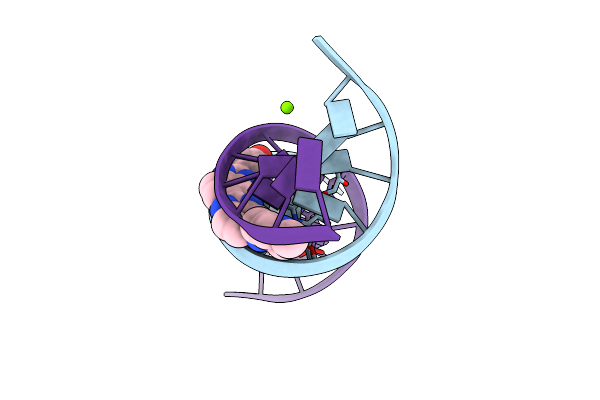
Structures Of Dna Duplexes Containing O6-Carboxymethylguanine, A Lesion Associated With Gastrointestinal Cancer, Reveal A Mechanism For Inducing Transition Mutation
Method: X-RAY DIFFRACTION
Resolution:1.54 Å Release Date: 2013-05-08 Classification: DNA Ligands: SR, HT |
Method: X-RAY DIFFRACTION
Release Date: 2013-05-08
Ligands: SR, HT
 |
Structures Of Dna Duplexes Containing O6-Carboxymethylguanine, A Lesion Associated With Gastrointestinal Cancer, Reveal A Mechanism For Inducing Transition Mutation
Method: X-RAY DIFFRACTION
Resolution:1.94 Å Release Date: 2013-05-08 Classification: DNA Ligands: MG, HT |
Method: X-RAY DIFFRACTION
Release Date: 2013-05-08
Ligands: MG, HT