

Search Count: 26
 |
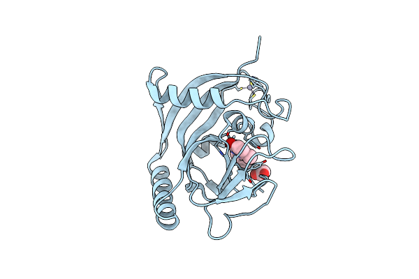
Crystal Structure Of Mouse Vps29 Bound To Dennd4C Peptide (Re-Refinement)
Organism: Mus musculus, Homo sapiens
Method: X-RAY DIFFRACTION Resolution:2.35 Å Release Date: 2025-08-13 Classification: PROTEIN TRANSPORT Ligands: CAC |
Organism: Mus musculus, Homo sapiens
Method: X-RAY DIFFRACTION
Release Date: 2025-08-13
Ligands: CAC
 |
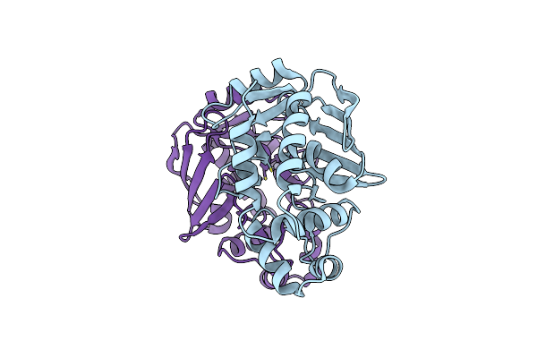
Human Tnks1 In Complex With 3-[4-(1-Hydroxy-1-Methyl-Ethyl)-Phenyl]-6-Methyl-2H-Pyrrolo[1,2-A]Pyrazin-1-One
Organism: Homo sapiens
Method: X-RAY DIFFRACTION Resolution:1.43 Å Release Date: 2021-07-28 Classification: TRANSFERASE Ligands: ACT, V8B, ZN |
Organism: Homo sapiens
Method: X-RAY DIFFRACTION
Release Date: 2021-07-28
Ligands: ACT, V8B, ZN
 |
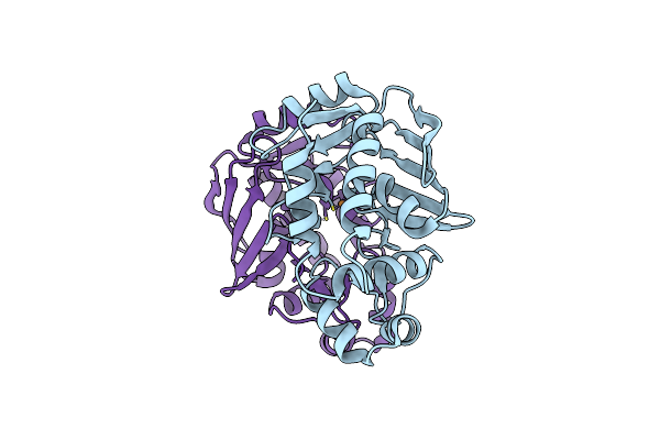
Crystal Structure Of The Mercury-Bound Form Of Merb Mutant D99S
Organism: Escherichia coli
Method: X-RAY DIFFRACTION Resolution:1.96 Å Release Date: 2016-02-03 Classification: LYASE,METAL BINDING PROTEIN Ligands: HG, BR |
Organism: Escherichia coli
Method: X-RAY DIFFRACTION
Release Date: 2016-02-03
Ligands: HG, BR
 |
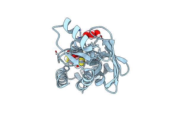
Crystal Structure Of The Copper-Bound Form Of Merb Mutant D99S
Organism: Escherichia coli
Method: X-RAY DIFFRACTION Resolution:1.87 Å Release Date: 2016-02-03 Classification: LYASE,METAL BINDING PROTEIN Ligands: CU, BR |
Organism: Escherichia coli
Method: X-RAY DIFFRACTION
Release Date: 2016-02-03
Ligands: CU, BR
 |
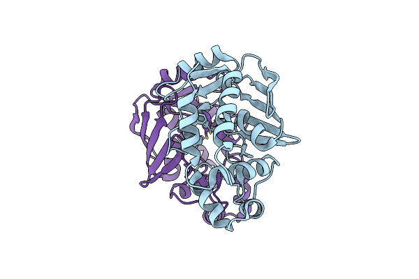
Crystal Structure Of The Mercury-Bound Form Of Merb2
Organism: Bacillus megaterium
Method: X-RAY DIFFRACTION Resolution:1.24 Å Release Date: 2016-02-03 Classification: LYASE Ligands: HG, GOL, DTV |
Organism: Bacillus megaterium
Method: X-RAY DIFFRACTION
Release Date: 2016-02-03
Ligands: HG, GOL, DTV
 |
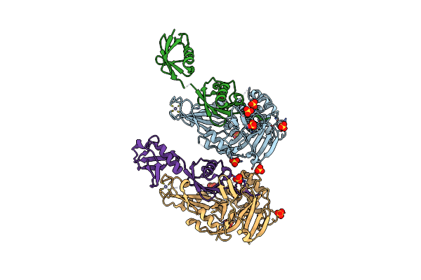
Crystal Structure Of The Mercury-Bound Form Of Merb Mutant D99S
Organism: Escherichia coli
Method: X-RAY DIFFRACTION Resolution:1.95 Å Release Date: 2016-02-03 Classification: LYASE,METAL BINDING PROTEIN Ligands: HG, BR |
Organism: Escherichia coli
Method: X-RAY DIFFRACTION
Release Date: 2016-02-03
Ligands: HG, BR
 |
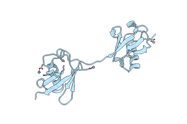
Structure Of Usp21 In Complex With Linear Diubiquitin-Aldehyde
Organism: Homo sapiens
Method: X-RAY DIFFRACTION Resolution:2.70 Å Release Date: 2012-01-25 Classification: PROTEIN BINDING/HYDROLASE Ligands: ZN, SO4 |
Organism: Homo sapiens
Method: X-RAY DIFFRACTION
Release Date: 2012-01-25
Ligands: ZN, SO4
 |
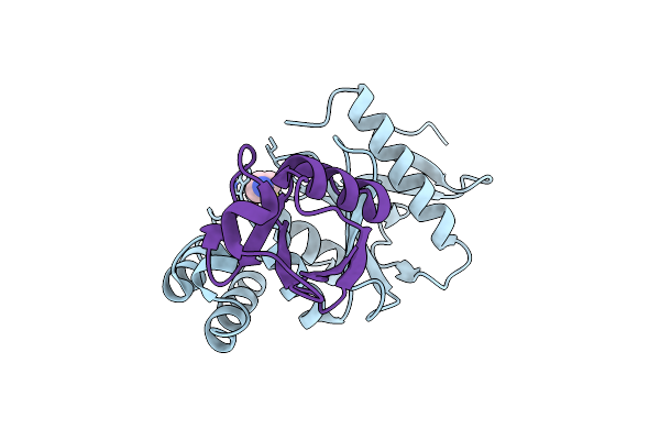
Crystal Structure Of Linear Di-Ubiquitin
Organism: Homo sapiens
Method: X-RAY DIFFRACTION Resolution:2.25 Å Release Date: 2009-04-28 Classification: CELL CYCLE Ligands: ZN, CL |
Organism: Homo sapiens
Method: X-RAY DIFFRACTION
Release Date: 2009-04-28
Ligands: ZN, CL
 |
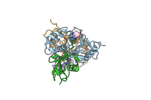
Structure Of Ovarian Tumor (Otu) Domain In Complex With Ubiquitin
Organism: Saccharomyces cerevisiae, Homo sapiens
Method: X-RAY DIFFRACTION Resolution:1.55 Å Release Date: 2008-02-19 Classification: CELL CYCLE, HYDROLASE Ligands: 3CN |
Organism: Saccharomyces cerevisiae, Homo sapiens
Method: X-RAY DIFFRACTION
Release Date: 2008-02-19
Ligands: 3CN
 |
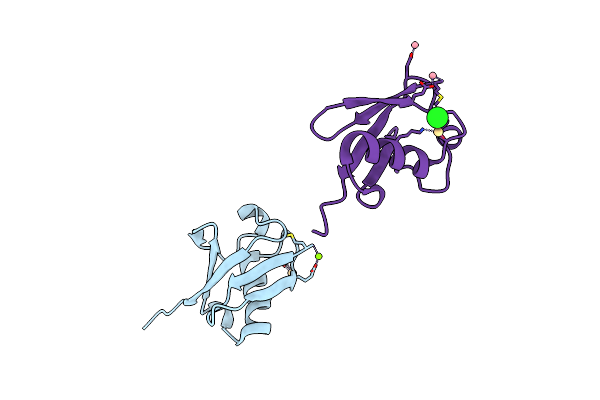
Structure Of Ovarian Tumor (Otu) Domain In Complex With Ubiquitin
Organism: Saccharomyces cerevisiae, Homo sapiens
Method: X-RAY DIFFRACTION Resolution:2.32 Å Release Date: 2008-02-19 Classification: CELL CYCLE, HYDROLASE Ligands: 3CN |
Organism: Saccharomyces cerevisiae, Homo sapiens
Method: X-RAY DIFFRACTION
Release Date: 2008-02-19
Ligands: 3CN
 |
Crystal Structure Of Lys63-Linked Di-Ubiquitin
Organism: Homo sapiens
Method: X-RAY DIFFRACTION Resolution:1.95 Å Release Date: 2008-02-05 Classification: SIGNALING PROTEIN Ligands: CD, MG, CO, CL |
Organism: Homo sapiens
Method: X-RAY DIFFRACTION
Release Date: 2008-02-05
Ligands: CD, MG, CO, CL
 |
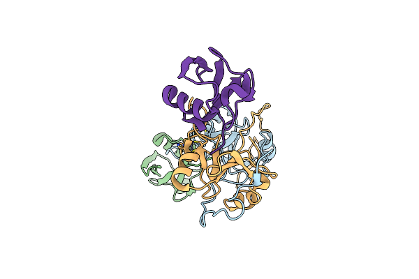
Structure Of The Znf Ubp Domain From Deubiquitinating Enzyme Isopeptidase T (Isot)
Organism: Homo sapiens
Method: X-RAY DIFFRACTION Resolution:2.09 Å Release Date: 2006-04-04 Classification: HYDROLASE Ligands: ZN, UNX |
Organism: Homo sapiens
Method: X-RAY DIFFRACTION
Release Date: 2006-04-04
Ligands: ZN, UNX
 |
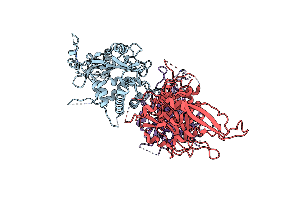
Co-Crystal Structure Of Znf Ubp Domain From The Deubiquitinating Enzyme Isopeptidase T (Isot) In Complex With Ubiquitin
Organism: Homo sapiens
Method: X-RAY DIFFRACTION Resolution:1.99 Å Release Date: 2006-04-04 Classification: HYDROLASE Ligands: ZN, CL |
Organism: Homo sapiens
Method: X-RAY DIFFRACTION
Release Date: 2006-04-04
Ligands: ZN, CL
 |
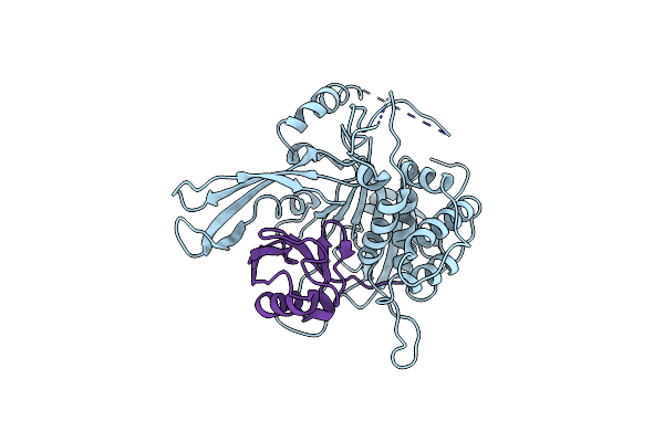
Structure Of Usp14, A Proteasome-Associated Deubiquitinating Enzyme
Organism: Homo sapiens
Method: X-RAY DIFFRACTION Resolution:3.20 Å Release Date: 2005-10-18 Classification: HYDROLASE |
Organism: Homo sapiens
Method: X-RAY DIFFRACTION
Release Date: 2005-10-18
 |
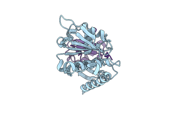
Structure Of Usp14 Bound To Ubquitin Aldehyde
Organism: Homo sapiens
Method: X-RAY DIFFRACTION Resolution:3.50 Å Release Date: 2005-10-18 Classification: HYDROLASE |
Organism: Homo sapiens
Method: X-RAY DIFFRACTION
Release Date: 2005-10-18
 |
Crystal Structure Of Den1 In Complex With Nedd8
Organism: Homo sapiens
Method: X-RAY DIFFRACTION Resolution:2.20 Å Release Date: 2004-12-21 Classification: HYDROLASE/HYDROLASE INHIBITOR |
Organism: Homo sapiens
Method: X-RAY DIFFRACTION
Release Date: 2004-12-21
 |
Comparison Of The Structure And Dna Binding Properties Of The E2 Proteins From An Oncogenic And A Non-Oncogenic Human Papillomavirus
Organism: Human papillomavirus type 6a
Method: X-RAY DIFFRACTION Resolution:1.90 Å Release Date: 2003-12-23 Classification: TRANSCRIPTION, REPLICATION Ligands: PO4 |
Organism: Human papillomavirus type 6a
Method: X-RAY DIFFRACTION
Release Date: 2003-12-23
Ligands: PO4
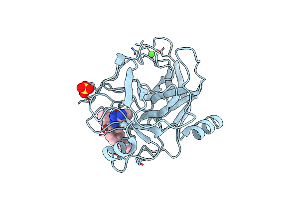 |
Trypsin Inhibitor Complex (Bpo)
Organism: Bos taurus
Method: X-RAY DIFFRACTION Resolution:2.00 Å Release Date: 2002-02-11 Classification: HYDROLASE Ligands: SO4, CA, BPO |
Organism: Bos taurus
Method: X-RAY DIFFRACTION
Release Date: 2002-02-11
Ligands: SO4, CA, BPO
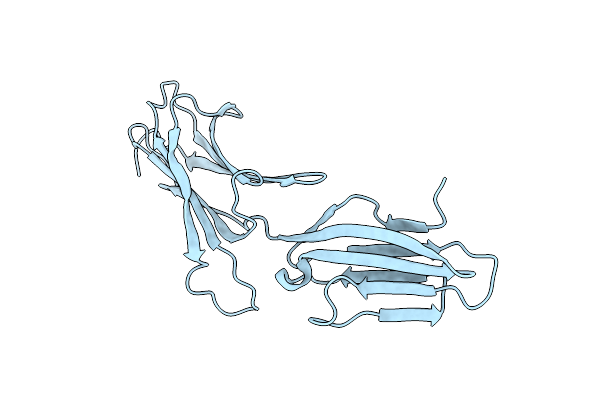 |
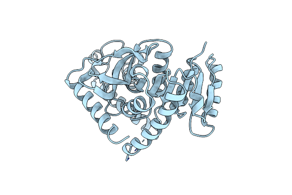
A Reassessment Of The Madcam-1 Structure And Its Role In Integrin Recognition.
Organism: Homo sapiens
Method: X-RAY DIFFRACTION Resolution:1.90 Å Release Date: 2002-01-29 Classification: CELL ADHESION PROTEIN |
Organism: Homo sapiens
Method: X-RAY DIFFRACTION
Release Date: 2002-01-29
 |
Chloroquine Binds In The Cofactor Binding Site Of Plasmodium Falciparum Lactate Dehydrogenase.
Organism: Plasmodium falciparum
Method: X-RAY DIFFRACTION Resolution:2.00 Å Release Date: 1999-03-19 Classification: OXIDOREDUCTASE |
Organism: Plasmodium falciparum
Method: X-RAY DIFFRACTION
Release Date: 1999-03-19