

Search Count: 13
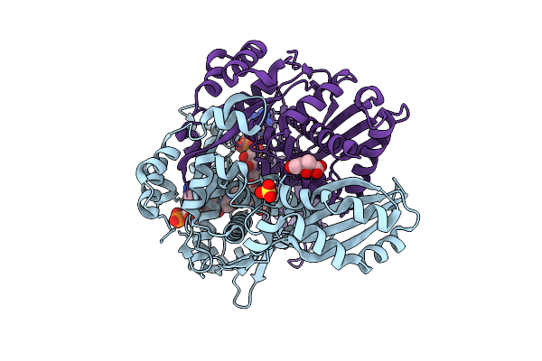 |
Structure Of Pmhmgr Bound To Mevalonate, Coa And Nad 2 Minutes 20 Seconds After Reaction Initiation At Ph 9
Organism: Pseudomonas sp. 'mevalonii'
Method: X-RAY DIFFRACTION Resolution:2.50 Å Release Date: 2026-04-08 Classification: OXIDOREDUCTASE/SUBSTRATE/INTERMEDIATE Ligands: COA, SO4, A1AFY, ZKE, NAI, MEV |
Organism: Pseudomonas sp. 'mevalonii'
Method: X-RAY DIFFRACTION
Release Date: 2026-04-08
Ligands: COA, SO4, A1AFY, ZKE, NAI, MEV
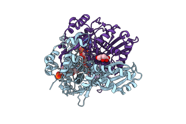 |
Structure Of Pmhmgr Bound To Mevalonate, Coa And Nad 1 Minute After Reaction Initiation At Ph 9
Organism: Pseudomonas sp. 'mevalonii'
Method: X-RAY DIFFRACTION Resolution:2.06 Å Release Date: 2026-03-18 Classification: OXIDOREDUCTASE/SUBSTRATE/INTERMEDIATE Ligands: COA, A1AFY, NAI, NAD, MEV |
Organism: Pseudomonas sp. 'mevalonii'
Method: X-RAY DIFFRACTION
Release Date: 2026-03-18
Ligands: COA, A1AFY, NAI, NAD, MEV
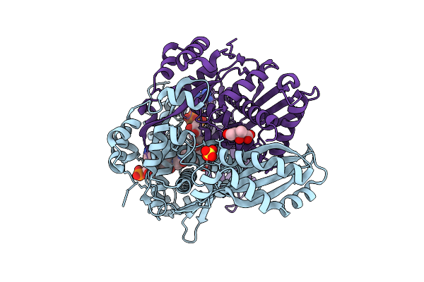 |
Pmhmgr Bound To Mevalonate, Coa, And Nad, Buffer-Exchanged To Ammonium Acetate Environment At Ph 6.7
Organism: Pseudomonas sp. 'mevalonii'
Method: X-RAY DIFFRACTION Resolution:1.93 Å Release Date: 2026-03-18 Classification: OXIDOREDUCTASE Ligands: COA, MEV, SO4, NAD |
Organism: Pseudomonas sp. 'mevalonii'
Method: X-RAY DIFFRACTION
Release Date: 2026-03-18
Ligands: COA, MEV, SO4, NAD
 |
Structure Of Pmhmgr Bound To Mevalonate, Coa And Nad.
Organism: Pseudomonas sp. 'mevalonii'
Method: X-RAY DIFFRACTION Resolution:1.99 Å Release Date: 2024-03-20 Classification: STRUCTURAL PROTEIN Ligands: COA, MEV, SO4, NAD |
Organism: Pseudomonas sp. 'mevalonii'
Method: X-RAY DIFFRACTION
Release Date: 2024-03-20
Ligands: COA, MEV, SO4, NAD
 |
Pmhmgr Bound To Mevaldehyde And Coa
Organism: Pseudomonas sp. 'mevalonii'
Method: X-RAY DIFFRACTION Resolution:1.65 Å Release Date: 2024-03-20 Classification: OXIDOREDUCTASE Ligands: ZKE, SO4, XII |
Organism: Pseudomonas sp. 'mevalonii'
Method: X-RAY DIFFRACTION
Release Date: 2024-03-20
Ligands: ZKE, SO4, XII
 |
Structure Of Pmhmgr Bound To Mevalonate, Coa And Nad 5 Minutes After Reaction Initiation At Ph 9
Organism: Pseudomonas sp. 'mevalonii'
Method: X-RAY DIFFRACTION Resolution:2.07 Å Release Date: 2024-03-20 Classification: OXIDOREDUCTASE Ligands: ZKE, COA, A1AFY, SO4, NAD, NAI, MEV |
Organism: Pseudomonas sp. 'mevalonii'
Method: X-RAY DIFFRACTION
Release Date: 2024-03-20
Ligands: ZKE, COA, A1AFY, SO4, NAD, NAI, MEV
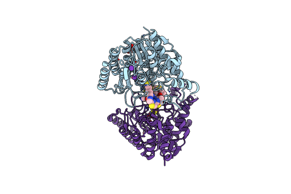 |
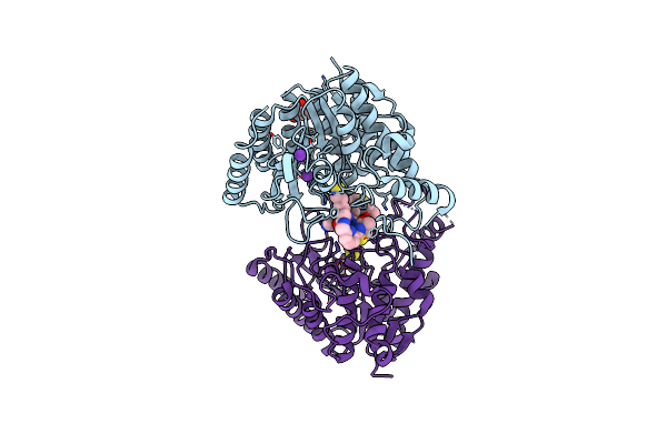
Crystal Structure Of S39D Hdac8 In Complex With A Largazole Analogue.
Organism: Homo sapiens
Method: X-RAY DIFFRACTION Resolution:1.76 Å Release Date: 2015-04-08 Classification: HYDROLASE/HYDROLASE INHIBITOR Ligands: L6G, ZN, K, IMD, GOL |
Organism: Homo sapiens
Method: X-RAY DIFFRACTION
Release Date: 2015-04-08
Ligands: L6G, ZN, K, IMD, GOL
 |
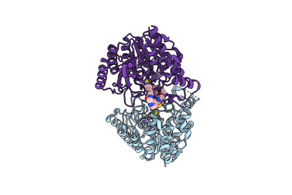
Crystal Structure Of S39D Hdac8 In Complex With A Largazole Analogue.
Organism: Homo sapiens
Method: X-RAY DIFFRACTION Resolution:2.18 Å Release Date: 2015-04-08 Classification: HYDROLASE/HYDROLASE INHIBITOR Ligands: ZN, K, L8G, IMD, GOL |
Organism: Homo sapiens
Method: X-RAY DIFFRACTION
Release Date: 2015-04-08
Ligands: ZN, K, L8G, IMD, GOL
 |
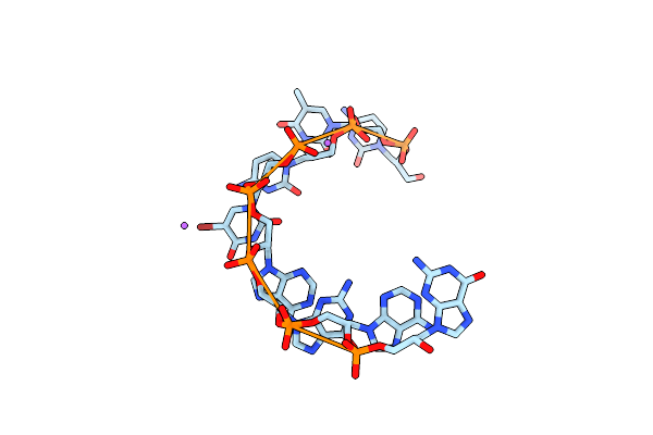
Crystal Structure Of S39D Hdac8 In Complex With A Largazole Analogue.
Organism: Homo sapiens
Method: X-RAY DIFFRACTION Resolution:2.39 Å Release Date: 2015-04-08 Classification: HYDROLASE/HYDROLASE INHIBITOR Ligands: ZN, K, L7G |
Organism: Homo sapiens
Method: X-RAY DIFFRACTION
Release Date: 2015-04-08
Ligands: ZN, K, L7G
 |
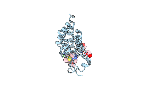
Crystal Structure Of The Gna 3'-Ctc(Br)Uagag-2'
|
Method: X-RAY DIFFRACTION
Release Date: 2011-05-04
 |
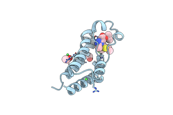
Crystal Structure Of The First Bromodomain Of Human Brd4 In Complex With The Inhibitor Jq1
Organism: Homo sapiens
Method: X-RAY DIFFRACTION Resolution:1.60 Å Release Date: 2010-10-06 Classification: CELL CYCLE Ligands: JQ1, IOD, EDO, DMS |
Organism: Homo sapiens
Method: X-RAY DIFFRACTION
Release Date: 2010-10-06
Ligands: JQ1, IOD, EDO, DMS
 |
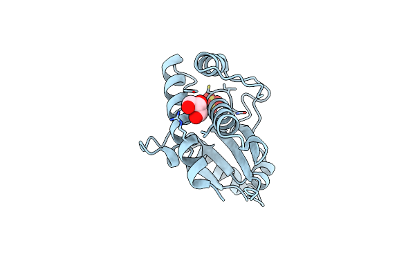
Crystal Structure Of The Second Bromodomain Of Human Brd2 In Complex With The Inhibitor Jq1
Organism: Homo sapiens
Method: X-RAY DIFFRACTION Resolution:1.61 Å Release Date: 2010-10-06 Classification: CELL CYCLE Ligands: JQ1, NI, EDO |
Organism: Homo sapiens
Method: X-RAY DIFFRACTION
Release Date: 2010-10-06
Ligands: JQ1, NI, EDO
 |
Crystal Structure Of Human B-Form Low Molecular Weight Phosphotyrosyl Phosphatase At 1.6 Angstrom Resolution
Organism: Homo sapiens
Method: X-RAY DIFFRACTION Resolution:1.63 Å Release Date: 2005-11-01 Classification: HYDROLASE Ligands: SO4, GOL |
Organism: Homo sapiens
Method: X-RAY DIFFRACTION
Release Date: 2005-11-01
Ligands: SO4, GOL