

Search Count: 11
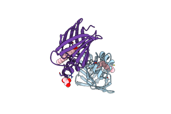 |
Structural Comparison Of Cellular Retinoic Acid Binding Protein I And Ii In The Presence And Absence Of Natural And Synthetic Ligands
Organism: Homo sapiens
Method: X-RAY DIFFRACTION Resolution:1.64 Å Release Date: 2021-02-17 Classification: SIGNALING PROTEIN Ligands: MYR, GOL, TDA |
Organism: Homo sapiens
Method: X-RAY DIFFRACTION
Release Date: 2021-02-17
Ligands: MYR, GOL, TDA
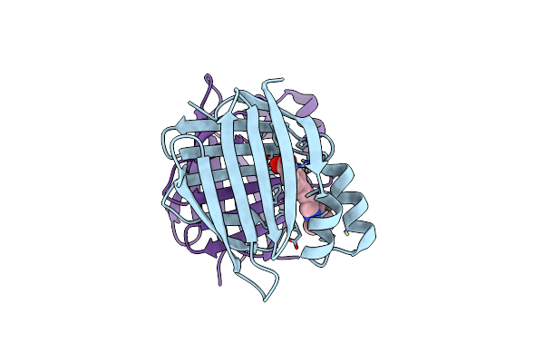 |
Structural Comparison Of Cellular Retinoic Acid Binding Protein I And Ii In The Presence And Absence Of Natural And Synthetic Ligands
Organism: Homo sapiens
Method: X-RAY DIFFRACTION Resolution:2.41 Å Release Date: 2021-02-17 Classification: SIGNALING PROTEIN Ligands: R62 |
Organism: Homo sapiens
Method: X-RAY DIFFRACTION
Release Date: 2021-02-17
Ligands: R62
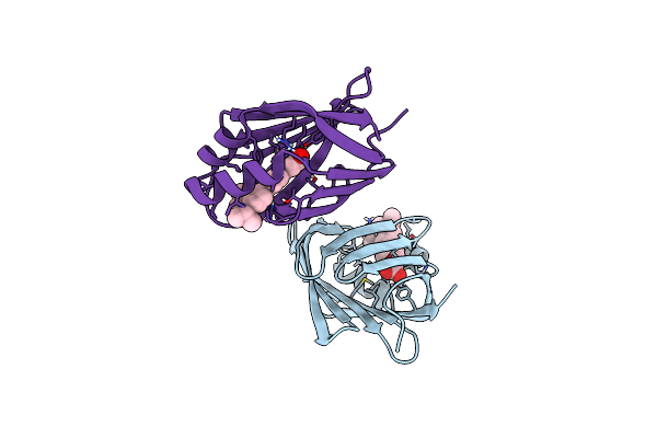 |
Structural Comparison Of Cellular Retinoic Acid Binding Protein I And Ii In The Presence And Absence Of Natural And Synthetic Ligands
Organism: Homo sapiens
Method: X-RAY DIFFRACTION Resolution:1.82 Å Release Date: 2021-02-17 Classification: SIGNALING PROTEIN Ligands: R6B |
Organism: Homo sapiens
Method: X-RAY DIFFRACTION
Release Date: 2021-02-17
Ligands: R6B
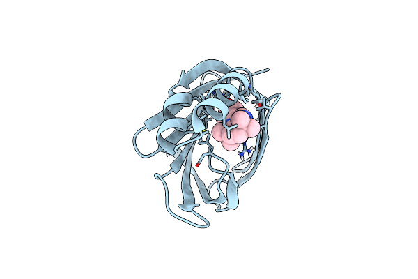 |
Structural Comparison Of Cellular Retinoic Acid Binding Proteins I And Ii In The Presence And Absence Of Natural And Synthetic Ligands
Organism: Homo sapiens
Method: X-RAY DIFFRACTION Resolution:1.71 Å Release Date: 2021-02-17 Classification: SIGNALING PROTEIN Ligands: R62 |
Organism: Homo sapiens
Method: X-RAY DIFFRACTION
Release Date: 2021-02-17
Ligands: R62
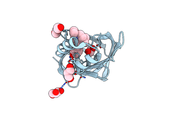 |
Human Cellular Retinoic Acid Binding Protein Ii (Crabpii) With Bound Synthetic Retinoid Dc271.
Organism: Homo sapiens
Method: X-RAY DIFFRACTION Resolution:1.50 Å Release Date: 2018-11-28 Classification: SIGNALING PROTEIN Ligands: EDO, G9Q, GOL, PG4 |
Organism: Homo sapiens
Method: X-RAY DIFFRACTION
Release Date: 2018-11-28
Ligands: EDO, G9Q, GOL, PG4
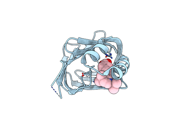 |
Human Cellular Retinoic Acid Binding Protein Ii (Crabpii) With Bound Synthetic Retinoid Dc360.
Organism: Homo sapiens
Method: X-RAY DIFFRACTION Resolution:1.80 Å Release Date: 2018-10-24 Classification: SIGNALING PROTEIN Ligands: 9U5 |
Organism: Homo sapiens
Method: X-RAY DIFFRACTION
Release Date: 2018-10-24
Ligands: 9U5
 |
Crystal Structure Of Hetero-Trimeric Core Of Lubac: Hoip Double-Uba Complexed With Hoil-1L Ubl And Sharpin Ubl
Organism: Mus musculus
Method: X-RAY DIFFRACTION Resolution:2.40 Å Release Date: 2018-05-02 Classification: LIGASE Ligands: GOL |
Organism: Mus musculus
Method: X-RAY DIFFRACTION
Release Date: 2018-05-02
Ligands: GOL
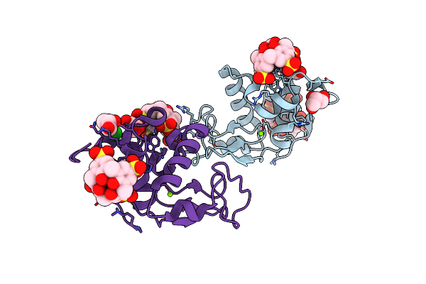 |
Crystal Structure Of Dimethyllysine Hen Egg-White Lysozyme In Complex With Sclx4 At 1.9 A Resolution
Organism: Gallus gallus
Method: X-RAY DIFFRACTION Resolution:1.90 Å Release Date: 2014-11-12 Classification: HYDROLASE Ligands: T3Y, CL, GOL, NA, MG |
Organism: Gallus gallus
Method: X-RAY DIFFRACTION
Release Date: 2014-11-12
Ligands: T3Y, CL, GOL, NA, MG
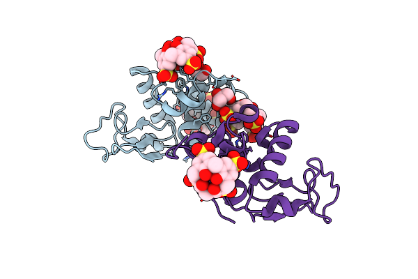 |
Crystal Structure Of Dimethyllysine Hen Egg-White Lysozyme In Complex With Sclx4 At 2.2 A Resolution
Organism: Gallus gallus
Method: X-RAY DIFFRACTION Resolution:2.20 Å Release Date: 2014-11-12 Classification: HYDROLASE Ligands: T3Y, GOL |
Organism: Gallus gallus
Method: X-RAY DIFFRACTION
Release Date: 2014-11-12
Ligands: T3Y, GOL
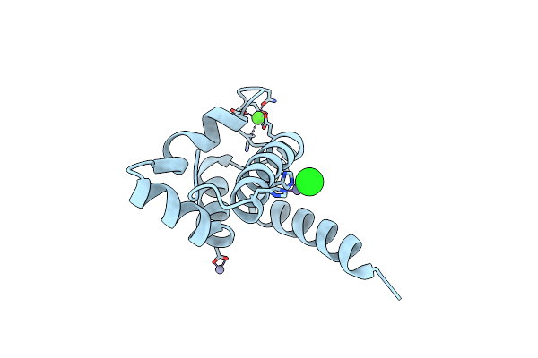 |
Structure Of Human S100A15 Bound To Zinc And Calcium
Organism: Homo sapiens
Method: X-RAY DIFFRACTION Resolution:1.70 Å Release Date: 2012-10-17 Classification: METAL BINDING PROTEIN Ligands: CA, ZN, CL |
Organism: Homo sapiens
Method: X-RAY DIFFRACTION
Release Date: 2012-10-17
Ligands: CA, ZN, CL
 |
Structure Of Human S100A7 D24G Bound To Zinc And Calcium
Organism: Homo sapiens
Method: X-RAY DIFFRACTION Resolution:1.60 Å Release Date: 2012-10-17 Classification: METAL BINDING PROTEIN Ligands: CA, CL, ZN |
Organism: Homo sapiens
Method: X-RAY DIFFRACTION
Release Date: 2012-10-17
Ligands: CA, CL, ZN