

Search Count: 42
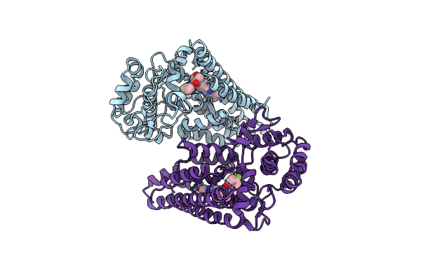 |
Ido1 In Complex With Compound S-1
Organism: Homo sapiens
Method: X-RAY DIFFRACTION Resolution:2.76 Å Release Date: 2022-08-24 Classification: OXIDOREDUCTASE Ligands: 6IZ |
Organism: Homo sapiens
Method: X-RAY DIFFRACTION
Release Date: 2022-08-24
Ligands: 6IZ
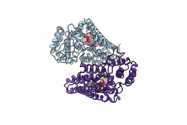 |
Ido1 In Complex With Compound 9
Organism: Homo sapiens
Method: X-RAY DIFFRACTION Resolution:2.69 Å Release Date: 2022-03-16 Classification: OXIDOREDUCTASE Ligands: 6ZI |
Organism: Homo sapiens
Method: X-RAY DIFFRACTION
Release Date: 2022-03-16
Ligands: 6ZI
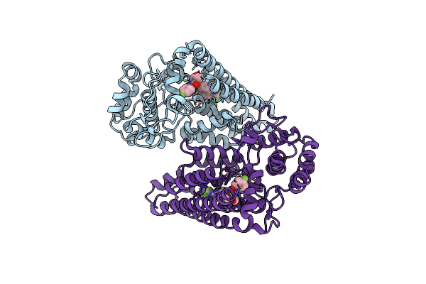 |
Ido1 In Complex With Compound 14
Organism: Homo sapiens
Method: X-RAY DIFFRACTION Resolution:2.18 Å Release Date: 2022-03-16 Classification: OXIDOREDUCTASE Ligands: 6RI |
Organism: Homo sapiens
Method: X-RAY DIFFRACTION
Release Date: 2022-03-16
Ligands: 6RI
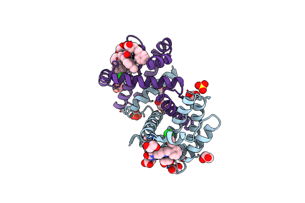 |
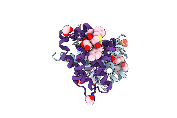
Crystal Structure Of Bcl-Xl Bound To Compound 8: (R)-3-(Benzylthio)-2-(3-(2-((4'-Chloro-[1,1'-Biphenyl]-2-Yl)Methyl)-1,2,3,4-Tetrahydroisoquinoline-6-Carbonyl)-3-(4-Methylbenzyl)Ureido)Propanoic Acid
Organism: Homo sapiens
Method: X-RAY DIFFRACTION Resolution:1.90 Å Release Date: 2021-05-05 Classification: Apoptosis/INHIBITOR Ligands: XOY, SO4, EDO |
Organism: Homo sapiens
Method: X-RAY DIFFRACTION
Release Date: 2021-05-05
Ligands: XOY, SO4, EDO
 |
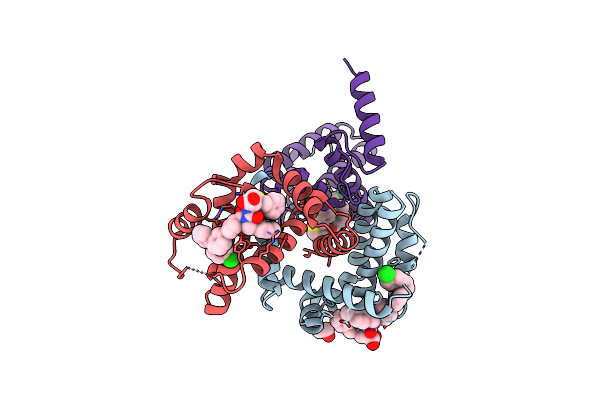
Crystal Structure Of Bcl-Xl Bound To Compound 2: (2R)-3-(Benzylsulfanyl)-2-({[(4-Methylphenyl)Methyl] [(4 Phenylphenyl)Carbonyl] Carbamoyl}Amino) Propanoic Acid
Organism: Homo sapiens
Method: X-RAY DIFFRACTION Resolution:2.15 Å Release Date: 2021-05-05 Classification: Apoptosis/INHIBITOR Ligands: XOU, SO4, EDO |
Organism: Homo sapiens
Method: X-RAY DIFFRACTION
Release Date: 2021-05-05
Ligands: XOU, SO4, EDO
 |
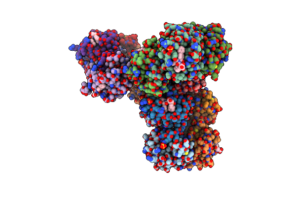
Crystal Structure Of Bcl-Xl Bound To Compound 7: (R)-3-(Benzylthio)-2-(3-(4-Chloro-[1,1':2',1'':3'',1'''-Quaterphenyl]-4'''-Carbonyl)-3-(4-Methylbenzyl)Ureido)Propanoic Acid
Organism: Homo sapiens
Method: X-RAY DIFFRACTION Resolution:2.87 Å Release Date: 2021-05-05 Classification: Apoptosis/INHIBITOR Ligands: QHV, EDO |
Organism: Homo sapiens
Method: X-RAY DIFFRACTION
Release Date: 2021-05-05
Ligands: QHV, EDO
 |
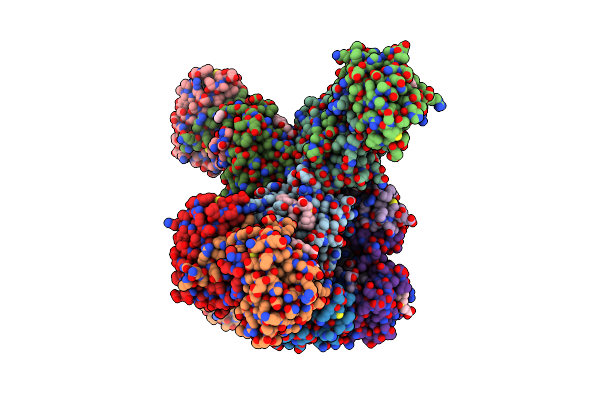
Crystal Structure Of Bcl-Xl Bound To Compound 12: (R)-2-(3-([1,1'-Biphenyl]-4-Carbonyl)-3-(4-Methylbenzyl)Ureido)-3-((Cyclohexylmethyl)Sulfonyl)Propanoic Acid
Organism: Homo sapiens
Method: X-RAY DIFFRACTION Resolution:2.24 Å Release Date: 2021-05-05 Classification: Apoptosis/INHIBITOR Ligands: SO4, EDO, QHS |
Organism: Homo sapiens
Method: X-RAY DIFFRACTION
Release Date: 2021-05-05
Ligands: SO4, EDO, QHS
 |
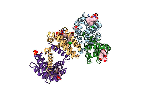
Crystal Structure Of Bcl-Xl Bound To Compound 13: (R)-2-(3-([1,1'-Biphenyl]-4-Carbonyl)-3-(4-Methylbenzyl)Ureido)-3-(((3R,5R,7R)-Adamantan-1-Ylmethyl)Sulfonyl)Propanoic Acid
Organism: Homo sapiens
Method: X-RAY DIFFRACTION Resolution:2.10 Å Release Date: 2021-05-05 Classification: Apoptosis/INHIBITOR Ligands: QHP, SO4, EDO |
Organism: Homo sapiens
Method: X-RAY DIFFRACTION
Release Date: 2021-05-05
Ligands: QHP, SO4, EDO
 |
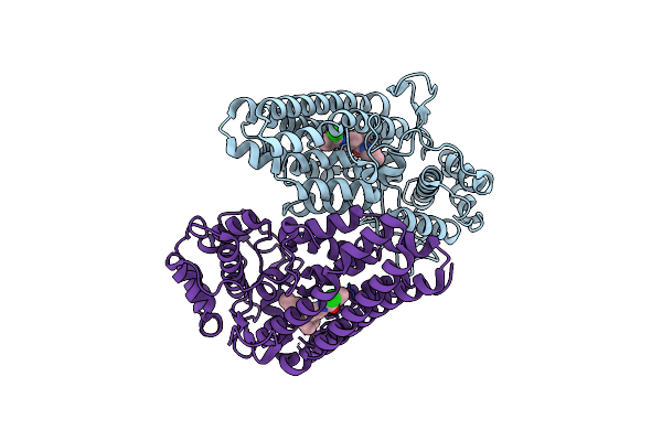
Crystal Structure Of Bcl-Xl Bound To Compound 15: (R)-2-(3-(2-((4'-Chloro-[1,1'-Biphenyl]-2-Yl)Methyl)-1,2,3,4-Tetrahydroisoquinoline-6-Carbonyl)-3-(4-Methylbenzyl)Ureido)-3-((Cyclohexylmethyl)Sulfonyl)Propanoic Acid
Organism: Homo sapiens
Method: X-RAY DIFFRACTION Resolution:2.19 Å Release Date: 2021-05-05 Classification: Apoptosis/INHIBITOR Ligands: EDO, SO4, QHJ |
Organism: Homo sapiens
Method: X-RAY DIFFRACTION
Release Date: 2021-05-05
Ligands: EDO, SO4, QHJ
 |
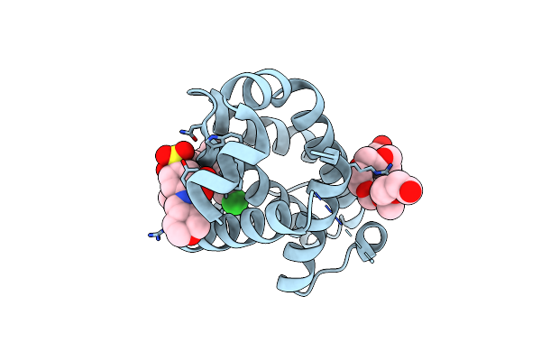
Ido1 In Complex With Compound 1
Organism: Homo sapiens
Method: X-RAY DIFFRACTION Resolution:1.78 Å Release Date: 2020-04-08 Classification: OXIDOREDUCTASE Ligands: QPV |
Organism: Homo sapiens
Method: X-RAY DIFFRACTION
Release Date: 2020-04-08
Ligands: QPV
 |
Crystal Structure Of Bcl-2 With Venetoclax
Organism: Homo sapiens
Method: X-RAY DIFFRACTION Resolution:1.62 Å Release Date: 2019-05-22 Classification: APOPTOSIS Ligands: LBM, 2PE |
Organism: Homo sapiens
Method: X-RAY DIFFRACTION
Release Date: 2019-05-22
Ligands: LBM, 2PE
 |
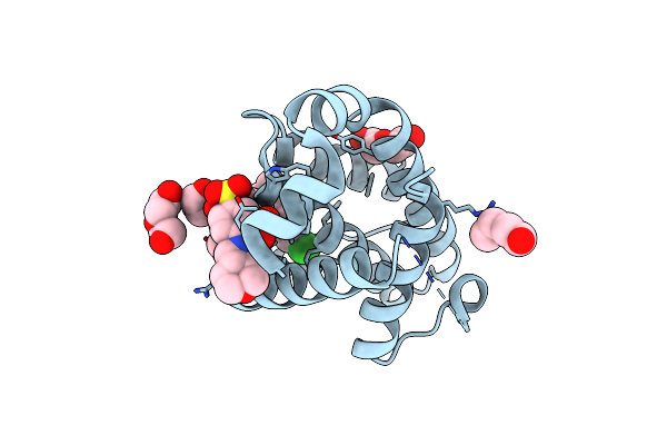
Crystal Structure Of Bcl-2 G101V Mutation With Venetoclax
Organism: Homo sapiens
Method: X-RAY DIFFRACTION Resolution:2.20 Å Release Date: 2019-05-22 Classification: APOPTOSIS Ligands: LBM, PEG, CL |
Organism: Homo sapiens
Method: X-RAY DIFFRACTION
Release Date: 2019-05-22
Ligands: LBM, PEG, CL
 |
Crystal Structure Of Bcl-2 F104L Mutation With Venetoclax
Organism: Homo sapiens
Method: X-RAY DIFFRACTION Resolution:1.75 Å Release Date: 2019-05-22 Classification: APOPTOSIS Ligands: LBM, PEG |
Organism: Homo sapiens
Method: X-RAY DIFFRACTION
Release Date: 2019-05-22
Ligands: LBM, PEG
 |
Crystal Structure Of Bcl-2 G101V Mutation With S55746
Organism: Homo sapiens
Method: X-RAY DIFFRACTION Resolution:2.00 Å Release Date: 2019-05-22 Classification: APOPTOSIS Ligands: F3Q |
Organism: Homo sapiens
Method: X-RAY DIFFRACTION
Release Date: 2019-05-22
Ligands: F3Q
 |
Crystal Structure Of Bcl-2 G101A Mutation With Venetoclax
Organism: Homo sapiens
Method: X-RAY DIFFRACTION Resolution:1.80 Å Release Date: 2019-05-22 Classification: APOPTOSIS Ligands: LBM, PEG |
Organism: Homo sapiens
Method: X-RAY DIFFRACTION
Release Date: 2019-05-22
Ligands: LBM, PEG
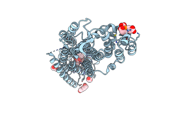 |
Structure Of Constitutively Active Neurotensin Receptor
Organism: Rattus norvegicus, Enterobacteria phage t4
Method: X-RAY DIFFRACTION Resolution:3.30 Å Release Date: 2016-12-21 Classification: SIGNALING PROTEIN Ligands: TCE, GOL, PEG |
Organism: Rattus norvegicus, Enterobacteria phage t4
Method: X-RAY DIFFRACTION
Release Date: 2016-12-21
Ligands: TCE, GOL, PEG
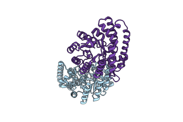 |
Crystal Structure Of Hypd, A 1-Pyrroline-4-Hydroxy-2-Carboxylate Deaminase From Sinorhizobium Meliloti
Organism: Rhizobium meliloti (strain 1021)
Method: X-RAY DIFFRACTION Resolution:1.92 Å Release Date: 2016-02-03 Classification: HYDROLASE |
Organism: Rhizobium meliloti (strain 1021)
Method: X-RAY DIFFRACTION
Release Date: 2016-02-03
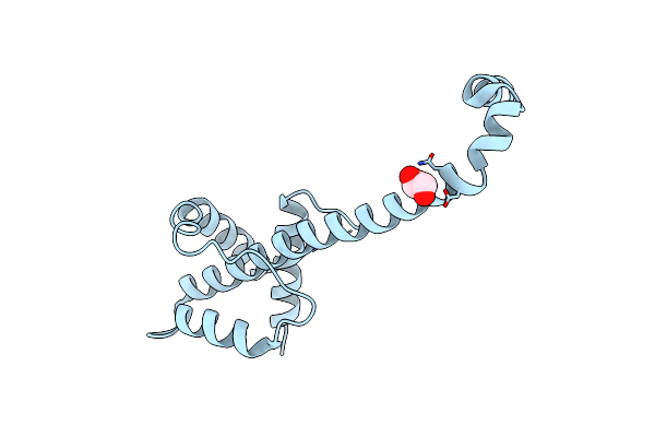 |
Crystal Structure Of Bcl-Xl Hexamer
Organism: Homo sapiens
Method: X-RAY DIFFRACTION Resolution:2.85 Å Release Date: 2015-03-04 Classification: APOPTOSIS Ligands: GOL |
Organism: Homo sapiens
Method: X-RAY DIFFRACTION
Release Date: 2015-03-04
Ligands: GOL
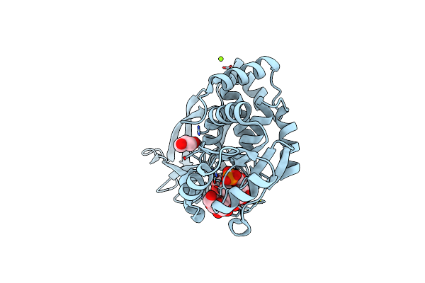 |
Phospho-Glucan Bound Structure Of Starch Phosphatase Starch Excess4 Reveals The Mechanism For C6-Specificty
Organism: Arabidopsis thaliana
Method: X-RAY DIFFRACTION Resolution:1.65 Å Release Date: 2014-04-23 Classification: HYDROLASE Ligands: MG, PO4, GOL |
Organism: Arabidopsis thaliana
Method: X-RAY DIFFRACTION
Release Date: 2014-04-23
Ligands: MG, PO4, GOL
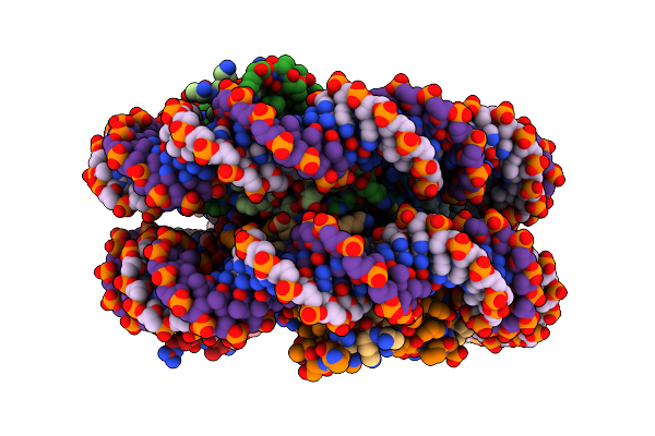 |
Crystal Structure Of Nucleosome Core Particle Containing A Poly (Da.Dt) Sequence Element
Organism: Xenopus laevis
Method: X-RAY DIFFRACTION Resolution:3.20 Å Release Date: 2006-09-26 Classification: STRUCTURAL PROTEIN/DNA |
Organism: Xenopus laevis
Method: X-RAY DIFFRACTION
Release Date: 2006-09-26