

Search Count: 177
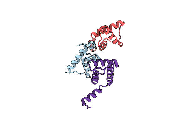 |
Structure Of C-Terminal Domain Of Telomere Resolvase, Rest, From Borrelia Garinii
Organism: Borreliella garinii
Method: X-RAY DIFFRACTION Resolution:2.26 Å Release Date: 2024-07-17 Classification: DNA BINDING PROTEIN |
Organism: Borreliella garinii
Method: X-RAY DIFFRACTION
Release Date: 2024-07-17
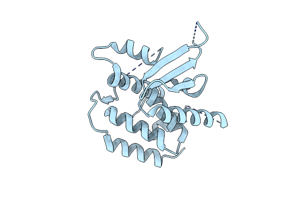 |
Structure Of Catalytic Domain Of Telomere Resolvase, Rest, From Borrelia Garinii
Organism: Borreliella garinii
Method: X-RAY DIFFRACTION Resolution:2.80 Å Release Date: 2024-07-17 Classification: DNA BINDING PROTEIN |
Organism: Borreliella garinii
Method: X-RAY DIFFRACTION
Release Date: 2024-07-17
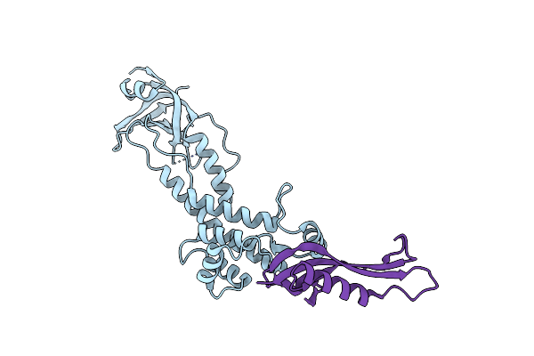 |
Crystal Structure Of The Periplasmic Domain Of Igaa From Escherichia Coli
Organism: Escherichia coli
Method: X-RAY DIFFRACTION Resolution:1.80 Å Release Date: 2024-07-17 Classification: SIGNALING PROTEIN |
Organism: Escherichia coli
Method: X-RAY DIFFRACTION
Release Date: 2024-07-17
 |
Crystal Structure Of The Periplasmic Domain Of Igaa From Klebsiella Pneumoniae
Organism: Klebsiella pneumoniae
Method: X-RAY DIFFRACTION Resolution:2.50 Å Release Date: 2024-07-17 Classification: SIGNALING PROTEIN |
Organism: Klebsiella pneumoniae
Method: X-RAY DIFFRACTION
Release Date: 2024-07-17
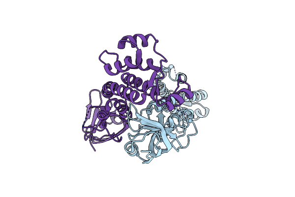 |
Crystal Structure Of The Periplasmic Domain Of Igaa From Escherichia Coli
Organism: Escherichia coli
Method: X-RAY DIFFRACTION Resolution:2.64 Å Release Date: 2024-07-17 Classification: SIGNALING PROTEIN |
Organism: Escherichia coli
Method: X-RAY DIFFRACTION
Release Date: 2024-07-17
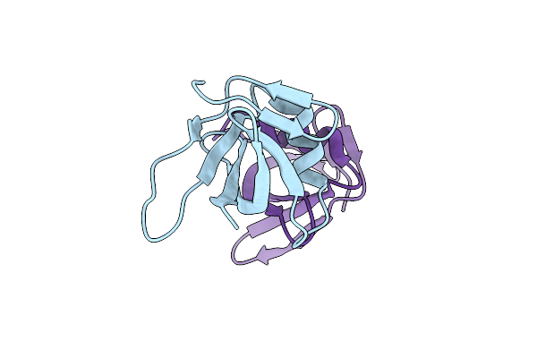 |
Structure Of Duf1480 Domain-Containing Protein From Klebsiella Pneumoniae Subsp. Pneumoniae Sa1
Organism: Klebsiella pneumoniae
Method: X-RAY DIFFRACTION Resolution:1.75 Å Release Date: 2024-06-19 Classification: UNKNOWN FUNCTION |
Organism: Klebsiella pneumoniae
Method: X-RAY DIFFRACTION
Release Date: 2024-06-19
 |
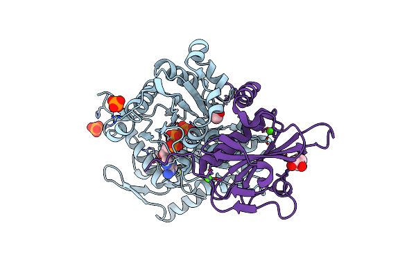
Crystal Structure Of Fragmin Domain-1 In Complex With Actin (Amppnp-Form)
Organism: Physarum polycephalum, Gallus gallus
Method: X-RAY DIFFRACTION Resolution:1.15 Å Release Date: 2022-10-26 Classification: CONTRACTILE PROTEIN Ligands: ANP, MG, PO4, EDO, CA |
Organism: Physarum polycephalum, Gallus gallus
Method: X-RAY DIFFRACTION
Release Date: 2022-10-26
Ligands: ANP, MG, PO4, EDO, CA
 |
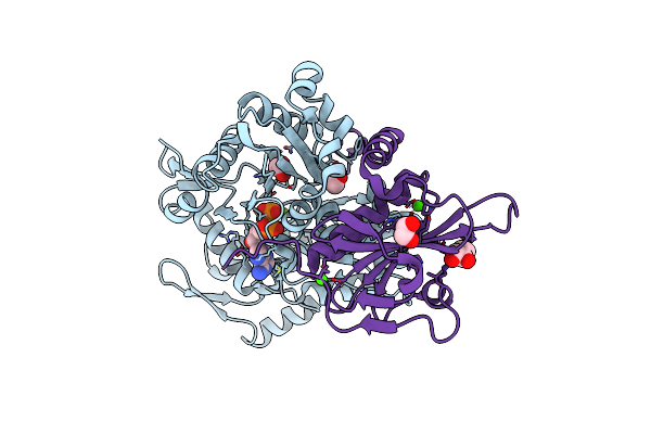
Crystal Structure Of Fragmin Domain-1 In Complex With Actin (Adp-Pi-Form)
Organism: Physarum polycephalum, Gallus gallus
Method: X-RAY DIFFRACTION Resolution:1.15 Å Release Date: 2022-10-26 Classification: CONTRACTILE PROTEIN Ligands: ADP, PO4, MG, EDO, CA |
Organism: Physarum polycephalum, Gallus gallus
Method: X-RAY DIFFRACTION
Release Date: 2022-10-26
Ligands: ADP, PO4, MG, EDO, CA
 |
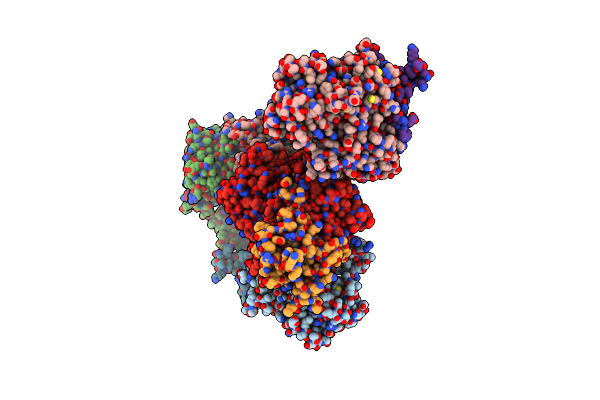
Crystal Structure Of Fragmin Domain-1 In Complex With Actin (Adp-Form)
Organism: Physarum polycephalum, Gallus gallus
Method: X-RAY DIFFRACTION Resolution:1.20 Å Release Date: 2022-10-26 Classification: CONTRACTILE PROTEIN Ligands: ADP, MG, EDO, CA |
Organism: Physarum polycephalum, Gallus gallus
Method: X-RAY DIFFRACTION
Release Date: 2022-10-26
Ligands: ADP, MG, EDO, CA
 |
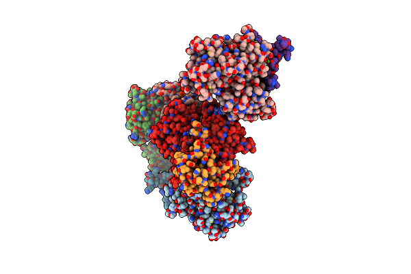
Crystal Structure Of Fragmin Domain-1 (15-160) In Complex With Actin
Organism: Physarum polycephalum, Gallus gallus
Method: X-RAY DIFFRACTION Resolution:2.00 Å Release Date: 2022-10-26 Classification: CONTRACTILE PROTEIN Ligands: ATP, CA, ACT, NA, EDO |
Organism: Physarum polycephalum, Gallus gallus
Method: X-RAY DIFFRACTION
Release Date: 2022-10-26
Ligands: ATP, CA, ACT, NA, EDO
 |
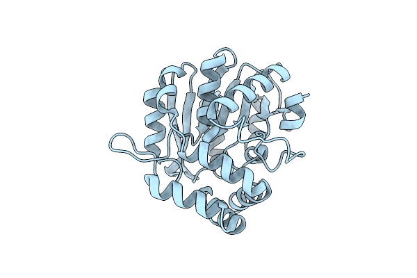
Crystal Structure Of Fragmin Domain-1 (1-160) In Complex With G-Form Actin
Organism: Physarum polycephalum, Gallus gallus
Method: X-RAY DIFFRACTION Resolution:2.70 Å Release Date: 2022-10-26 Classification: CONTRACTILE PROTEIN Ligands: ADP, CA, ACT, NA, EDO, PO4 |
Organism: Physarum polycephalum, Gallus gallus
Method: X-RAY DIFFRACTION
Release Date: 2022-10-26
Ligands: ADP, CA, ACT, NA, EDO, PO4
 |
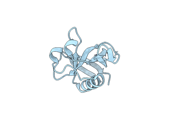
Crystal Structure Of Arabidopsis Thaliana Dlk2
Organism: Arabidopsis thaliana
Method: X-RAY DIFFRACTION Resolution:1.48 Å Release Date: 2022-09-28 Classification: HYDROLASE |
Organism: Arabidopsis thaliana
Method: X-RAY DIFFRACTION
Release Date: 2022-09-28
 |
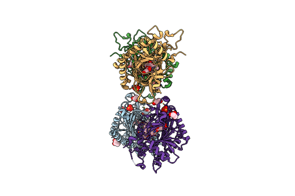
Crystal Structure Of Nsp1 From Sars-Cov-2
Organism: Severe acute respiratory syndrome coronavirus 2
Method: X-RAY DIFFRACTION Resolution:1.65 Å Release Date: 2020-09-30 Classification: VIRAL PROTEIN |
Organism: Severe acute respiratory syndrome coronavirus 2
Method: X-RAY DIFFRACTION
Release Date: 2020-09-30
 |
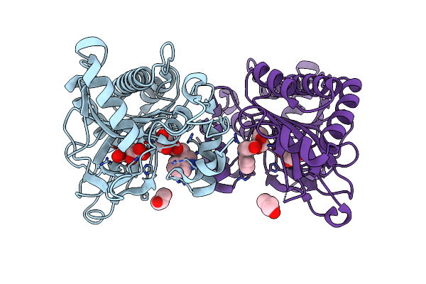
Crystal Structure Of Gibberellin 2-Oxidase3 (Ga2Ox3)In Rice
Organism: Oryza sativa subsp. japonica
Method: X-RAY DIFFRACTION Resolution:2.15 Å Release Date: 2020-05-13 Classification: BIOSYNTHETIC PROTEIN Ligands: GA4, AKG, GOL, SO4 |
Organism: Oryza sativa subsp. japonica
Method: X-RAY DIFFRACTION
Release Date: 2020-05-13
Ligands: GA4, AKG, GOL, SO4
 |
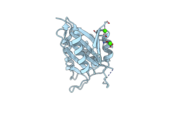
Crystal Structure Of Dioxygenase For Auxin Oxidation (Dao) In Rice
Organism: Oryza sativa subsp. indica
Method: X-RAY DIFFRACTION Resolution:2.00 Å Release Date: 2020-05-13 Classification: BIOSYNTHETIC PROTEIN Ligands: AKG, IAC, GOL |
Organism: Oryza sativa subsp. indica
Method: X-RAY DIFFRACTION
Release Date: 2020-05-13
Ligands: AKG, IAC, GOL
 |
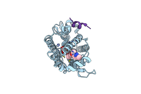
Crystal Structure Of Type Vi Secretion System Effector, Tseh (Vca0285)
Organism: Vibrio cholerae
Method: X-RAY DIFFRACTION Resolution:1.80 Å Release Date: 2020-01-15 Classification: HYDROLASE Ligands: CA |
Organism: Vibrio cholerae
Method: X-RAY DIFFRACTION
Release Date: 2020-01-15
Ligands: CA
 |
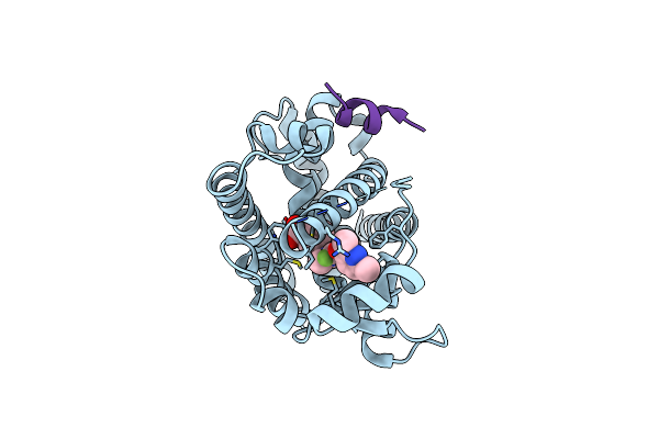
Crystal Structure Of The Ppargamma-Lbd Complexed With Compound 1L
Organism: Homo sapiens
Method: X-RAY DIFFRACTION Resolution:1.80 Å Release Date: 2019-03-27 Classification: TRANSCRIPTION Ligands: B0X |
Organism: Homo sapiens
Method: X-RAY DIFFRACTION
Release Date: 2019-03-27
Ligands: B0X
 |
Crystal Structure Of The Ppargamma-Lbd Complexed With Compound 3G
Organism: Homo sapiens
Method: X-RAY DIFFRACTION Resolution:1.75 Å Release Date: 2019-03-27 Classification: TRANSCRIPTION Ligands: B1O |
Organism: Homo sapiens
Method: X-RAY DIFFRACTION
Release Date: 2019-03-27
Ligands: B1O
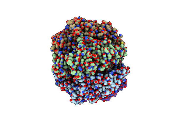 |
Structure Of The Assembled Atpase Escn From The Enteropathogenic E. Coli (Epec) Type Iii Secretion System
Organism: Escherichia coli o127:h6 (strain e2348/69 / epec)
Method: ELECTRON MICROSCOPY Release Date: 2019-02-20 Classification: HYDROLASE Ligands: ADP, MG, AF3 |
Organism: Escherichia coli o127:h6 (strain e2348/69 / epec)
Method: ELECTRON MICROSCOPY
Release Date: 2019-02-20
Ligands: ADP, MG, AF3
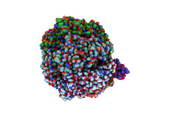 |
Structure Of The Assembled Atpase Escn In Complex With Its Central Stalk Esco From The Enteropathogenic E. Coli (Epec) Type Iii Secretion System
Organism: Escherichia coli o127:h6 (strain e2348/69 / epec)
Method: ELECTRON MICROSCOPY Release Date: 2019-02-20 Classification: HYDROLASE Ligands: ADP, MG, AF3 |
Organism: Escherichia coli o127:h6 (strain e2348/69 / epec)
Method: ELECTRON MICROSCOPY
Release Date: 2019-02-20
Ligands: ADP, MG, AF3