

Search Count: 29
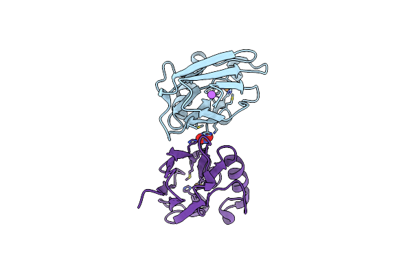 |
All Phe-Azurin Variant - F15Y
Organism: Pseudomonas aeruginosa
Method: X-RAY DIFFRACTION Resolution:1.45 Å Release Date: 2022-06-22 Classification: ELECTRON TRANSPORT Ligands: CU, NA, NO3 |
Organism: Pseudomonas aeruginosa
Method: X-RAY DIFFRACTION
Release Date: 2022-06-22
Ligands: CU, NA, NO3
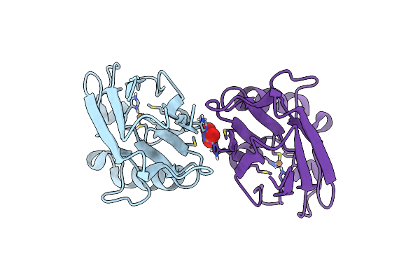 |
All Phe-Azurin Variant - F15W
Organism: Pseudomonas aeruginosa
Method: X-RAY DIFFRACTION Resolution:1.85 Å Release Date: 2022-06-22 Classification: ELECTRON TRANSPORT Ligands: CU, NO3 |
Organism: Pseudomonas aeruginosa
Method: X-RAY DIFFRACTION
Release Date: 2022-06-22
Ligands: CU, NO3
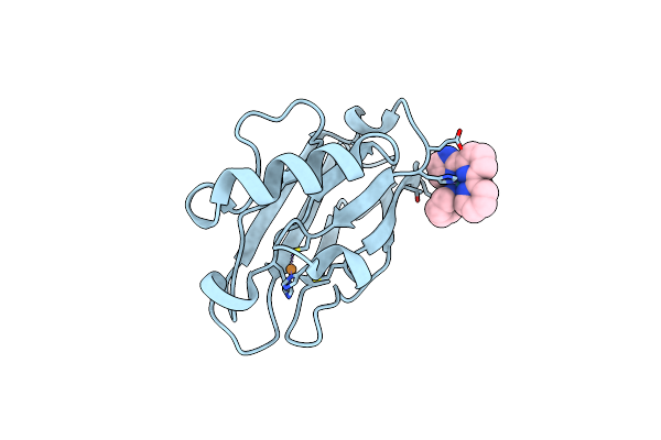 |
Crystal Structure Of The Pseudomonas Aeruginosa Azurin, Ruh107No Yoh109
Organism: Pseudomonas aeruginosa
Method: X-RAY DIFFRACTION Resolution:1.60 Å Release Date: 2012-11-21 Classification: ELECTRON TRANSPORT Ligands: CU, DRU |
Organism: Pseudomonas aeruginosa
Method: X-RAY DIFFRACTION
Release Date: 2012-11-21
Ligands: CU, DRU
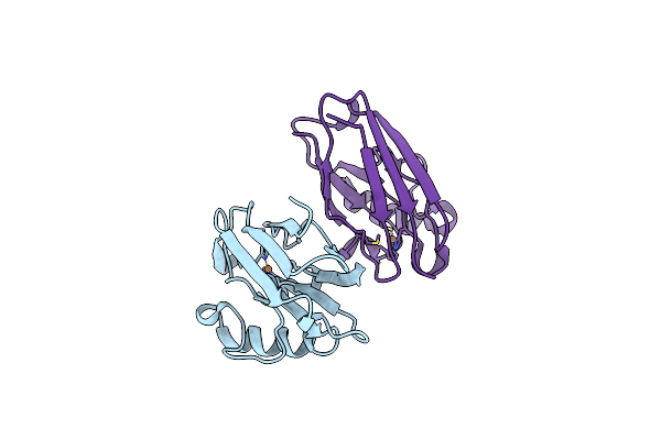 |
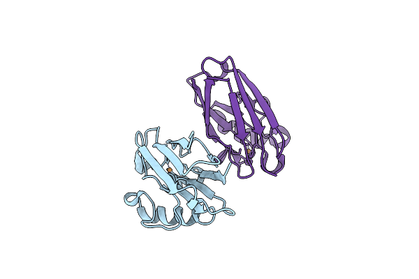
Crystal Structure Of The Pseudomonas Aeruginosa Azurin, H124No Yoh122
Organism: Pseudomonas aeruginosa
Method: X-RAY DIFFRACTION Resolution:2.00 Å Release Date: 2012-11-21 Classification: ELECTRON TRANSPORT Ligands: CU |
Organism: Pseudomonas aeruginosa
Method: X-RAY DIFFRACTION
Release Date: 2012-11-21
Ligands: CU
 |
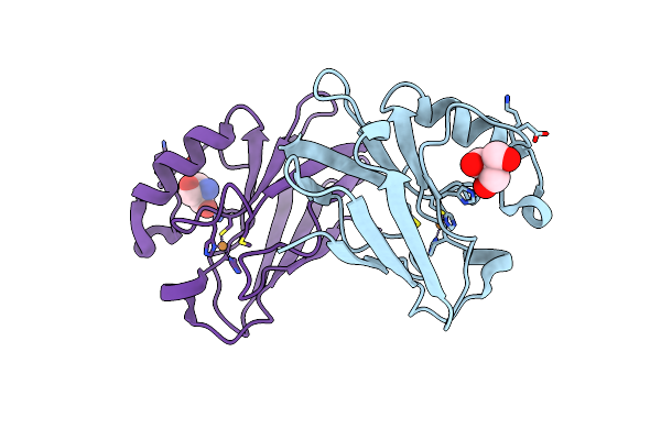
Crystal Structure Of The Pseudomonas Aeruginosa Azurin, H126No Yoh109
Organism: Pseudomonas aeruginosa
Method: X-RAY DIFFRACTION Resolution:1.90 Å Release Date: 2012-11-21 Classification: ELECTRON TRANSPORT Ligands: CU |
Organism: Pseudomonas aeruginosa
Method: X-RAY DIFFRACTION
Release Date: 2012-11-21
Ligands: CU
 |
Crystal Structure Of P. Aeruginoas Azurin Containing A Tyr-His Hydrogen Bonded Pair
Organism: Pseudomonas aeruginosa
Method: X-RAY DIFFRACTION Resolution:1.18 Å Release Date: 2011-12-28 Classification: ELECTRON TRANSPORT Ligands: CU, TRS |
Organism: Pseudomonas aeruginosa
Method: X-RAY DIFFRACTION
Release Date: 2011-12-28
Ligands: CU, TRS
 |
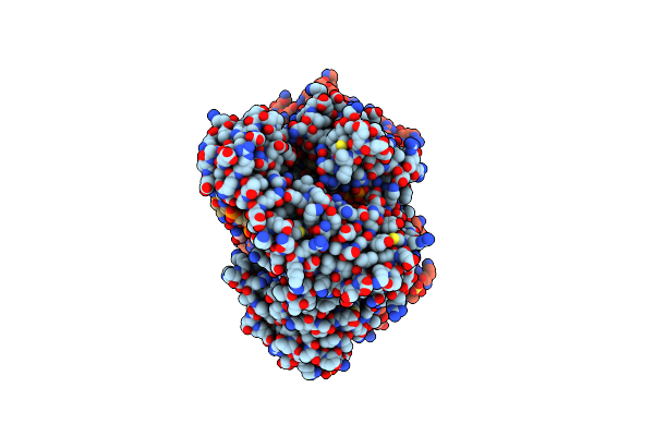
Cocrystal Structure Of Bacillus Fragment Dna Polymerase I With Duplex Dna (Open Form)
Organism: Bacillus stearothermophilus
Method: X-RAY DIFFRACTION Resolution:2.10 Å Release Date: 2009-11-10 Classification: TRANSFERASE/DNA Ligands: SO4 |
Organism: Bacillus stearothermophilus
Method: X-RAY DIFFRACTION
Release Date: 2009-11-10
Ligands: SO4
 |
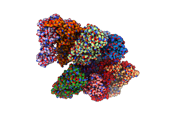
Cocrystal Structure Of Bacillus Fragment Dna Polymerase I With Duplex Dna , Dctp, And Zinc (Closed Form).
Organism: Bacillus stearothermophilus
Method: X-RAY DIFFRACTION Resolution:1.90 Å Release Date: 2009-11-10 Classification: TRANSFERASE/DNA Ligands: SO4, ZN, DAD |
Organism: Bacillus stearothermophilus
Method: X-RAY DIFFRACTION
Release Date: 2009-11-10
Ligands: SO4, ZN, DAD
 |
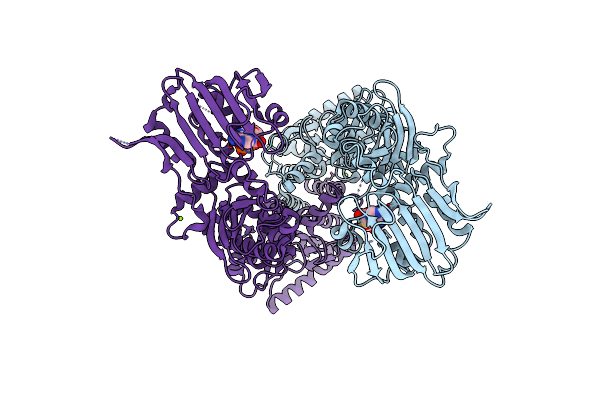
Structure Of Middle Plus C-Terminal Domains (M+C) Of Grp94
Organism: Canis lupus familiaris
Method: X-RAY DIFFRACTION Resolution:3.20 Å Release Date: 2007-10-23 Classification: CHAPERONE |
Organism: Canis lupus familiaris
Method: X-RAY DIFFRACTION
Release Date: 2007-10-23
 |
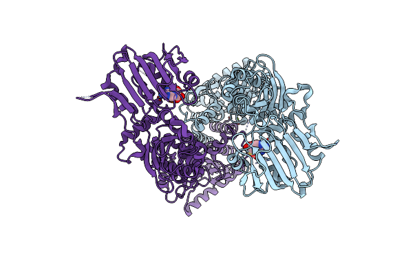
Structure Of Full Length Grp94 With Amp-Pnp Bound
Organism: Canis lupus familiaris
Method: X-RAY DIFFRACTION Resolution:2.40 Å Release Date: 2007-10-23 Classification: CHAPERONE Ligands: MG, ANP |
Organism: Canis lupus familiaris
Method: X-RAY DIFFRACTION
Release Date: 2007-10-23
Ligands: MG, ANP
 |
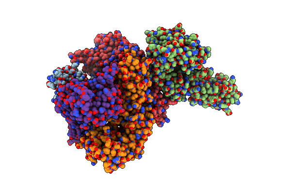
Structure Of Full Length Grp94 With Adp Bound
Organism: Canis lupus familiaris
Method: X-RAY DIFFRACTION Resolution:2.45 Å Release Date: 2007-10-23 Classification: CHAPERONE Ligands: MG, ADP |
Organism: Canis lupus familiaris
Method: X-RAY DIFFRACTION
Release Date: 2007-10-23
Ligands: MG, ADP
 |
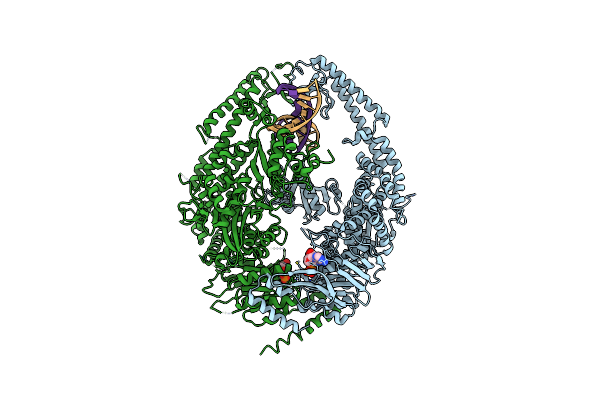
Structure Of N-Terminal Plus Middle Domains (N+M) Of Grp94
Organism: Canis lupus familiaris
Method: X-RAY DIFFRACTION Resolution:3.40 Å Release Date: 2007-10-23 Classification: CHAPERONE |
Organism: Canis lupus familiaris
Method: X-RAY DIFFRACTION
Release Date: 2007-10-23
 |
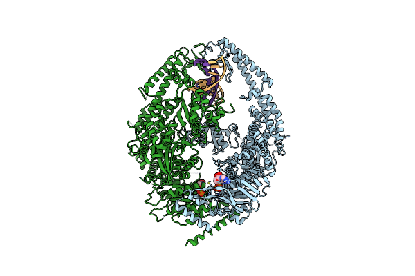
Human Mutsalpha (Msh2/Msh6) Bound To Adp And A G T Mispair
Organism: Homo sapiens
Method: X-RAY DIFFRACTION Resolution:2.75 Å Release Date: 2007-06-05 Classification: DNA BINDING PROTEIN/DNA Ligands: MG, ADP |
Organism: Homo sapiens
Method: X-RAY DIFFRACTION
Release Date: 2007-06-05
Ligands: MG, ADP
 |
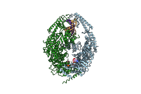
Human Mutsalpha (Msh2/Msh6) Bound To Adp And An O6-Methyl-Guanine T Mispair
Organism: Homo sapiens
Method: X-RAY DIFFRACTION Resolution:3.37 Å Release Date: 2007-06-05 Classification: DNA BINDING PROTEIN/DNA Ligands: MG, ADP |
Organism: Homo sapiens
Method: X-RAY DIFFRACTION
Release Date: 2007-06-05
Ligands: MG, ADP
 |
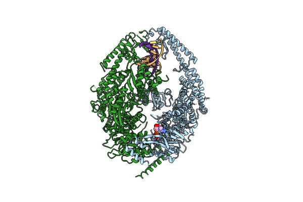
Human Mutsalpha (Msh2/Msh6) Bound To Adp And A G Du Mispair
Organism: Homo sapiens
Method: X-RAY DIFFRACTION Resolution:3.00 Å Release Date: 2007-06-05 Classification: DNA BINDING PROTEIN/DNA Ligands: MG, ADP |
Organism: Homo sapiens
Method: X-RAY DIFFRACTION
Release Date: 2007-06-05
Ligands: MG, ADP
 |
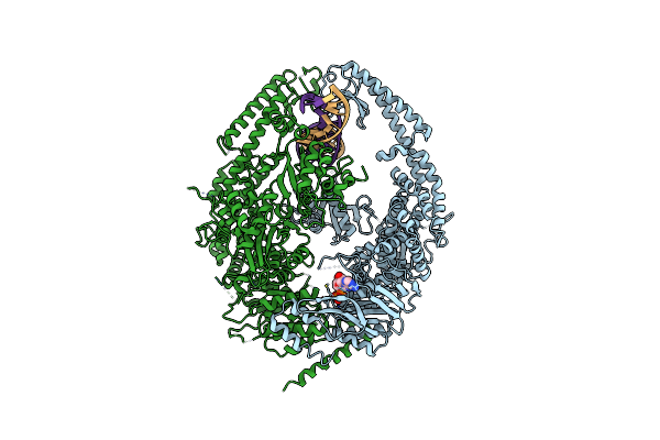
Human Mutsalpha (Msh2/Msh6) Bound To A G T Mispair, With Adp Bound To Msh2 Only
Organism: Homo sapiens
Method: X-RAY DIFFRACTION Resolution:3.30 Å Release Date: 2007-06-05 Classification: DNA BINDING PROTEIN/DNA Ligands: MG, ADP |
Organism: Homo sapiens
Method: X-RAY DIFFRACTION
Release Date: 2007-06-05
Ligands: MG, ADP
 |
Human Mutsalpha (Msh2/Msh6) Bound To Dna With A Single Base T Insert
Organism: Homo sapiens
Method: X-RAY DIFFRACTION Resolution:3.25 Å Release Date: 2007-06-05 Classification: DNA BINDING PROTEIN/DNA Ligands: MG, ADP |
Organism: Homo sapiens
Method: X-RAY DIFFRACTION
Release Date: 2007-06-05
Ligands: MG, ADP
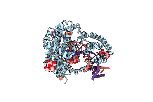 |
O6-Methyl-Guanine:T Pair In The Polymerase-10 Basepair Position
Organism: Geobacillus stearothermophilus
Method: X-RAY DIFFRACTION Resolution:1.80 Å Release Date: 2006-12-12 Classification: Transferase/DNA Ligands: SO4, MG |
Organism: Geobacillus stearothermophilus
Method: X-RAY DIFFRACTION
Release Date: 2006-12-12
Ligands: SO4, MG
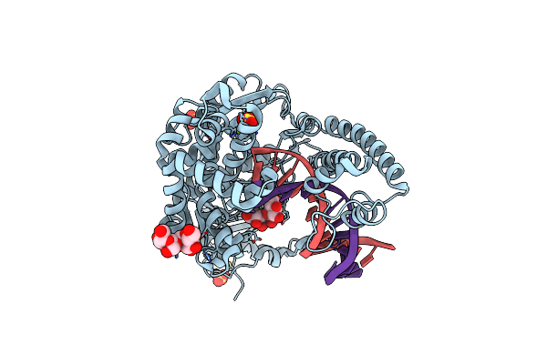 |
O6-Methyl:C Pair In The Polymerase-10 Basepair Position
Organism: Geobacillus stearothermophilus
Method: X-RAY DIFFRACTION Resolution:1.80 Å Release Date: 2006-12-12 Classification: Transferase/DNA Ligands: SO4, MG |
Organism: Geobacillus stearothermophilus
Method: X-RAY DIFFRACTION
Release Date: 2006-12-12
Ligands: SO4, MG
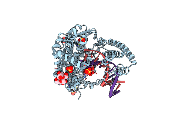 |
C:O6-Methyl-Guanine Pair In The Polymerase-2 Basepair Position
Organism: Geobacillus stearothermophilus
Method: X-RAY DIFFRACTION Resolution:2.05 Å Release Date: 2006-12-12 Classification: Transferase/DNA Ligands: SO4, MG |
Organism: Geobacillus stearothermophilus
Method: X-RAY DIFFRACTION
Release Date: 2006-12-12
Ligands: SO4, MG