
Search Count: 1,515
All
Selected
 |
Organism: Homo sapiens
Method: ELECTRON MICROSCOPY Release Date: 2026-05-13 Classification: MEMBRANE PROTEIN Ligands: TRP |
 |
Organism: Homo sapiens
Method: ELECTRON MICROSCOPY Release Date: 2026-05-13 Classification: MEMBRANE PROTEIN Ligands: NAG, PHE |
 |
Organism: Porphyromonas gingivalis atcc 33277
Method: X-RAY DIFFRACTION Resolution:2.60 Å Release Date: 2026-05-13 Classification: CYTOSOLIC PROTEIN Ligands: MN, PO4 |
 |
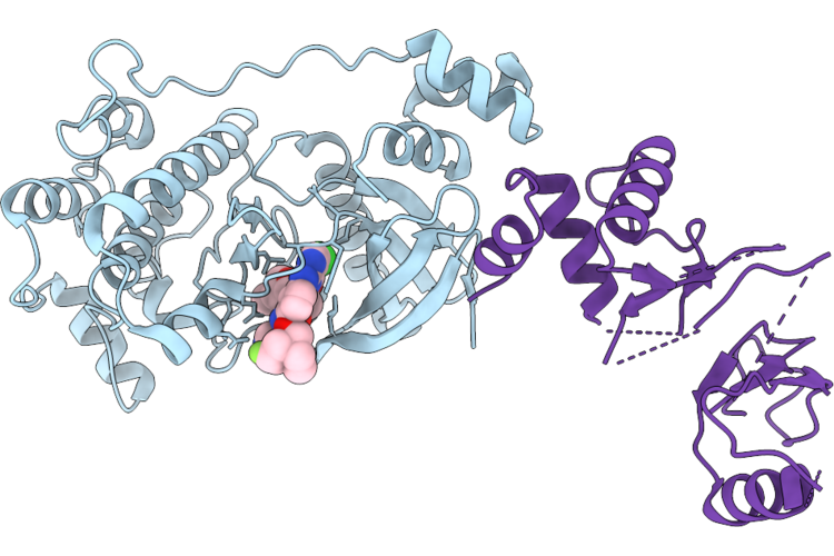
Crystal Structure Of The Nuak1-Mark3 Kinase Domain Chimera Bound With Small Molecule Inhibitor 2-Amino-N-(5-((5-Chloro-4-(((3R,3Ar,6R,6Ar)-6-Methoxyhexahydrofuro[3,2-B]Furan-3-Yl)Oxy)Pyrimidin-2-Yl)Amino)-2-((2-(Dimethylamino)Ethyl)(Methyl)Amino)Phenyl)Acetamide
Organism: Homo sapiens
Method: X-RAY DIFFRACTION Resolution:2.50 Å Release Date: 2026-04-22 Classification: TRANSFERASE Ligands: A1ER0 |
 |
Crystal Structure Of The Nuak1-Mark3 Kinase Domain Chimera Bound With Small Molecule Inhibitor 4-((5-((5-Chloro-4-(((3R,3Ar,6R,6Ar)-6-Methoxyhexahydrofuro[3,2-B]Furan-3-Yl)Oxy)Pyrimidin-2-Yl)Amino)-2-((2-(Dimethylamino)Ethyl)(Methyl)Amino)Phenyl)Carbamoyl)-1-Methyl-3H-Pyrazol-1-Ium-3-Ide
Organism: Homo sapiens
Method: X-RAY DIFFRACTION Resolution:2.00 Å Release Date: 2026-04-22 Classification: CYTOSOLIC PROTEIN Ligands: A1ER9 |
 |
Crystal Structure Of The Nuak1-Mark3 Kinase Domain Chimera Bound With Small Molecule Inhibitor N-(5-((5-Chloro-4-(((3As,6R,6Ar)-6-Methoxy-3A,5,6,6A-Tetrahydrofuro[3,2-B]Furan-3-Yl)Oxy)Pyrimidin-2-Yl)Amino)-2-(((2R,7Ar)-2-Fluorotetrahydro-1H-Pyrrolizin-7A(5H)-Yl)Methoxy)Phenyl)-1-Methyl-1H-Pyrazole-4-Carboxamide
Organism: Homo sapiens
Method: X-RAY DIFFRACTION Resolution:2.80 Å Release Date: 2026-04-22 Classification: CYTOSOLIC PROTEIN Ligands: A1ESF |
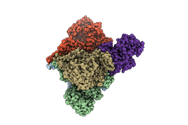 |
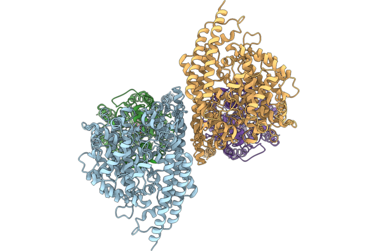
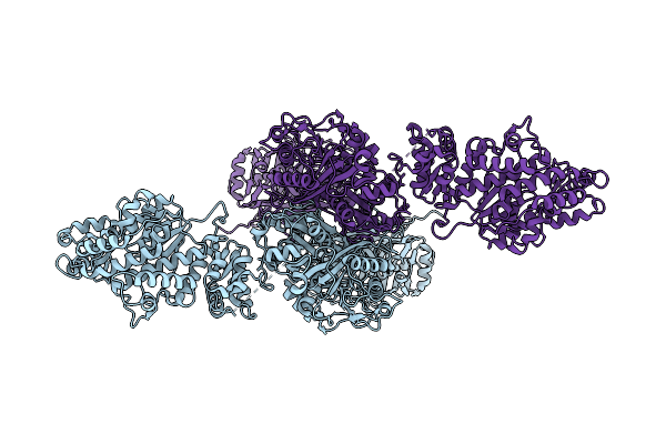
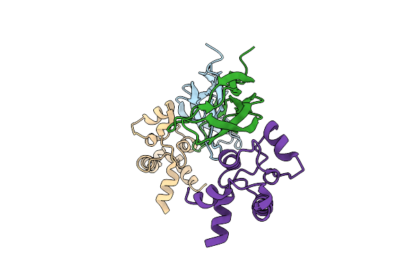
Structural Basis For The Assembly And Translocation Of The Vip1-Vip2 Insecticidal Binary Toxin From Bacillus Thuringiensis
Organism: Bacillus thuringiensis
Method: ELECTRON MICROSCOPY Resolution:3.05 Å Release Date: 2026-04-15 Classification: TOXIN Ligands: CA |
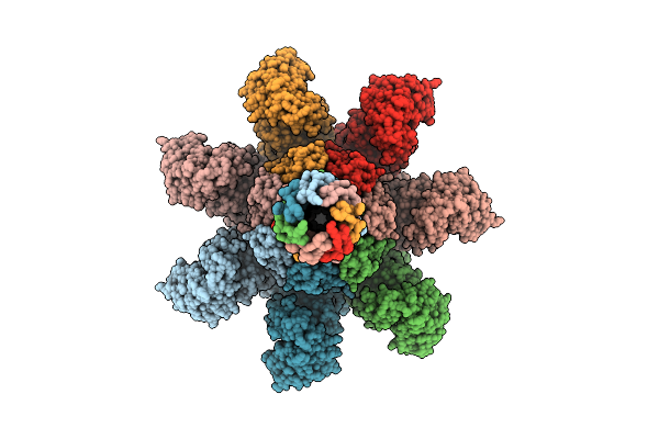 |
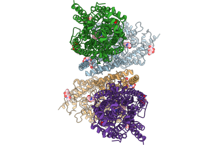
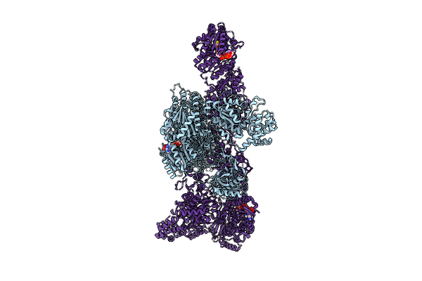
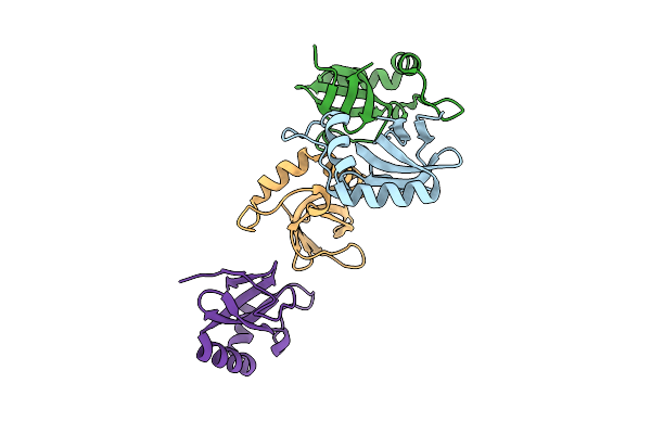
Structural Basis For The Assembly And Translocation Of The Vip1-Vip2 Insecticidal Binary Toxin From Bacillus Thuringiensis
Organism: Bacillus thuringiensis
Method: ELECTRON MICROSCOPY Resolution:2.40 Å Release Date: 2026-04-15 Classification: TOXIN Ligands: CA |
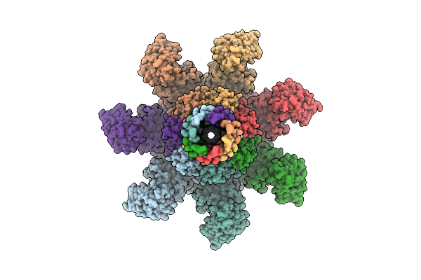 |
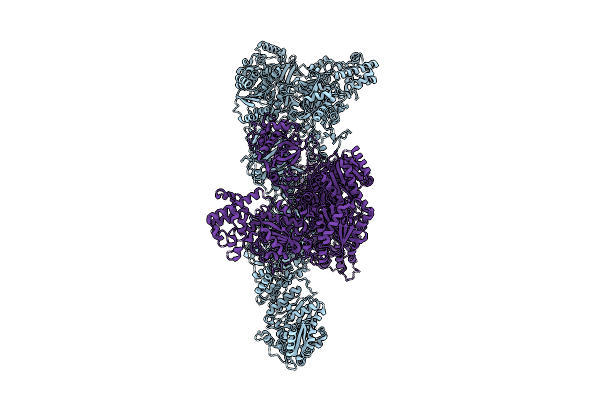
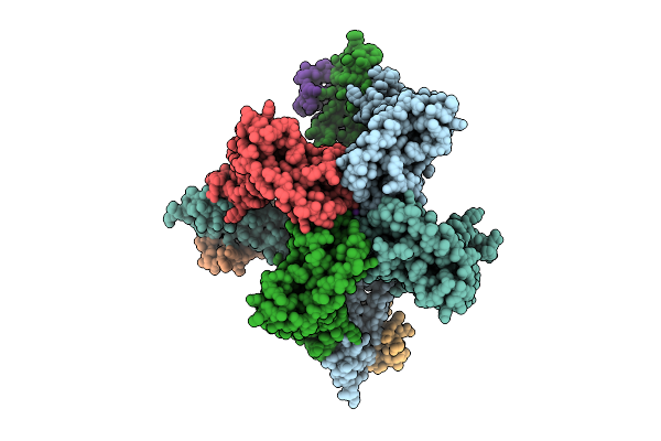
Structural Basis For The Assembly And Translocation Of The Vip1-Vip2 Insecticidal Binary Toxin From Bacillus Thuringiensis
Organism: Bacillus thuringiensis
Method: ELECTRON MICROSCOPY Resolution:3.31 Å Release Date: 2026-04-15 Classification: TOXIN Ligands: CA |
 |
Organism: Aspergillus fumigatus (strain atcc mya-4609 / cbs 101355 / fgsc a1100 / af293)
Method: ELECTRON MICROSCOPY Resolution:2.99 Å Release Date: 2026-04-15 Classification: BIOSYNTHETIC PROTEIN |
 |
Organism: Aspergillus fumigatus af293
Method: ELECTRON MICROSCOPY Resolution:3.16 Å Release Date: 2026-04-08 Classification: BIOSYNTHETIC PROTEIN Ligands: MLC |
 |
Organism: Aspergillus fumigatus af293
Method: ELECTRON MICROSCOPY Resolution:3.36 Å Release Date: 2026-04-08 Classification: BIOSYNTHETIC PROTEIN Ligands: COZ, NDP |
 |
Organism: Aspergillus fumigatus af293
Method: ELECTRON MICROSCOPY Resolution:3.39 Å Release Date: 2026-04-08 Classification: BIOSYNTHETIC PROTEIN |
 |
Organism: Pseudomonas aeruginosa pao1
Method: ELECTRON MICROSCOPY Release Date: 2026-04-01 Classification: BIOSYNTHETIC PROTEIN Ligands: DAB, ATP |
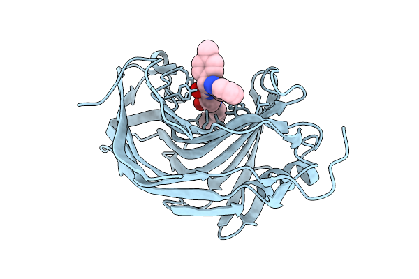 |
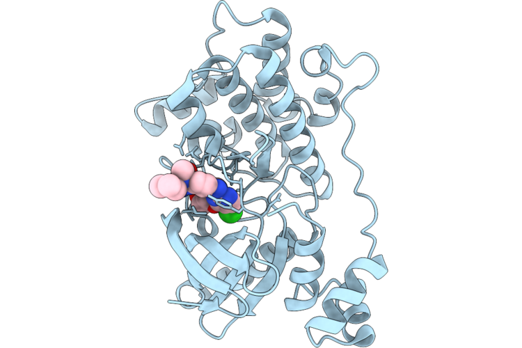
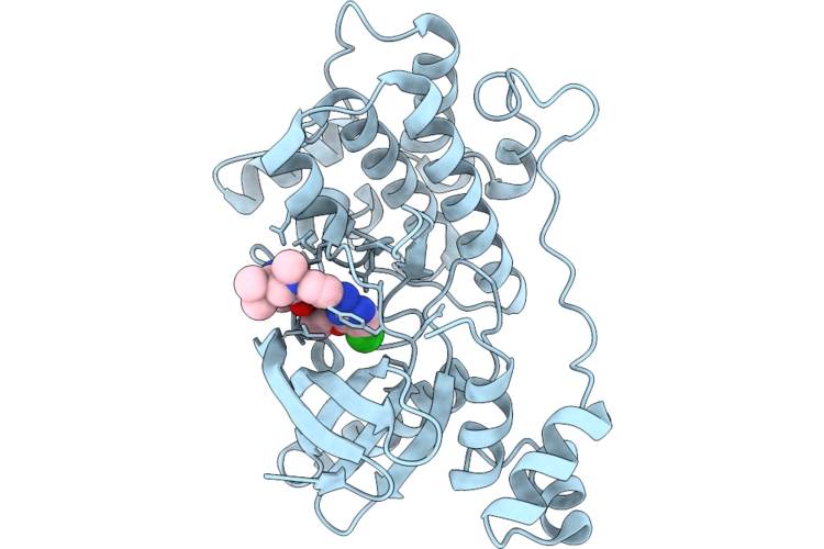
Crystal Structure Of The Pathogen-Secreted Apoplastic Gh12 Xyloglucan-Specific Endoglucanase Xeg1
Organism: Phytophthora sojae strain p6497
Method: X-RAY DIFFRACTION Resolution:2.59 Å Release Date: 2026-03-18 Classification: HYDROLASE Ligands: A1ENU |
 |
Structure Of Norrin In Complex With Human Tspan12 Large Extracellular Loop (Tspan12 Lel)
Organism: Escherichia coli o157:h7, Homo sapiens
Method: ELECTRON MICROSCOPY Release Date: 2026-03-18 Classification: SIGNALING PROTEIN |
 |
Organism: Streptomyces fagopyri
Method: X-RAY DIFFRACTION Resolution:1.87 Å Release Date: 2026-03-18 Classification: BIOSYNTHETIC PROTEIN |
 |
Organism: Bacteriophage sp.
Method: X-RAY DIFFRACTION Resolution:2.49 Å Release Date: 2026-03-11 Classification: HYDROLASE |
 |
Organism: Homo sapiens
Method: ELECTRON MICROSCOPY Resolution:3.34 Å Release Date: 2026-03-11 Classification: MEMBRANE PROTEIN Ligands: A1ETX, K |
 |
Organism: Homo sapiens, Apis mellifera
Method: ELECTRON MICROSCOPY Resolution:3.23 Å Release Date: 2026-03-11 Classification: MEMBRANE PROTEIN Ligands: K, POV |