

Search Count: 20
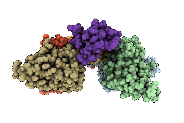 |
D11 Mabs Bound To Alpha-Bungarotoxin At Ph 7.5
Organism: Homo sapiens, Bungarus multicinctus
Method: X-RAY DIFFRACTION Resolution:1.33 Å Release Date: 2025-11-19 Classification: TOXIN Ligands: EDO |
Organism: Homo sapiens, Bungarus multicinctus
Method: X-RAY DIFFRACTION
Release Date: 2025-11-19
Ligands: EDO
 |
A01 Mabs Bound To Cobratoxin At Ph 5.5
Organism: Homo sapiens, Naja kaouthia
Method: X-RAY DIFFRACTION Resolution:1.60 Å Release Date: 2025-11-19 Classification: TOXIN Ligands: GOL, CL, K, NA, SO4 |
Organism: Homo sapiens, Naja kaouthia
Method: X-RAY DIFFRACTION
Release Date: 2025-11-19
Ligands: GOL, CL, K, NA, SO4
 |
A01 Mabs Bound To Cobratoxin At Ph 6.0
Organism: Homo sapiens, Naja kaouthia
Method: X-RAY DIFFRACTION Resolution:1.49 Å Release Date: 2025-11-19 Classification: TOXIN Ligands: CL, GOL, TRS, SO4, NA |
Organism: Homo sapiens, Naja kaouthia
Method: X-RAY DIFFRACTION
Release Date: 2025-11-19
Ligands: CL, GOL, TRS, SO4, NA
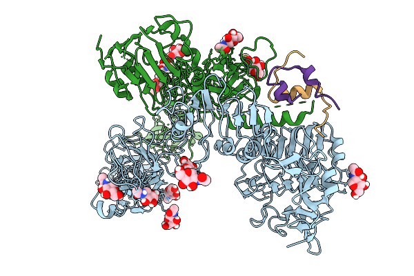 |
Cryo-Em Structure Of Drosophila Melanogaster Insulin Receptor (Dmir) Bound With One Dilp1, Asymmetric Conformation
Organism: Drosophila melanogaster
Method: ELECTRON MICROSCOPY Resolution:4.40 Å Release Date: 2025-10-01 Classification: STRUCTURAL PROTEIN Ligands: NAG |
Organism: Drosophila melanogaster
Method: ELECTRON MICROSCOPY
Release Date: 2025-10-01
Ligands: NAG
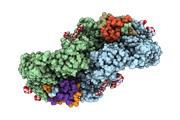 |
Cryo-Em Structure Of Drosophila Melanogaster Insulin Receptor (Dmir) Bound With Two Dilp1, Symmetric Conformation
Organism: Drosophila melanogaster
Method: ELECTRON MICROSCOPY Resolution:3.90 Å Release Date: 2025-10-01 Classification: STRUCTURAL PROTEIN Ligands: NAG |
Organism: Drosophila melanogaster
Method: ELECTRON MICROSCOPY
Release Date: 2025-10-01
Ligands: NAG
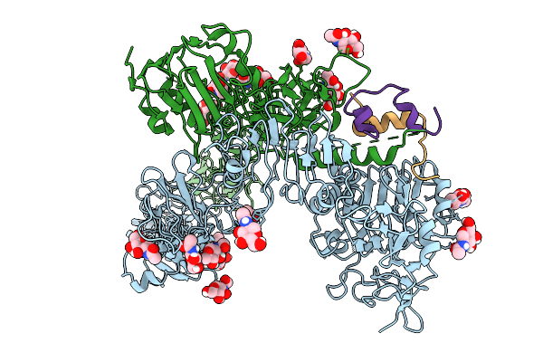 |
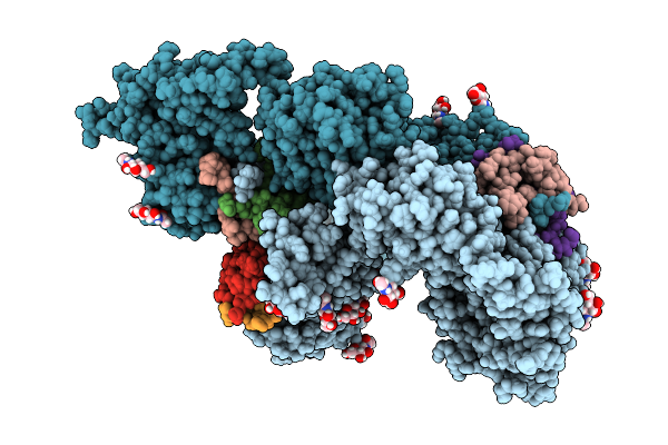
Cryo-Em Structure Of Drosophila Melanogaster Insulin Receptor (Dmir) Bound With One Dilp2, Asymmetric Conformation
Organism: Drosophila melanogaster
Method: ELECTRON MICROSCOPY Resolution:3.70 Å Release Date: 2025-10-01 Classification: STRUCTURAL PROTEIN Ligands: NAG |
Organism: Drosophila melanogaster
Method: ELECTRON MICROSCOPY
Release Date: 2025-10-01
Ligands: NAG
 |
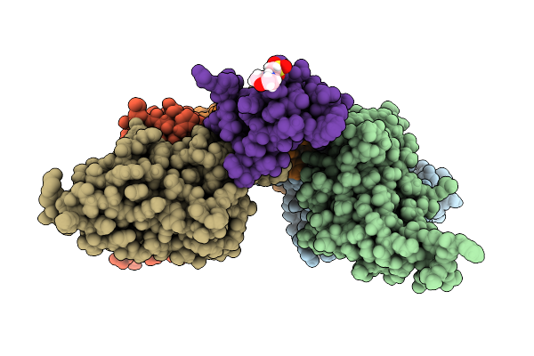
Cryo-Em Structure Of Drosophila Melanogaster Insulin Receptor (Dmir) Bound With Three Dilp5, Asymmetric Conformation
Organism: Drosophila melanogaster
Method: ELECTRON MICROSCOPY Resolution:3.60 Å Release Date: 2025-10-01 Classification: STRUCTURAL PROTEIN Ligands: NAG |
Organism: Drosophila melanogaster
Method: ELECTRON MICROSCOPY
Release Date: 2025-10-01
Ligands: NAG
 |
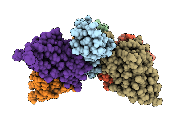
D11 Mabs Bound To Alpha-Bungarotoxin
Organism: Homo sapiens, Bungarus multicinctus
Method: X-RAY DIFFRACTION Resolution:1.32 Å Release Date: 2025-07-16 Classification: TOXIN Ligands: MES |
Organism: Homo sapiens, Bungarus multicinctus
Method: X-RAY DIFFRACTION
Release Date: 2025-07-16
Ligands: MES
 |
Mabs In Complex With Cobratoxin At Ph 4.5
Organism: Naja kaouthia, Homo sapiens
Method: X-RAY DIFFRACTION Resolution:1.55 Å Release Date: 2025-07-16 Classification: TOXIN Ligands: CL, GOL, SO4, NA |
Organism: Naja kaouthia, Homo sapiens
Method: X-RAY DIFFRACTION
Release Date: 2025-07-16
Ligands: CL, GOL, SO4, NA
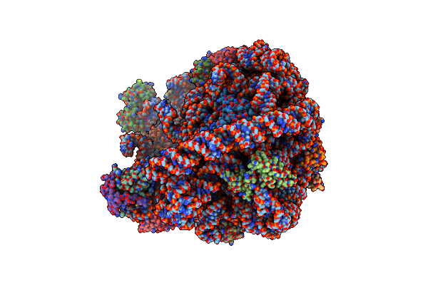 |
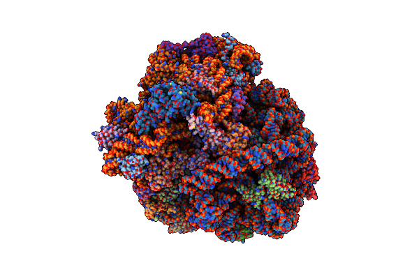
Cryo-Em Structure Of Mycobacterium Smegmatis C(Minus) 50S Ribosomal Subunit
Organism: Mycobacterium smegmatis str. mc2 155, Mycobacterium smegmatis (strain atcc 700084 / mc(2)155)
Method: ELECTRON MICROSCOPY Release Date: 2018-10-03 Classification: RIBOSOME |
 |
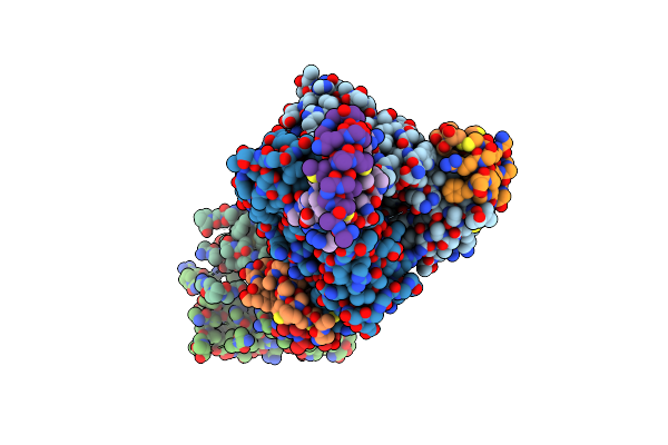
Cryo-Em Structure Of Mycobacterium Smegmatis 70S C(Minus) Ribosome 70S-Mpy Complex
Organism: Mycobacterium smegmatis str. mc2 155, Mycobacterium smegmatis (strain atcc 700084 / mc(2)155), Mycobacterium smegmatis
Method: ELECTRON MICROSCOPY Release Date: 2018-09-26 Classification: RIBOSOME |
 |
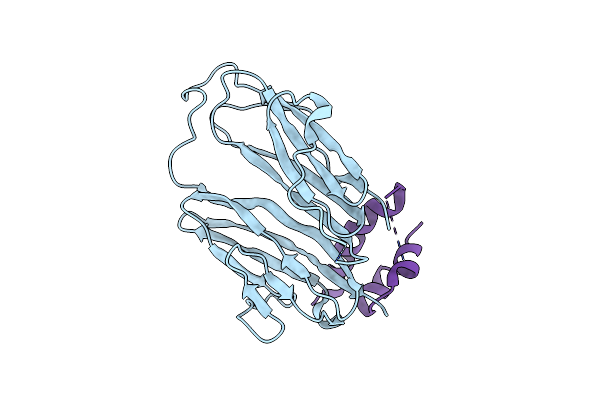
Crystal Structure Of Drosophila Neural Ectodermal Development Factor Imp-L2 With Drosophila Dilp5 Insulin
Organism: Drosophila melanogaster
Method: X-RAY DIFFRACTION Resolution:3.48 Å Release Date: 2018-09-26 Classification: PEPTIDE BINDING PROTEIN |
Organism: Drosophila melanogaster
Method: X-RAY DIFFRACTION
Release Date: 2018-09-26
 |
Crystal Structure Of Drosophila Neural Ectodermal Development Factor Imp-L1 With Human Igf-I
Organism: Drosophila melanogaster, Homo sapiens
Method: X-RAY DIFFRACTION Resolution:2.57 Å Release Date: 2018-09-26 Classification: PEPTIDE BINDING PROTEIN |
Organism: Drosophila melanogaster, Homo sapiens
Method: X-RAY DIFFRACTION
Release Date: 2018-09-26
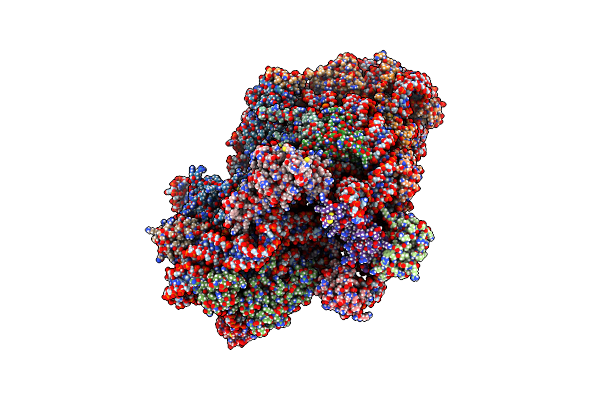 |
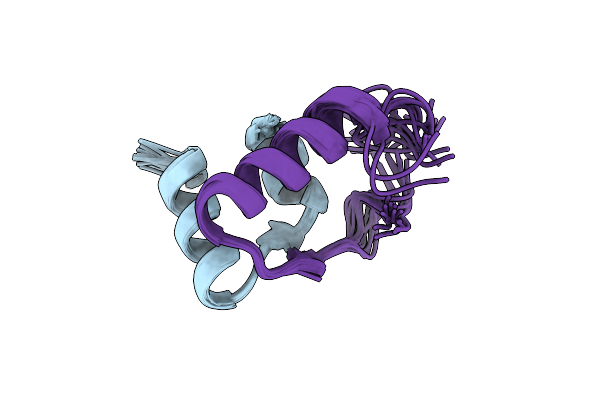
Cryo-Em Structure Of Mycobacterium Smegmatis C(Minus) 30S Ribosomal Subunit With Mpy
Organism: Mycobacterium smegmatis str. mc2 155, Mycobacterium smegmatis (strain atcc 700084 / mc(2)155), Mycobacterium smegmatis
Method: ELECTRON MICROSCOPY Release Date: 2018-09-19 Classification: RIBOSOME |
 |
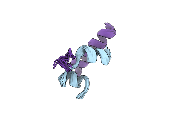
Solution Nmr Structure Of Human Relaxin-2
Organism: Homo sapiens
Method: SOLUTION NMR Release Date: 2015-02-04 Classification: SIGNALING PROTEIN |
Organism: Homo sapiens
Method: SOLUTION NMR
Release Date: 2015-02-04
 |
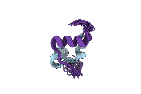
Solution Structure Of Human Insulin-Like Peptide 5 (Insl5)
|
Organism: Synthetic construct
Method: SOLUTION NMR
Release Date: 2009-03-24
 |
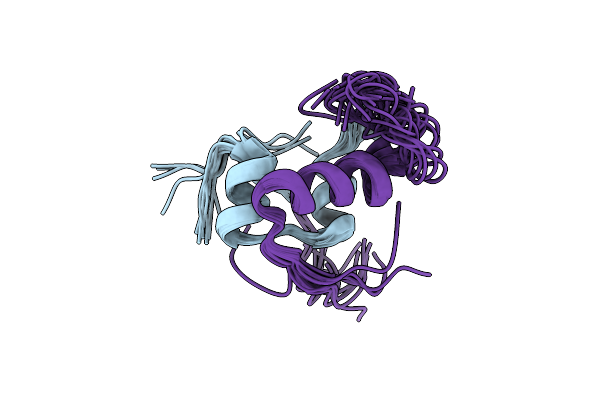
R3/I5 Relaxin Chimera
|
Method: SOLUTION NMR
Release Date: 2008-04-08
 |
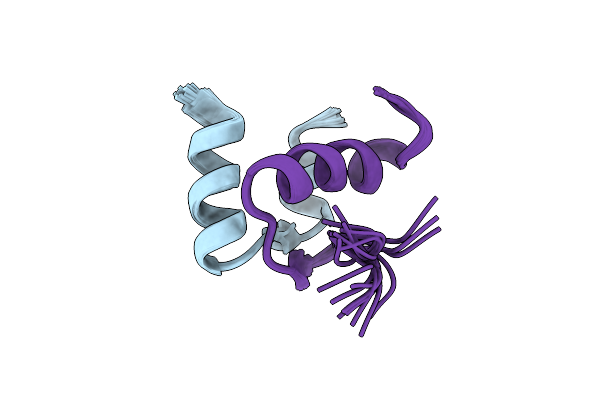
Solution Structure Of Insl3
|
Method: SOLUTION NMR
Release Date: 2006-08-01
 |
Solution Structure Of Human Relaxin-3
|
Method: SOLUTION NMR
Release Date: 2006-01-24
 |
The Three-Dimensional Solution Structure Of The Rabbit Kidney Defensin, Rk-1
Organism: Oryctolagus cuniculus
Method: SOLUTION NMR Release Date: 2001-05-02 Classification: ANTIMICROBIAL PROTEIN |
Organism: Oryctolagus cuniculus
Method: SOLUTION NMR
Release Date: 2001-05-02