

Search Count: 23
 |
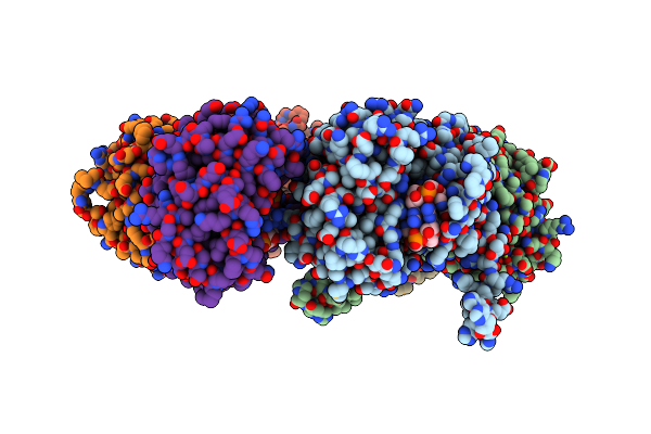
X-Ray Crystal Structure Of The Formyltransferase/Hydrolase Complex (Fhcabcd) From Methylorubrum Extorquens In Complex With Methylofuran
|
 |
Crystal Structure Of Smba In Complex With C-Di-Gmp
Organism: Caulobacter vibrioides (strain atcc 19089 / cb15)
Method: X-RAY DIFFRACTION Resolution:2.80 Å Release Date: 2019-07-31 Classification: SIGNALING PROTEIN Ligands: C2E, MG |
Organism: Caulobacter vibrioides (strain atcc 19089 / cb15)
Method: X-RAY DIFFRACTION
Release Date: 2019-07-31
Ligands: C2E, MG
 |
Crystal Structure Of Smba In Complex With Ppgpp.
Organism: Caulobacter vibrioides (strain atcc 19089 / cb15)
Method: X-RAY DIFFRACTION Resolution:3.30 Å Release Date: 2019-07-03 Classification: SIGNALING PROTEIN Ligands: G4P |
Organism: Caulobacter vibrioides (strain atcc 19089 / cb15)
Method: X-RAY DIFFRACTION
Release Date: 2019-07-03
Ligands: G4P
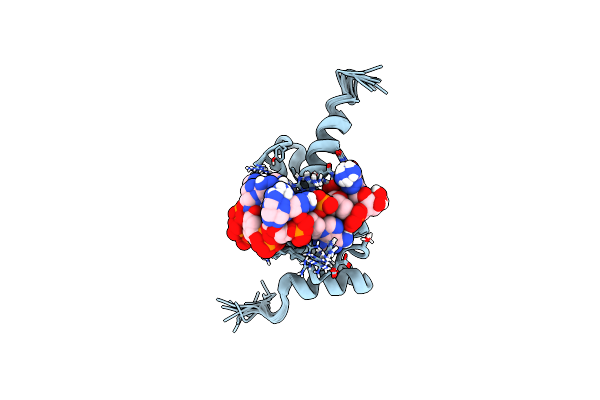 |
Nmr Structure Of The Complex Formed By An Engineered Region 2 Of Sigmae In Complex With Gtaaaa
Organism: Escherichia coli (strain k12), Bacillus subtilis, Synthetic construct
Method: SOLUTION NMR Release Date: 2018-06-27 Classification: TRANSCRIPTION |
Organism: Escherichia coli (strain k12), Bacillus subtilis, Synthetic construct
Method: SOLUTION NMR
Release Date: 2018-06-27
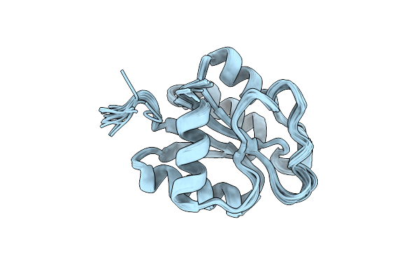 |
Solution Structure Of Sdrg From Sphingomonas Melonis Fr1
Organism: Sphingomonas melonis fr1
Method: SOLUTION NMR Release Date: 2016-07-20 Classification: PROTEIN |
Organism: Sphingomonas melonis fr1
Method: SOLUTION NMR
Release Date: 2016-07-20
 |
Solution Structure Of The Bef3-Activated Conformation Of Sdrg From Pseudomonas Melonis Fr1
Organism: Sphingomonas melonis fr1
Method: SOLUTION NMR Release Date: 2016-07-20 Classification: PROTEIN |
Organism: Sphingomonas melonis fr1
Method: SOLUTION NMR
Release Date: 2016-07-20
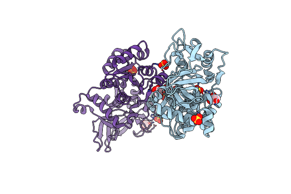 |
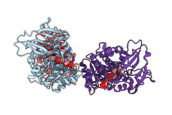
Apo-Structure Of The Y79F,W322E-Double Mutant Of Etr1P
Organism: Candida tropicalis
Method: X-RAY DIFFRACTION Resolution:2.00 Å Release Date: 2015-03-18 Classification: OXIDOREDUCTASE Ligands: GOL, SO4 |
Organism: Candida tropicalis
Method: X-RAY DIFFRACTION
Release Date: 2015-03-18
Ligands: GOL, SO4
 |
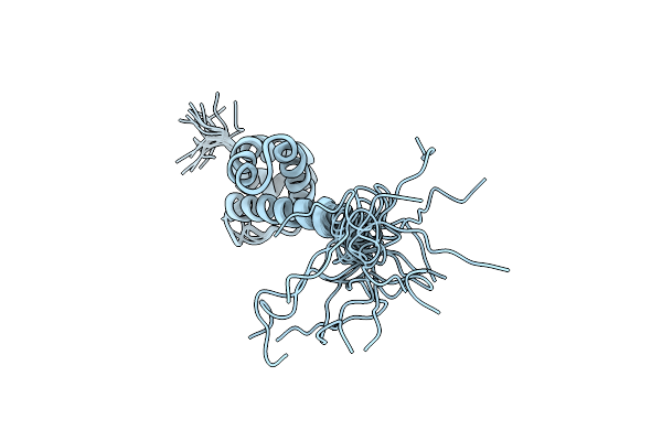
Structure Of The Etr1P/Nadp/Crotonyl-Coa Complex
Organism: Candida tropicalis
Method: X-RAY DIFFRACTION Resolution:1.70 Å Release Date: 2015-03-18 Classification: OXIDOREDUCTASE Ligands: NAP, COO |
Organism: Candida tropicalis
Method: X-RAY DIFFRACTION
Release Date: 2015-03-18
Ligands: NAP, COO
 |
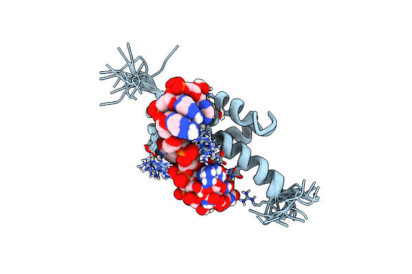
Nmr Structure Of Region 2 Of E. Coli Sigmae
Organism: Escherichia coli
Method: SOLUTION NMR Release Date: 2014-02-19 Classification: TRANSCRIPTION |
Organism: Escherichia coli
Method: SOLUTION NMR
Release Date: 2014-02-19
 |
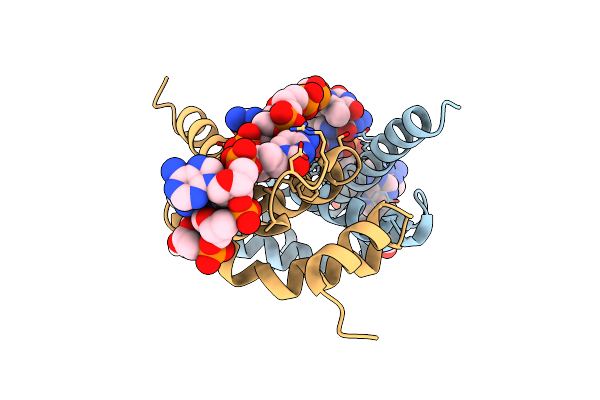
Solution Structure Of The Complex Formed By The Region 2 Of E. Coli Sigmae And Its Cognate -10 Promoter Element Non Template Strand Tgtcaaa.
Organism: Escherichia coli
Method: SOLUTION NMR Release Date: 2014-02-19 Classification: TRANSCRIPTION/DNA |
Organism: Escherichia coli
Method: SOLUTION NMR
Release Date: 2014-02-19
 |
Crystal Structure Of The Complex Formed By Region Of E. Coli Sigmae Bound To Its -10 Element Non Template Strand
Organism: Escherichia coli
Method: X-RAY DIFFRACTION Resolution:1.20 Å Release Date: 2014-02-19 Classification: TRANSCRIPTION/DNA Ligands: EDO |
Organism: Escherichia coli
Method: X-RAY DIFFRACTION
Release Date: 2014-02-19
Ligands: EDO
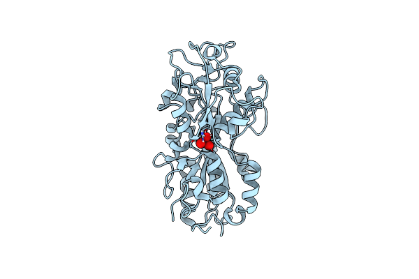 |
Subatomic Resolution Structure Of A High Affinity Periplasmic Phosphate-Binding Protein (Pfluding) Bound With Arsenate At Ph 8.5
Organism: Pseudomonas fluorescens
Method: X-RAY DIFFRACTION Resolution:0.96 Å Release Date: 2012-09-05 Classification: phosphate-binding protein Ligands: 8AR |
Organism: Pseudomonas fluorescens
Method: X-RAY DIFFRACTION
Release Date: 2012-09-05
Ligands: 8AR
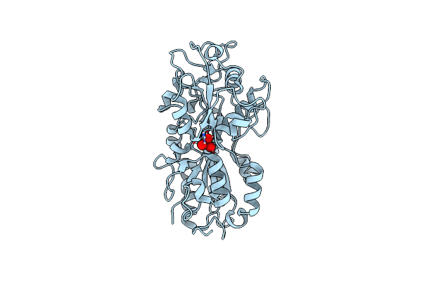 |
Subatomic Resolution Structure Of A High Affinity Periplasmic Phosphate-Binding Protein (Pfluding) Bound With Arsenate At Ph 4.5
Organism: Pseudomonas fluorescens
Method: X-RAY DIFFRACTION Resolution:0.95 Å Release Date: 2012-09-05 Classification: phosphate-binding protein Ligands: 8AR |
Organism: Pseudomonas fluorescens
Method: X-RAY DIFFRACTION
Release Date: 2012-09-05
Ligands: 8AR
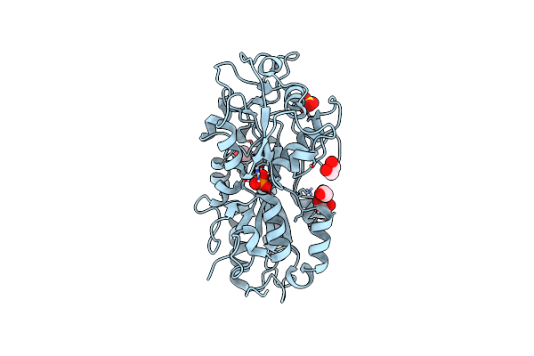 |
Subatomic Resolution Structure Of A High Affinity Periplasmic Phosphate-Binding Protein (Pfluding) Bound With Phosphate At Ph 4.5
Organism: Pseudomonas fluorescens
Method: X-RAY DIFFRACTION Resolution:0.98 Å Release Date: 2012-05-23 Classification: PHOSPHATE-BINDING PROTEIN Ligands: PI, EDO, SO4 |
Organism: Pseudomonas fluorescens
Method: X-RAY DIFFRACTION
Release Date: 2012-05-23
Ligands: PI, EDO, SO4
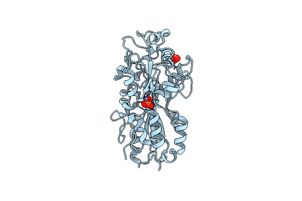 |
Subatomic Resolution Structure Of A High Affinity Periplasmic Phosphate-Binding Protein (Pfluding) Bound With Phosphate At Ph 8.5
Organism: Pseudomonas fluorescens
Method: X-RAY DIFFRACTION Resolution:0.88 Å Release Date: 2012-05-23 Classification: PHOSPHATE-BINDING PROTEIN Ligands: PI, SO4 |
Organism: Pseudomonas fluorescens
Method: X-RAY DIFFRACTION
Release Date: 2012-05-23
Ligands: PI, SO4
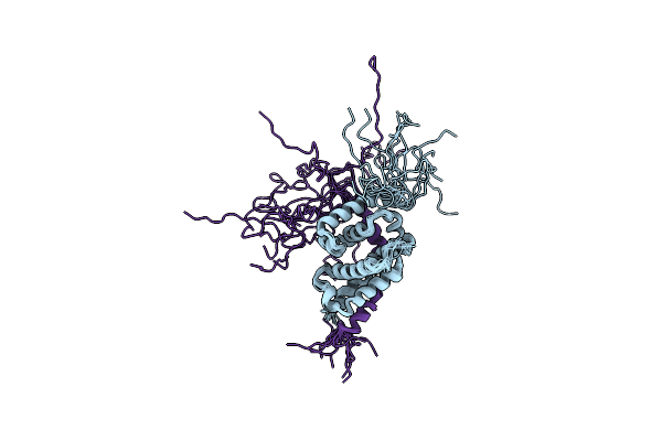 |
Nmr Structure Of The Phyrsl-Nepr Complex From Sphingomonas Sp. Fr1
Organism: Sphingomonas sp. fr1
Method: SOLUTION NMR Release Date: 2012-04-25 Classification: SIGNALING PROTEIN |
Organism: Sphingomonas sp. fr1
Method: SOLUTION NMR
Release Date: 2012-04-25
 |
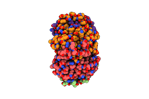
Structure Of The Tetrahydromethanopterin Dependent Formaldehyde-Activating Enzyme (Fae) From Methylobacterium Extorquens Am1
Organism: Methylobacterium extorquens
Method: X-RAY DIFFRACTION Resolution:2.00 Å Release Date: 2005-01-11 Classification: LYASE Ligands: CA, NA |
Organism: Methylobacterium extorquens
Method: X-RAY DIFFRACTION
Release Date: 2005-01-11
Ligands: CA, NA
 |
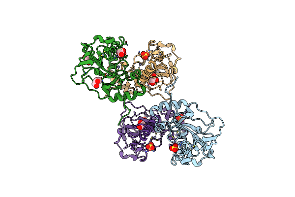
Structure Of The Tetrahydromethanopterin Dependent Formaldehyde-Activating Enzyme (Fae) From Methylobacterium Extorquens Am1 With Bound 5,10-Methylene Tetrahydromethanopterin
Organism: Methylobacterium extorquens
Method: X-RAY DIFFRACTION Resolution:1.90 Å Release Date: 2005-01-11 Classification: LYASE Ligands: H4M |
Organism: Methylobacterium extorquens
Method: X-RAY DIFFRACTION
Release Date: 2005-01-11
Ligands: H4M
 |
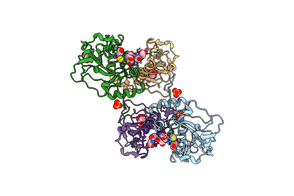
Crystal Structure Of The Glutathione-Dependent Formaldehyde-Activating Enzyme (Gfa)
Organism: Paracoccus denitrificans
Method: X-RAY DIFFRACTION Resolution:2.35 Å Release Date: 2004-11-23 Classification: LYASE Ligands: ZN, SO4, GOL |
Organism: Paracoccus denitrificans
Method: X-RAY DIFFRACTION
Release Date: 2004-11-23
Ligands: ZN, SO4, GOL
 |
Crystal Structure Analysis Of Glutathione-Dependent Formaldehyde-Activating Enzyme (Gfa)
Organism: Paracoccus denitrificans
Method: X-RAY DIFFRACTION Resolution:2.40 Å Release Date: 2004-11-23 Classification: LYASE Ligands: ZN, SO4, GSH, GOL |
Organism: Paracoccus denitrificans
Method: X-RAY DIFFRACTION
Release Date: 2004-11-23
Ligands: ZN, SO4, GSH, GOL