

Search Count: 25
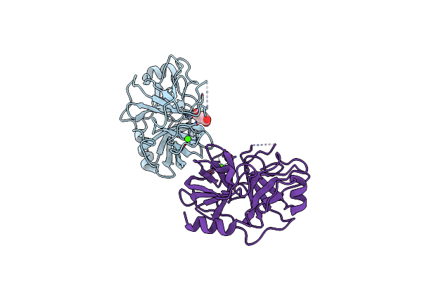 |
Succinic Acid Bound Trypsin Crystallized As Dimer
Organism: Bos taurus
Method: X-RAY DIFFRACTION Resolution:2.70 Å Release Date: 2016-08-10 Classification: HYDROLASE Ligands: SIN, CA |
Organism: Bos taurus
Method: X-RAY DIFFRACTION
Release Date: 2016-08-10
Ligands: SIN, CA
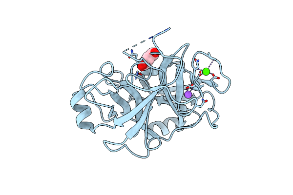 |
Trypsin Bound With Succinic Acid At 1.9A
Organism: Bos taurus
Method: X-RAY DIFFRACTION Resolution:1.91 Å Release Date: 2016-07-06 Classification: HYDROLASE Ligands: CA, SIN, NA |
Organism: Bos taurus
Method: X-RAY DIFFRACTION
Release Date: 2016-07-06
Ligands: CA, SIN, NA
 |
Crystal Structure Of Ttha1159 In Complex With Adp
Organism: Thermus thermophilus hb8
Method: X-RAY DIFFRACTION Resolution:2.10 Å Release Date: 2015-05-13 Classification: TRANSPORT PROTEIN Ligands: ADP, CL, MG |
Organism: Thermus thermophilus hb8
Method: X-RAY DIFFRACTION
Release Date: 2015-05-13
Ligands: ADP, CL, MG
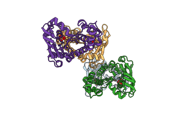 |
Crystal Structure Of Apo-Ttha1159
Organism: Thermus thermophilus hb8
Method: X-RAY DIFFRACTION Resolution:2.40 Å Release Date: 2015-05-13 Classification: TRANSPORT PROTEIN Ligands: SO4 |
Organism: Thermus thermophilus hb8
Method: X-RAY DIFFRACTION
Release Date: 2015-05-13
Ligands: SO4
 |
Use Of Europium For Sad Phasing At The Cu K Alpha Wavelength
Organism: Gallus gallus
Method: X-RAY DIFFRACTION Resolution:2.30 Å Release Date: 2012-10-24 Classification: HYDROLASE Ligands: EU, CL, NA |
Organism: Gallus gallus
Method: X-RAY DIFFRACTION
Release Date: 2012-10-24
Ligands: EU, CL, NA
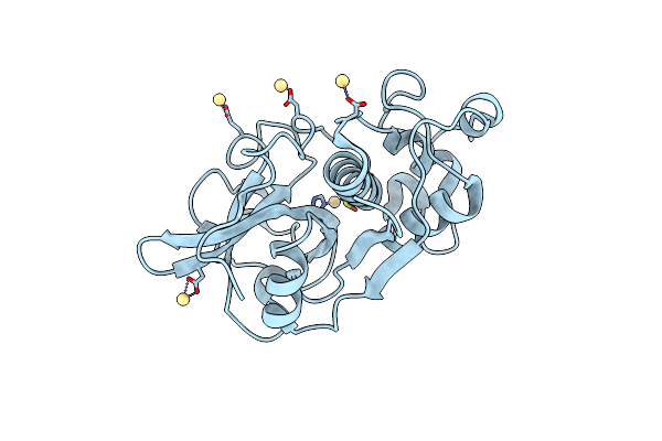 |
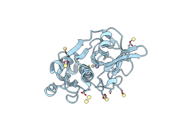
Actinidin From Actinidia Arguta Planch (Sarusashi)
Organism: Actinidia arguta
Method: X-RAY DIFFRACTION Resolution:1.50 Å Release Date: 2010-11-03 Classification: HYDROLASE Ligands: CD |
Organism: Actinidia arguta
Method: X-RAY DIFFRACTION
Release Date: 2010-11-03
Ligands: CD
 |
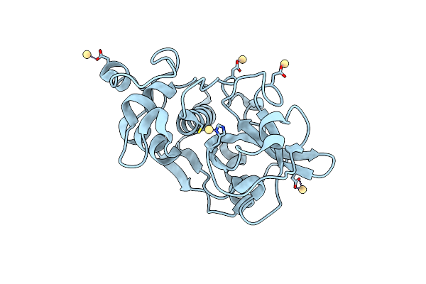
Actinidin From Actinidia Arguta Planch (Sarusashi)
Organism: Actinidia arguta
Method: X-RAY DIFFRACTION Resolution:1.90 Å Release Date: 2010-11-03 Classification: HYDROLASE Ligands: CD |
Organism: Actinidia arguta
Method: X-RAY DIFFRACTION
Release Date: 2010-11-03
Ligands: CD
 |
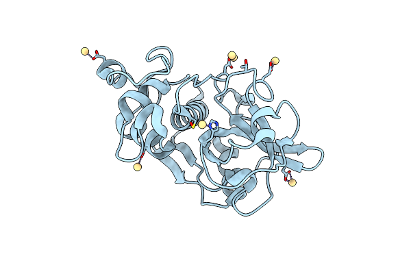
Actinidin From Actinidia Arguta Planch (Sarusashi)
Organism: Actinidia arguta
Method: X-RAY DIFFRACTION Resolution:2.20 Å Release Date: 2010-11-03 Classification: HYDROLASE Ligands: CD |
Organism: Actinidia arguta
Method: X-RAY DIFFRACTION
Release Date: 2010-11-03
Ligands: CD
 |
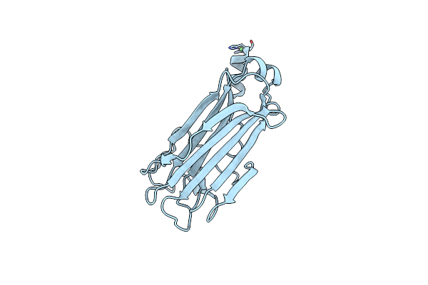
Actinidin From Actinidia Arguta Planch (Sarusashi)
Organism: Actinidia arguta
Method: X-RAY DIFFRACTION Resolution:2.20 Å Release Date: 2010-11-03 Classification: HYDROLASE Ligands: CD |
Organism: Actinidia arguta
Method: X-RAY DIFFRACTION
Release Date: 2010-11-03
Ligands: CD
 |
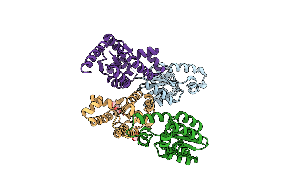
Crystal Structure Of The Sugar Recognizing Scf Ubiquitin Ligase At 1.7 Resolution
Organism: Mus musculus
Method: X-RAY DIFFRACTION Resolution:1.70 Å Release Date: 2008-10-14 Classification: LIGASE Ligands: NI, CL |
Organism: Mus musculus
Method: X-RAY DIFFRACTION
Release Date: 2008-10-14
Ligands: NI, CL
 |
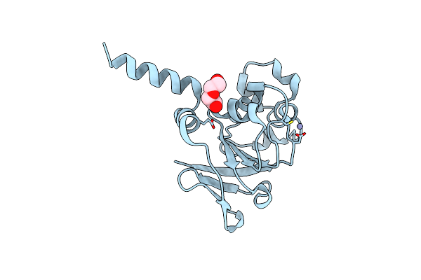
Crystal Structure Of Shikimate Kinase (Aq_2177) From Aquifex Aeolicus Vf5
Organism: Aquifex aeolicus
Method: X-RAY DIFFRACTION Resolution:2.10 Å Release Date: 2008-05-13 Classification: TRANSFERASE Ligands: EDO, PEG |
Organism: Aquifex aeolicus
Method: X-RAY DIFFRACTION
Release Date: 2008-05-13
Ligands: EDO, PEG
 |
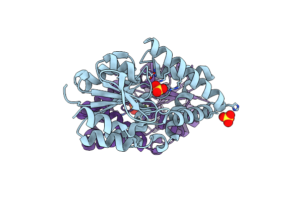
Crystal Structure Of 3-Isopropylmalate Dehydratase (Leud)From Methhanocaldococcus Jannaschii Dsm2661 (Mj1271)
Organism: Methanocaldococcus jannaschii
Method: X-RAY DIFFRACTION Resolution:2.10 Å Release Date: 2008-04-22 Classification: LYASE Ligands: ZN, PEG |
Organism: Methanocaldococcus jannaschii
Method: X-RAY DIFFRACTION
Release Date: 2008-04-22
Ligands: ZN, PEG
 |
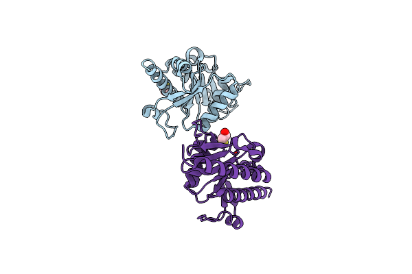
Hypothetical Protein (Probable Phosphoserine Phosph (Ph0253) From Pyrococcus Horikoshii Ot3
Organism: Pyrococcus horikoshii
Method: X-RAY DIFFRACTION Resolution:2.20 Å Release Date: 2008-01-29 Classification: HYDROLASE Ligands: SO4, MG, CL |
Organism: Pyrococcus horikoshii
Method: X-RAY DIFFRACTION
Release Date: 2008-01-29
Ligands: SO4, MG, CL
 |
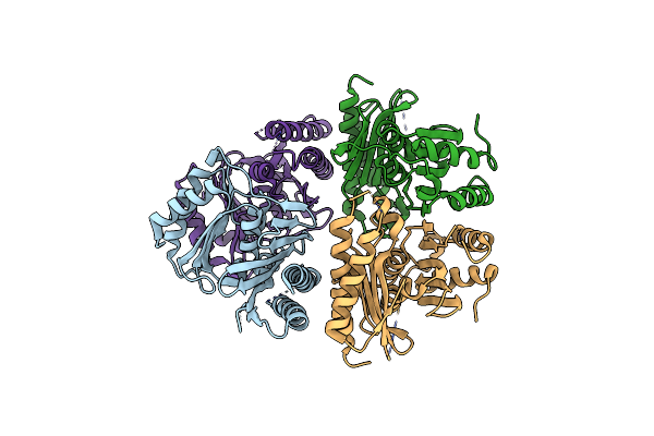
Crystal Structure Of The L-Fuculose-1-Phosphate Aldolase (Aq_1979) From Aquifex Aeolicus Vf5
Organism: Aquifex aeolicus
Method: X-RAY DIFFRACTION Resolution:2.40 Å Release Date: 2007-10-30 Classification: LYASE Ligands: CL, BME |
Organism: Aquifex aeolicus
Method: X-RAY DIFFRACTION
Release Date: 2007-10-30
Ligands: CL, BME
 |
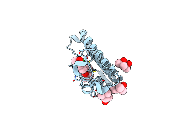
Crystal Structure Of Myo-Inositol-1(Or 4)-Monophosphatase (Aq_1983) From Aquifex Aeolicus Vf5
Organism: Aquifex aeolicus
Method: X-RAY DIFFRACTION Resolution:2.60 Å Release Date: 2007-10-02 Classification: HYDROLASE |
Organism: Aquifex aeolicus
Method: X-RAY DIFFRACTION
Release Date: 2007-10-02
 |
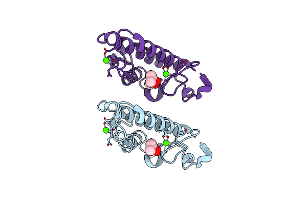
A Possible Of Second Calcium Ion In Interfacial Binding: Atomic And Medium Resolution Crystal Structures Of The Quadruple Mutant Of Phospholipase A2
Organism: Bos taurus
Method: X-RAY DIFFRACTION Resolution:1.10 Å Release Date: 2006-07-04 Classification: HYDROLASE Ligands: CA, CL, MPD |
Organism: Bos taurus
Method: X-RAY DIFFRACTION
Release Date: 2006-07-04
Ligands: CA, CL, MPD
 |
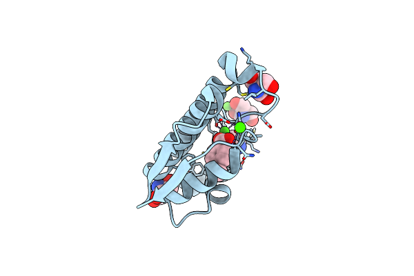
A Possible Role Of The Second Calcium Ion In Interfacial Binding: Atomic And Medium Resolution Crystal Structures Of The Quadruple Mutant Of Phospholipase A2
Organism: Bos taurus
Method: X-RAY DIFFRACTION Resolution:1.90 Å Release Date: 2006-07-04 Classification: HYDROLASE Ligands: CA, MPD |
Organism: Bos taurus
Method: X-RAY DIFFRACTION
Release Date: 2006-07-04
Ligands: CA, MPD
 |
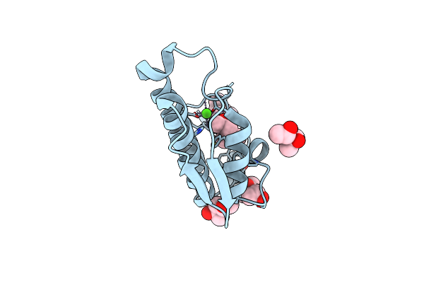
Third Calcium Ion Found In An Inhibitor Bound Phospholipase A2
Organism: Bos taurus
Method: X-RAY DIFFRACTION Resolution:1.70 Å Release Date: 2006-03-28 Classification: HYDROLASE Ligands: CA, CL, TRS, ANN |
Organism: Bos taurus
Method: X-RAY DIFFRACTION
Release Date: 2006-03-28
Ligands: CA, CL, TRS, ANN
 |
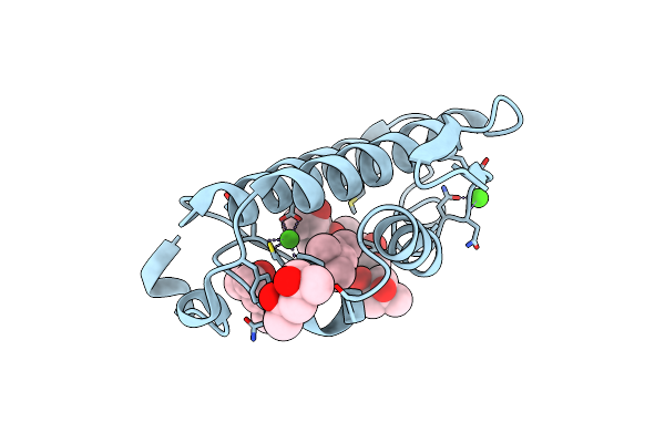
Atomic Resolution Structure Of The Double Mutant (K53,56M) Of Bovine Pancreatic Phospholipase A2
Organism: Bos taurus
Method: X-RAY DIFFRACTION Resolution:1.10 Å Release Date: 2005-10-25 Classification: HYDROLASE Ligands: CA, CL, MRD, MPD |
Organism: Bos taurus
Method: X-RAY DIFFRACTION
Release Date: 2005-10-25
Ligands: CA, CL, MRD, MPD
 |
Atomic Resolution (0.97A) Structure Of The Triple Mutant (K53,56,121M) Of Bovine Pancreatic Phospholipase A2
Organism: Bos taurus
Method: X-RAY DIFFRACTION Resolution:0.97 Å Release Date: 2004-10-19 Classification: HYDROLASE Ligands: CA, CL, MRD, MPD |
Organism: Bos taurus
Method: X-RAY DIFFRACTION
Release Date: 2004-10-19
Ligands: CA, CL, MRD, MPD