
Search Count: 19
All
Selected
 |
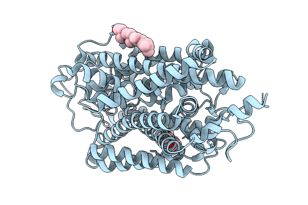
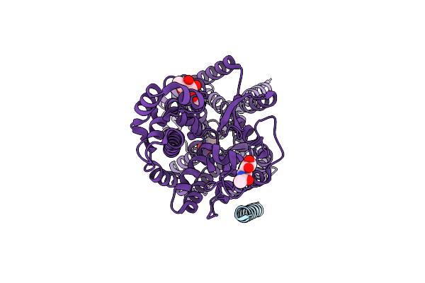
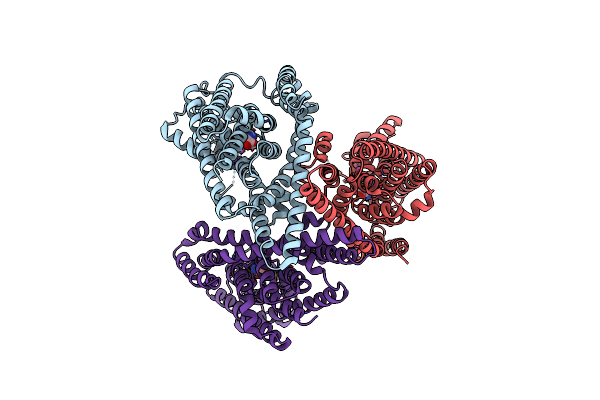
Outward-Open Structure Of Human Glycine Transporter 2 Bound To Allosteric Inhibitor Org25543
Organism: Homo sapiens
Method: ELECTRON MICROSCOPY Resolution:2.49 Å Release Date: 2026-02-25 Classification: MEMBRANE PROTEIN Ligands: A1EF1, Y01, CLR, CL, NA |
 |
Outward-Open Structure Of Human Glycine Transporter 2 Bound To Allosteric Inhibitor Rpi-Glyt2-82
Organism: Homo sapiens
Method: ELECTRON MICROSCOPY Release Date: 2026-02-25 Classification: MEMBRANE PROTEIN Ligands: CL, A1IXX, CLR, Y01, NA |
 |
Inward-Open Structure Of Human Glycine Transporter 2 In Substrate-Free State
Organism: Homo sapiens
Method: ELECTRON MICROSCOPY Release Date: 2026-02-25 Classification: MEMBRANE PROTEIN Ligands: CLR, CL |
 |
Inward-Occluded Structure Of Human Glycine Transporter 2 Bound To Substrate Glycine
Organism: Homo sapiens
Method: ELECTRON MICROSCOPY Release Date: 2026-02-25 Classification: MEMBRANE PROTEIN |
 |
Organism: Homo sapiens
Method: ELECTRON MICROSCOPY Release Date: 2024-06-12 Classification: MEMBRANE PROTEIN Ligands: NAG |
 |
Organism: Homo sapiens
Method: ELECTRON MICROSCOPY Release Date: 2024-06-12 Classification: MEMBRANE PROTEIN Ligands: ZN, NDG, NAG |
 |
Organism: Homo sapiens
Method: ELECTRON MICROSCOPY Release Date: 2024-06-12 Classification: MEMBRANE PROTEIN Ligands: ZN, NDG, NAG |
 |
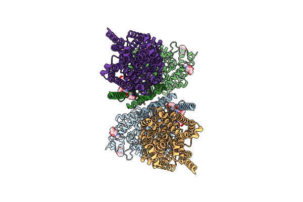
Organism: Homo sapiens
Method: ELECTRON MICROSCOPY Release Date: 2024-06-12 Classification: MEMBRANE PROTEIN Ligands: NAG, YCP, CL |
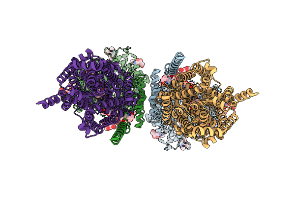 |
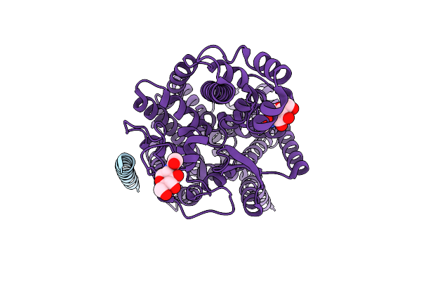
Structure Of Human Sit1:Ace2 Complex (Open Pd Conformation) Bound To L-Pipecolate
Organism: Homo sapiens
Method: ELECTRON MICROSCOPY Release Date: 2024-06-12 Classification: MEMBRANE PROTEIN Ligands: ZN, NDG, NAG, YCP, CL |
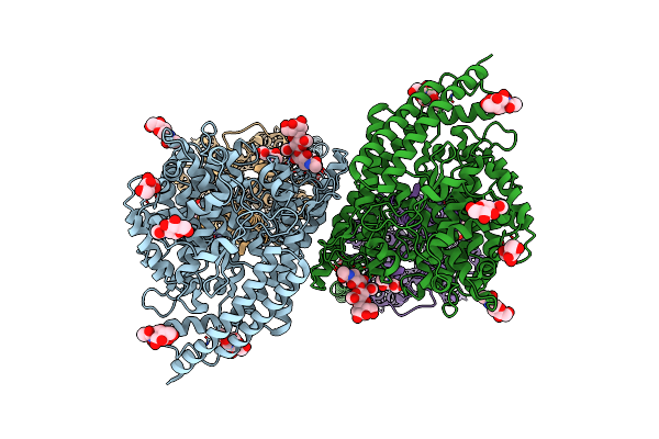 |
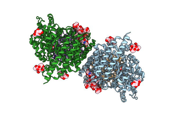
Structure Of Human Sit1:Ace2 Complex (Closed Pd Conformation) Bound To L-Pipecolate
Organism: Homo sapiens
Method: ELECTRON MICROSCOPY Release Date: 2024-06-12 Classification: MEMBRANE PROTEIN Ligands: ZN, NDG, NAG, YCP |
 |
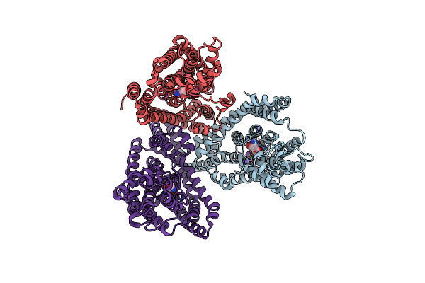
Cryo-Em Structure Of The Gltph L152C-G321C Mutant In The Intermediate State
Organism: Pyrococcus horikoshii (strain atcc 700860 / dsm 12428 / jcm 9974 / nbrc 100139 / ot-3)
Method: ELECTRON MICROSCOPY Release Date: 2021-02-17 Classification: TRANSPORT PROTEIN Ligands: ASP |
 |
Cryo-Em Structure Of The Gltph L152C-G321C Mutant In The Intermediate Chloride Conducting State.
Organism: Pyrococcus horikoshii (strain atcc 700860 / dsm 12428 / jcm 9974 / nbrc 100139 / ot-3)
Method: ELECTRON MICROSCOPY Release Date: 2021-02-17 Classification: TRANSPORT PROTEIN Ligands: ASP |
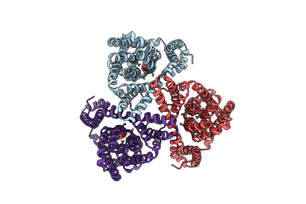 |
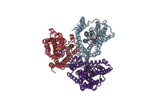
Cryo-Em Structure Of Gltph L152C-G351C Mutant In The Intermediate Outward-Facing State.
Organism: Pyrococcus horikoshii (strain atcc 700860 / dsm 12428 / jcm 9974 / nbrc 100139 / ot-3)
Method: ELECTRON MICROSCOPY Resolution:3.90 Å Release Date: 2021-02-17 Classification: TRANSPORT PROTEIN Ligands: ASP |
 |
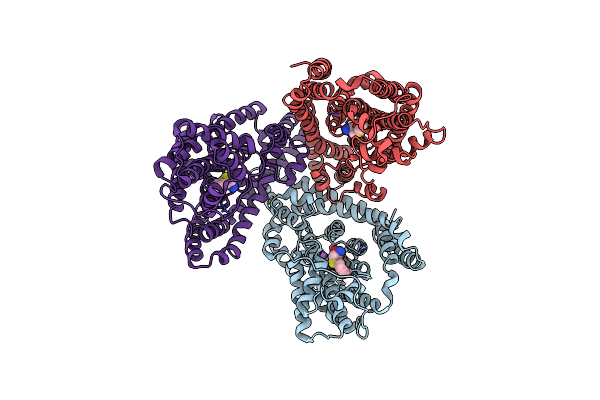
Crystal Structure Of The Gltph V216C-G388C Mutant Cross-Linked With Divalent Mercury
Organism: Pyrococcus horikoshii (strain atcc 700860 / dsm 12428 / jcm 9974 / nbrc 100139 / ot-3)
Method: X-RAY DIFFRACTION Resolution:3.45 Å Release Date: 2021-02-17 Classification: TRANSPORT PROTEIN Ligands: ASP, NA, HG |
 |
Crystal Structure Of The Gltph V216C-A391C Mutant Cross-Linked In Outward-Facing State
Organism: Pyrococcus horikoshii (strain atcc 700860 / dsm 12428 / jcm 9974 / nbrc 100139 / ot-3)
Method: X-RAY DIFFRACTION Resolution:3.65 Å Release Date: 2021-02-17 Classification: TRANSPORT PROTEIN Ligands: ASP, NA |
 |
Organism: Pyrococcus horikoshii
Method: X-RAY DIFFRACTION Resolution:3.40 Å Release Date: 2018-01-17 Classification: TRANSPORT PROTEIN |
 |
Organism: Pyrococcus horikoshii
Method: X-RAY DIFFRACTION Resolution:3.80 Å Release Date: 2018-01-17 Classification: TRANSPORT PROTEIN |
 |
Organism: Pyrococcus horikoshii
Method: X-RAY DIFFRACTION Resolution:3.70 Å Release Date: 2018-01-17 Classification: TRANSPORT PROTEIN |
 |
Organism: Pyrococcus horikoshii
Method: X-RAY DIFFRACTION Resolution:3.90 Å Release Date: 2018-01-17 Classification: TRANSPORT PROTEIN |