

Search Count: 37
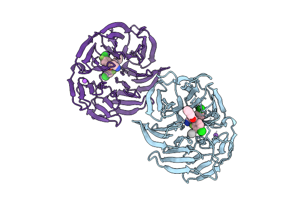 |
Crystal Structure Of The Human Dcaf1 Wdr Domain In Complex With Oicr-41109
Organism: Homo sapiens
Method: X-RAY DIFFRACTION Resolution:1.60 Å Release Date: 2025-11-26 Classification: TRANSPORT PROTEIN Ligands: A1CY9, NA, UNX |
Organism: Homo sapiens
Method: X-RAY DIFFRACTION
Release Date: 2025-11-26
Ligands: A1CY9, NA, UNX
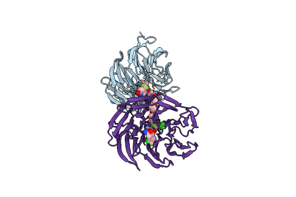 |
Crystal Structure Of The Ternary Complex Of Dcaf1 And Wdr5 With Protac, Oicr-40333
Organism: Homo sapiens
Method: X-RAY DIFFRACTION Resolution:2.06 Å Release Date: 2024-11-06 Classification: PROTEIN BINDING Ligands: A1AM2 |
Organism: Homo sapiens
Method: X-RAY DIFFRACTION
Release Date: 2024-11-06
Ligands: A1AM2
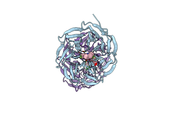 |
Crystal Structure Of The Ternary Complex Of Dcaf1 And Wdr5 With Protac, Oicr-40407
Organism: Homo sapiens
Method: X-RAY DIFFRACTION Resolution:2.05 Å Release Date: 2024-11-06 Classification: PROTEIN BINDING Ligands: A1ANM |
Organism: Homo sapiens
Method: X-RAY DIFFRACTION
Release Date: 2024-11-06
Ligands: A1ANM
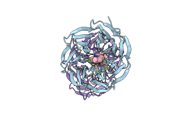 |
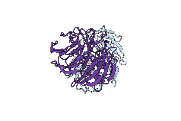
Crystal Structure Of The Ternary Complex Of Dcaf1 And Wdr5 With Protac, Oicr-40792
Organism: Homo sapiens
Method: X-RAY DIFFRACTION Resolution:1.92 Å Release Date: 2024-11-06 Classification: PROTEIN BINDING Ligands: A1ANN |
Organism: Homo sapiens
Method: X-RAY DIFFRACTION
Release Date: 2024-11-06
Ligands: A1ANN
 |
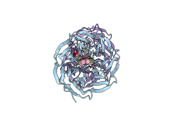
Crystal Structure Of The Binary Complex Of Dcaf1 And Wdr5
Organism: Homo sapiens
Method: X-RAY DIFFRACTION Resolution:2.97 Å Release Date: 2024-11-06 Classification: PROTEIN BINDING Ligands: IMD |
Organism: Homo sapiens
Method: X-RAY DIFFRACTION
Release Date: 2024-11-06
Ligands: IMD
 |
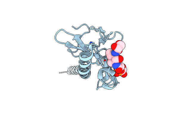
Crystal Structure Of The Ternary Complex Of Dcaf1 And Wdr5 With Protac, Oicr-41114
Organism: Homo sapiens
Method: X-RAY DIFFRACTION Resolution:2.07 Å Release Date: 2024-11-06 Classification: PROTEIN BINDING Ligands: A1BAF |
Organism: Homo sapiens
Method: X-RAY DIFFRACTION
Release Date: 2024-11-06
Ligands: A1BAF
 |
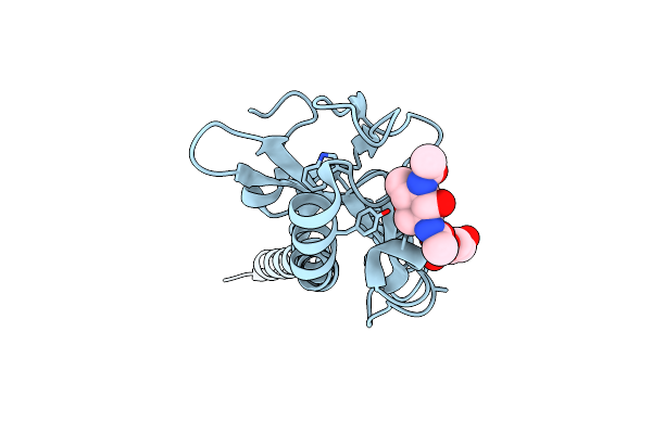
Neisseria Meningitidis Type Iv Pilus Sb-Gatdh Variant
Organism: Neisseria meningitidis 8013
Method: ELECTRON MICROSCOPY Release Date: 2024-04-03 Classification: PROTEIN FIBRIL Ligands: G3P, WKE |
Organism: Neisseria meningitidis 8013
Method: ELECTRON MICROSCOPY
Release Date: 2024-04-03
Ligands: G3P, WKE
 |
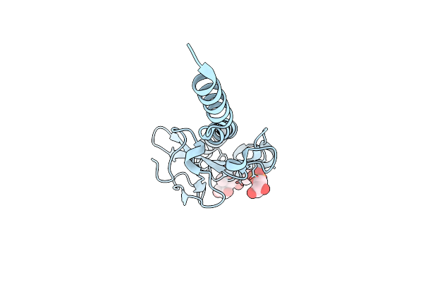
Neisseria Meningitidis Type Iv Pilus Sb-Datdh Variant
Organism: Neisseria meningitidis 8013
Method: ELECTRON MICROSCOPY Release Date: 2024-04-03 Classification: PROTEIN FIBRIL Ligands: G3P, B6D |
Organism: Neisseria meningitidis 8013
Method: ELECTRON MICROSCOPY
Release Date: 2024-04-03
Ligands: G3P, B6D
 |
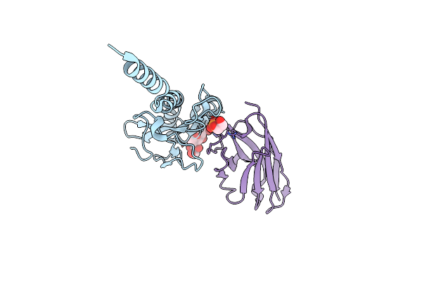
Neisseria Meningitidis Type Iv Pilus Sa-Gatdh Variant
Organism: Neisseria meningitidis 8013
Method: ELECTRON MICROSCOPY Release Date: 2024-04-03 Classification: PROTEIN FIBRIL Ligands: G3P, WKE |
Organism: Neisseria meningitidis 8013
Method: ELECTRON MICROSCOPY
Release Date: 2024-04-03
Ligands: G3P, WKE
 |
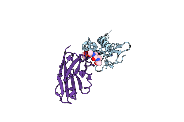
Neisseria Meningitidis Type Iv Pilus Sb-Gatdh Variant Bound To The C24 Nanobody
Organism: Neisseria meningitidis 8013, Vicugna pacos
Method: ELECTRON MICROSCOPY Release Date: 2024-04-03 Classification: PROTEIN FIBRIL Ligands: G3P, WKE |
Organism: Neisseria meningitidis 8013, Vicugna pacos
Method: ELECTRON MICROSCOPY
Release Date: 2024-04-03
Ligands: G3P, WKE
 |
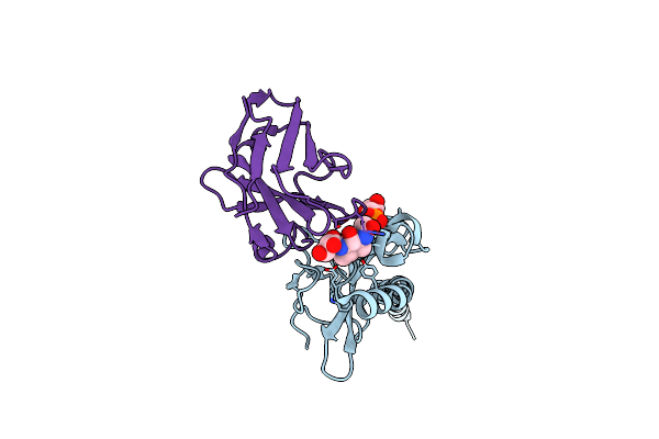
Neisseria Meningitidis Type Iv Pilus Sb-Datdh Variant Bound To The C24 Nanobody
Organism: Vicugna pacos, Neisseria meningitidis 8013
Method: ELECTRON MICROSCOPY Release Date: 2024-04-03 Classification: PROTEIN FIBRIL Ligands: G3P, B6D |
Organism: Vicugna pacos, Neisseria meningitidis 8013
Method: ELECTRON MICROSCOPY
Release Date: 2024-04-03
Ligands: G3P, B6D
 |
Neisseria Meningitidis Pile, Sb-Gatdh Variant, Bound To The F10 Nanobody
Organism: Vicugna pacos, Neisseria meningitidis 8013
Method: ELECTRON MICROSCOPY Release Date: 2024-04-03 Classification: PROTEIN FIBRIL Ligands: G3P, WKE |
Organism: Vicugna pacos, Neisseria meningitidis 8013
Method: ELECTRON MICROSCOPY
Release Date: 2024-04-03
Ligands: G3P, WKE
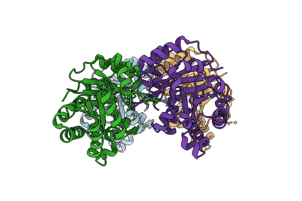 |
Crystal Structure Of A Novel Triose Phosphate Isomerase Identified On The Shrimp Transcriptome
Organism: Penaeus vannamei
Method: X-RAY DIFFRACTION Resolution:1.70 Å Release Date: 2023-12-06 Classification: ISOMERASE Ligands: CL, MG |
Organism: Penaeus vannamei
Method: X-RAY DIFFRACTION
Release Date: 2023-12-06
Ligands: CL, MG
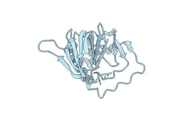 |
Crystal Structure Of The Ketc7 Antigen From Taenia Solium
Organism: Taenia solium
Method: X-RAY DIFFRACTION Resolution:1.72 Å Release Date: 2023-11-01 Classification: UNKNOWN FUNCTION |
Organism: Taenia solium
Method: X-RAY DIFFRACTION
Release Date: 2023-11-01
 |
Crystal Structure Of The Brown Dog Tick (Rhipicephalus Sanguineus) Arginine Kinase In Absence Of Substrate Or Ligands
Organism: Rhipicephalus sanguineus
Method: X-RAY DIFFRACTION Resolution:1.53 Å Release Date: 2021-11-17 Classification: TRANSFERASE |
Organism: Rhipicephalus sanguineus
Method: X-RAY DIFFRACTION
Release Date: 2021-11-17
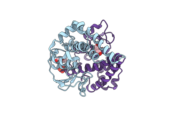 |
Crystal Structure Of A Glutathione S-Transferase Class Gtt2 Of Vibrio Parahaemolyticus (Vpgstt2)
Organism: Vibrio parahaemolyticus
Method: X-RAY DIFFRACTION Resolution:1.92 Å Release Date: 2021-07-07 Classification: TRANSFERASE Ligands: PEG, SO4 |
Organism: Vibrio parahaemolyticus
Method: X-RAY DIFFRACTION
Release Date: 2021-07-07
Ligands: PEG, SO4
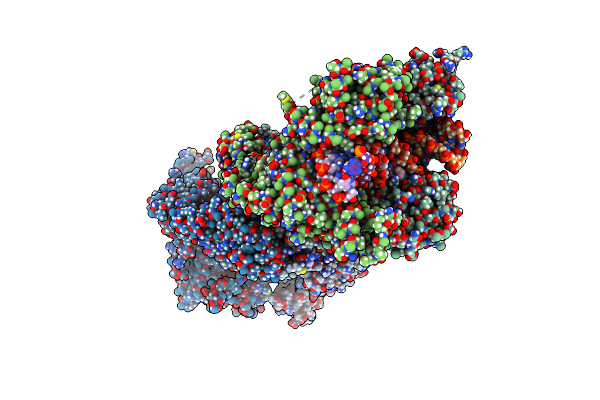 |
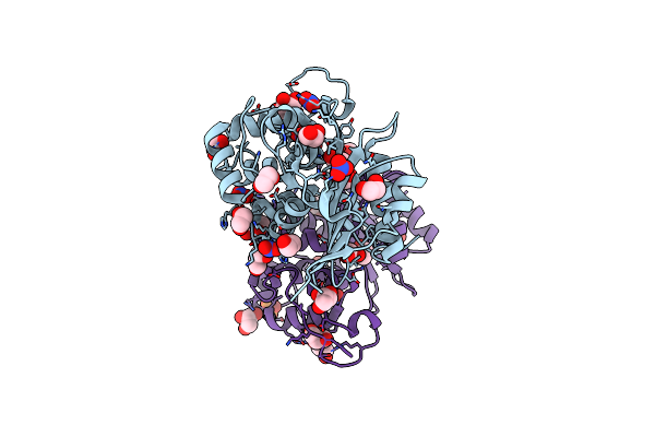
K. Pneumoniae Topoisomerase Iv (Pare-Parc) In Complex With Dna And Compound 34 (7-[(1S,5R)-1-Amino-3-Azabicyclo[3.1.0]Hexan-3-Yl]-4-(Aminomethyl)-1-Cyclopropyl-3,6-Difluoro-8-Methylquinolin-2(1H)-One)
Organism: Klebsiella pneumoniae 342
Method: X-RAY DIFFRACTION Resolution:3.20 Å Release Date: 2020-07-15 Classification: ISOMERASE/DNA/ISOMERASE INHIBITOR Ligands: MPD, MG, CL, TNJ |
Organism: Klebsiella pneumoniae 342
Method: X-RAY DIFFRACTION
Release Date: 2020-07-15
Ligands: MPD, MG, CL, TNJ
 |
Crystal Structure Of An Oxa-48 Beta-Lactamase Synthetic Mutant
Organism: Klebsiella pneumoniae
Method: X-RAY DIFFRACTION Resolution:1.75 Å Release Date: 2020-03-04 Classification: HYDROLASE Ligands: GOL, CL, NO3, EDO, PEG, PO4 |
Organism: Klebsiella pneumoniae
Method: X-RAY DIFFRACTION
Release Date: 2020-03-04
Ligands: GOL, CL, NO3, EDO, PEG, PO4
 |
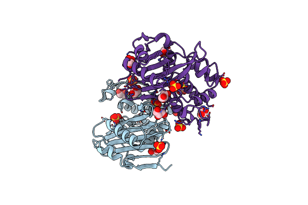
Crystal Structure Of Oxa-427 Class D Beta-Lactamase
Organism: Klebsiella pneumoniae
Method: X-RAY DIFFRACTION Resolution:2.78 Å Release Date: 2019-10-30 Classification: HYDROLASE Ligands: SO4 |
Organism: Klebsiella pneumoniae
Method: X-RAY DIFFRACTION
Release Date: 2019-10-30
Ligands: SO4
 |
Crystal Structure Of Rationally Designed Oxa-48Loop18 Beta-Lactamase
Organism: Klebsiella pneumoniae
Method: X-RAY DIFFRACTION Resolution:2.38 Å Release Date: 2019-10-09 Classification: HYDROLASE Ligands: SO4, GOL, F |
Organism: Klebsiella pneumoniae
Method: X-RAY DIFFRACTION
Release Date: 2019-10-09
Ligands: SO4, GOL, F