

Search Count: 25
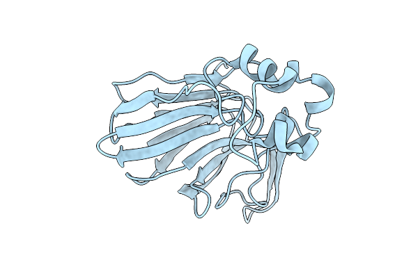 |
Acoustofluidic Sample Delivery System For Serial Crystallography, Thaumatin With Acoustic On
Organism: Thaumatococcus daniellii
Method: X-RAY DIFFRACTION Resolution:1.80 Å Release Date: 2026-04-15 Classification: PLANT PROTEIN |
Organism: Thaumatococcus daniellii
Method: X-RAY DIFFRACTION
Release Date: 2026-04-15
 |
Acoustofluidic Sample Delivery System For Serial Crystallography, Thaumatin With Acoustic Off
Organism: Thaumatococcus daniellii
Method: X-RAY DIFFRACTION Resolution:2.40 Å Release Date: 2026-04-15 Classification: PLANT PROTEIN |
Organism: Thaumatococcus daniellii
Method: X-RAY DIFFRACTION
Release Date: 2026-04-15
 |
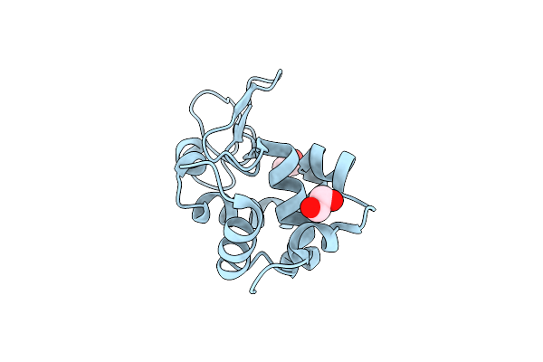
Room-Temperature Serial Synchrotron Crystallography Structure Of Spinacia Oleracea Rubisco
Organism: Spinacia oleracea
Method: X-RAY DIFFRACTION Resolution:2.30 Å Release Date: 2024-05-29 Classification: LYASE Ligands: MG |
Organism: Spinacia oleracea
Method: X-RAY DIFFRACTION
Release Date: 2024-05-29
Ligands: MG
 |
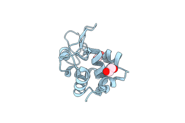
Lysozyme Structure Solved From Serial Crystallography Data Collected At 2 Khz For 5 Seconds With Jungfrau Detector At Maxiv
Organism: Gallus gallus
Method: X-RAY DIFFRACTION Resolution:2.05 Å Release Date: 2023-10-18 Classification: HYDROLASE Ligands: EDO, CL |
Organism: Gallus gallus
Method: X-RAY DIFFRACTION
Release Date: 2023-10-18
Ligands: EDO, CL
 |
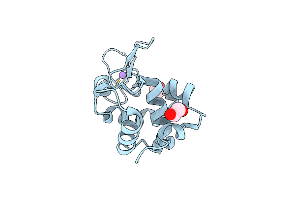
Lysozyme Structure Solved From Serial Crystallography Data Collected At 2 Khz With Jungfrau Detector At Maxiv
Organism: Gallus gallus
Method: X-RAY DIFFRACTION Resolution:1.70 Å Release Date: 2023-10-18 Classification: HYDROLASE Ligands: EDO, CL |
Organism: Gallus gallus
Method: X-RAY DIFFRACTION
Release Date: 2023-10-18
Ligands: EDO, CL
 |
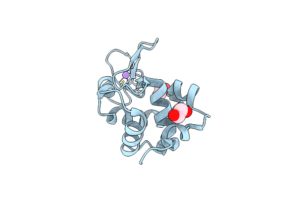
Lysozyme Structure Solved From Serial Crystallography Data Collected At 1 Khz With Jungfrau Detector At Maxiv
Organism: Gallus gallus
Method: X-RAY DIFFRACTION Resolution:1.58 Å Release Date: 2023-10-18 Classification: HYDROLASE Ligands: EDO, CL, NA |
Organism: Gallus gallus
Method: X-RAY DIFFRACTION
Release Date: 2023-10-18
Ligands: EDO, CL, NA
 |
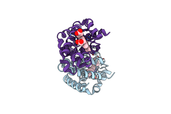
Lysozyme Structure Solved From Serial Crystallography Data Collected At 100 Hz With Jungfrau Detector At Maxiv
Organism: Gallus gallus
Method: X-RAY DIFFRACTION Resolution:1.60 Å Release Date: 2023-10-18 Classification: HYDROLASE Ligands: EDO, CL, NA |
Organism: Gallus gallus
Method: X-RAY DIFFRACTION
Release Date: 2023-10-18
Ligands: EDO, CL, NA
 |
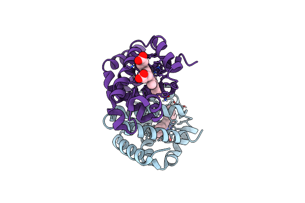
Methemoglobin Structure From Serial Synchrotron Crystallography With Fixed Target
Organism: Equus caballus
Method: X-RAY DIFFRACTION Resolution:1.95 Å Release Date: 2023-10-18 Classification: OXYGEN TRANSPORT Ligands: HEM |
Organism: Equus caballus
Method: X-RAY DIFFRACTION
Release Date: 2023-10-18
Ligands: HEM
 |
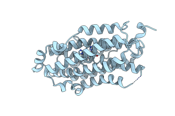
Dexyhemoglobin Structure From Serial Synchrotron Crystallography With Fixed Target
Organism: Equus caballus
Method: X-RAY DIFFRACTION Resolution:1.85 Å Release Date: 2023-10-18 Classification: OXYGEN TRANSPORT Ligands: HEM |
Organism: Equus caballus
Method: X-RAY DIFFRACTION
Release Date: 2023-10-18
Ligands: HEM
 |
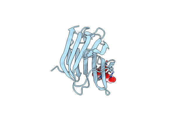
Crystal Structure Of Ribonucleotide Reductase R2 Subunit Solved By Serial Synchrotron Crystallography
Organism: Saccharopolyspora erythraea (strain atcc 11635 / dsm 40517 / jcm 4748 / nbrc 13426 / ncimb 8594 / nrrl 2338)
Method: X-RAY DIFFRACTION Resolution:2.40 Å Release Date: 2020-10-07 Classification: OXIDOREDUCTASE Ligands: MN3, FE |
 |
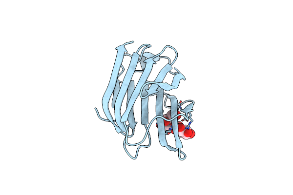
Structure Of Galectin-3C In Complex With Lactose Determined By Serial Crystallography Using A Silicon Nitride Membrane Support
Organism: Homo sapiens
Method: X-RAY DIFFRACTION Resolution:1.70 Å Release Date: 2020-07-29 Classification: SUGAR BINDING PROTEIN |
Organism: Homo sapiens
Method: X-RAY DIFFRACTION
Release Date: 2020-07-29
 |
Structure Of Galectin-3C In Complex With Lactose Determined By Serial Crystallography Using An Xtaltool Support
Organism: Homo sapiens
Method: X-RAY DIFFRACTION Resolution:1.70 Å Release Date: 2020-06-17 Classification: SUGAR BINDING PROTEIN Ligands: CL |
Organism: Homo sapiens
Method: X-RAY DIFFRACTION
Release Date: 2020-06-17
Ligands: CL
 |
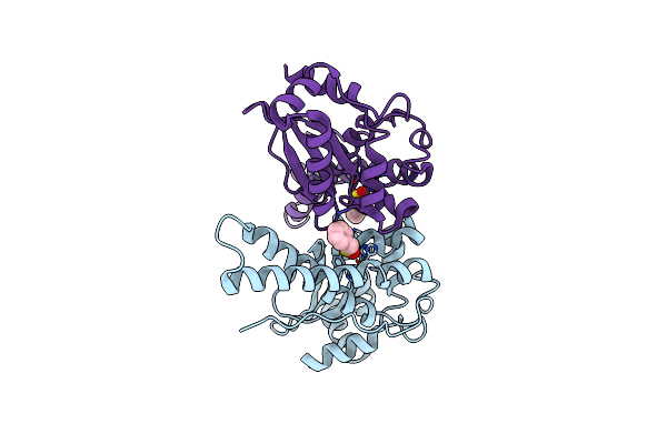
Iron Superoxide Dismutase (Fe-Sod) From The Hyperthermophile Sulfolobus Solfataricus. Crystal Structure Of The Y41F Mutant.
Organism: Sulfolobus solfataricus
Method: X-RAY DIFFRACTION Resolution:2.24 Å Release Date: 2004-11-08 Classification: OXIDOREDUCTASE Ligands: FE |
Organism: Sulfolobus solfataricus
Method: X-RAY DIFFRACTION
Release Date: 2004-11-08
Ligands: FE
 |
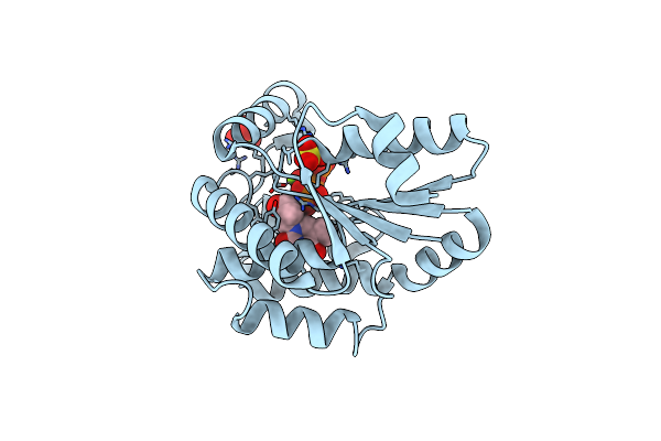
Iron Superoxide Dismutase (Fe-Sod) From The Hyperthermophile Sulfolobus Solfataricus. 2.3 A Resolution Structure Of Recombinant Protein With A Covalently Modified Tyrosine In The Active Site.
Organism: Sulfolobus solfataricus
Method: X-RAY DIFFRACTION Resolution:2.30 Å Release Date: 2004-11-08 Classification: OXIDOREDUCTASE Ligands: FE, PMS |
Organism: Sulfolobus solfataricus
Method: X-RAY DIFFRACTION
Release Date: 2004-11-08
Ligands: FE, PMS
 |
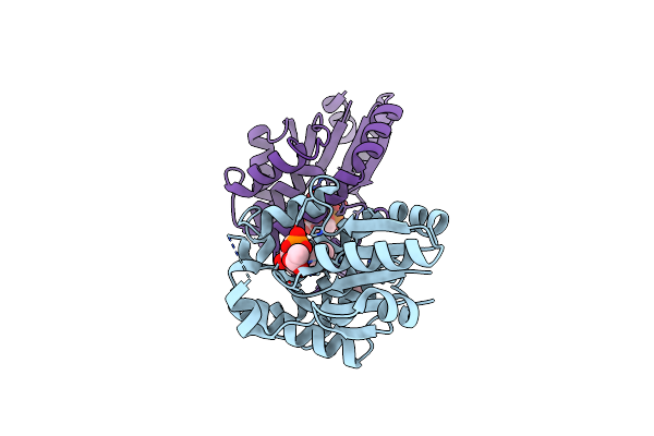
Crystal Structure Of Mycobacterium Tuberculosis Thymidylate Kinase Complexed With Thymidine Diphosphate (Tdp) And Thymidine Triphosphate (Ttp) At Ph 5.4 (1.85 A Resolution)
Organism: Mycobacterium tuberculosis
Method: X-RAY DIFFRACTION Resolution:1.85 Å Release Date: 2003-04-15 Classification: TRANSFERASE Ligands: SO4, MG, TYD, TTP |
Organism: Mycobacterium tuberculosis
Method: X-RAY DIFFRACTION
Release Date: 2003-04-15
Ligands: SO4, MG, TYD, TTP
 |
Crystal Structure Of Mycobacterium Tuberculosis Thymidylate Kinase Crystallized In Sodium Malonate (Resolution 2.1 A)
Organism: Mycobacterium tuberculosis
Method: X-RAY DIFFRACTION Resolution:2.10 Å Release Date: 2003-04-15 Classification: TRANSFERASE Ligands: ACT, TMP, MG |
Organism: Mycobacterium tuberculosis
Method: X-RAY DIFFRACTION
Release Date: 2003-04-15
Ligands: ACT, TMP, MG
 |
Crystal Structure Of Mycobacterium Tuberculosis Thymidylate Kinase Crystallized In Sodium Malonate, After Catalysis In The Crystal (2.3 A Resolution)
Organism: Mycobacterium tuberculosis
Method: X-RAY DIFFRACTION Resolution:2.30 Å Release Date: 2003-04-15 Classification: TRANSFERASE Ligands: ACT, TMP, MG, TYD, DPO |
Organism: Mycobacterium tuberculosis
Method: X-RAY DIFFRACTION
Release Date: 2003-04-15
Ligands: ACT, TMP, MG, TYD, DPO
 |
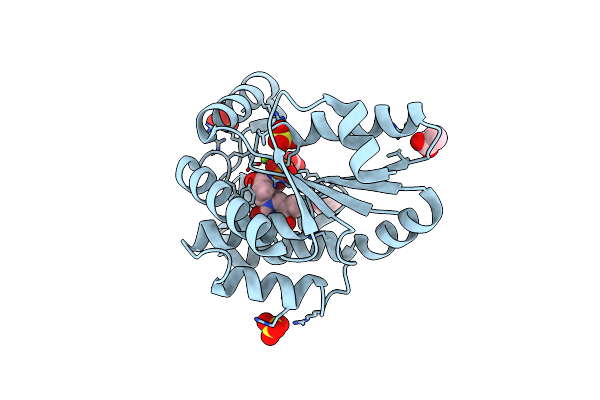
Crystal Structure Of Inactive Mycobacterium Tuberculosis Thymidylate Kinase Complexed With Thymidine Monophosphate (Tmp) At Ph 4.6 (Resolution 1.85 A)
Organism: Mycobacterium tuberculosis
Method: X-RAY DIFFRACTION Resolution:1.85 Å Release Date: 2003-04-08 Classification: TRANSFERASE Ligands: SO4, FLC, TMP, ATP |
Organism: Mycobacterium tuberculosis
Method: X-RAY DIFFRACTION
Release Date: 2003-04-08
Ligands: SO4, FLC, TMP, ATP
 |
Crystal Structure Of Mycobacterium Tuberculosis Thymidylate Kinase Complexed With Thymidine Monophosphate (Tmp)
Organism: Mycobacterium tuberculosis
Method: X-RAY DIFFRACTION Resolution:1.60 Å Release Date: 2002-03-28 Classification: TRANSFERASE Ligands: MG, TMP, SO4, ACT |
Organism: Mycobacterium tuberculosis
Method: X-RAY DIFFRACTION
Release Date: 2002-03-28
Ligands: MG, TMP, SO4, ACT
 |
Crystal Structure Of Mycobacterium Tuberculosis Thymidylate Kinase Complexed With Thymidine-5'-Diphosphate (Tdp)
Organism: Mycobacterium tuberculosis
Method: X-RAY DIFFRACTION Resolution:1.55 Å Release Date: 2002-03-28 Classification: TRANSFERASE Ligands: MG, TYD, SO4, ACT, TMP |
Organism: Mycobacterium tuberculosis
Method: X-RAY DIFFRACTION
Release Date: 2002-03-28
Ligands: MG, TYD, SO4, ACT, TMP