

Search Count: 30
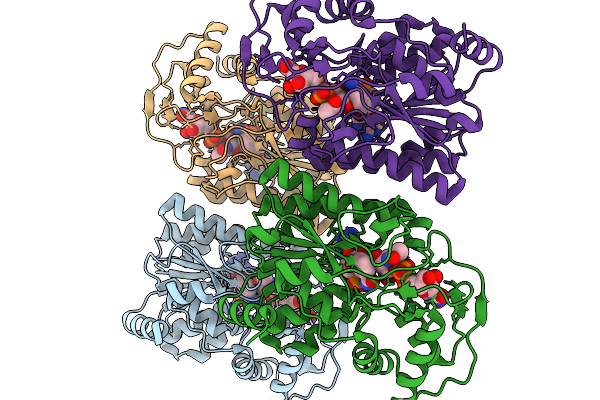 |
Crystal Structure Of Capsular Polysaccharide Biosynthesis Protein From Bordetella Pertussis In Complex With Nad And Uridine-Diphosphate-N-Acetylgalactosamine (Cocrystallization)
Organism: Bordetella pertussis tohama i
Method: X-RAY DIFFRACTION Resolution:1.70 Å Release Date: 2026-02-04 Classification: ISOMERASE Ligands: CL, NAD, UD2 |
Organism: Bordetella pertussis tohama i
Method: X-RAY DIFFRACTION
Release Date: 2026-02-04
Ligands: CL, NAD, UD2
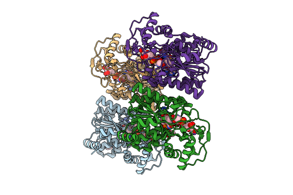 |
Crystal Structure Of Capsular Polysaccharide Biosynthesis Protein From Bordetella Pertussis In Complex With Nad And Uridine-Diphosphate-N-Acetylgalactosamine
Organism: Bordetella pertussis tohama i
Method: X-RAY DIFFRACTION Resolution:2.40 Å Release Date: 2025-08-06 Classification: ISOMERASE Ligands: CL, NAD, UD2, ACT |
Organism: Bordetella pertussis tohama i
Method: X-RAY DIFFRACTION
Release Date: 2025-08-06
Ligands: CL, NAD, UD2, ACT
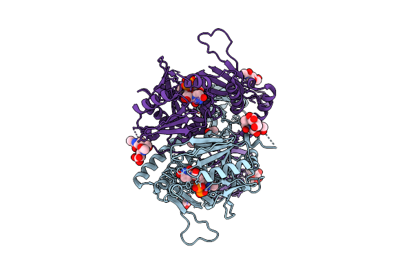 |
Human B4Galnt1 In Complex With Udp-Galnac
Organism: Homo sapiens
Method: X-RAY DIFFRACTION Resolution:2.75 Å Release Date: 2025-07-16 Classification: TRANSFERASE Ligands: NAG, MN, UD2, ACY |
Organism: Homo sapiens
Method: X-RAY DIFFRACTION
Release Date: 2025-07-16
Ligands: NAG, MN, UD2, ACY
 |
Structure Of Senb In Complex With Udp-Galnac At 1.64 Angstroms Resolution
Organism: Variovorax paradoxus
Method: X-RAY DIFFRACTION Resolution:1.64 Å Release Date: 2024-02-07 Classification: TRANSFERASE Ligands: UD2 |
Organism: Variovorax paradoxus
Method: X-RAY DIFFRACTION
Release Date: 2024-02-07
Ligands: UD2
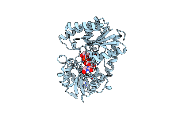 |
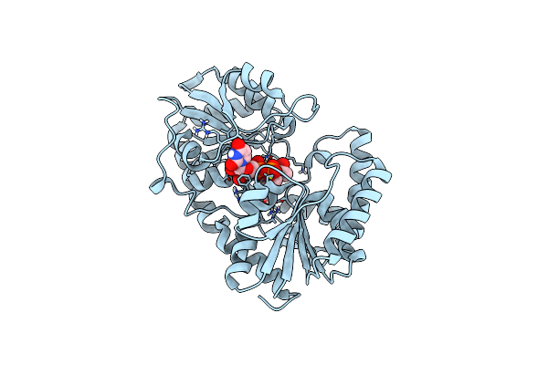
Structure Of The Campylobacter Concisus Glycosyltransferase Pgla
Organism: Campylobacter concisus
Method: X-RAY DIFFRACTION Resolution:1.78 Å Release Date: 2024-01-17 Classification: MEMBRANE PROTEIN Ligands: UD2 |
Organism: Campylobacter concisus
Method: X-RAY DIFFRACTION
Release Date: 2024-01-17
Ligands: UD2
 |
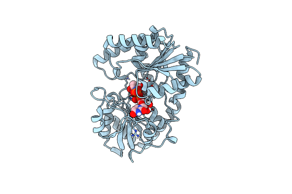
Structure Of The Campylobacter Concisus Glycosyltransferase Pgla R203Q
Organism: Campylobacter concisus
Method: X-RAY DIFFRACTION Resolution:2.29 Å Release Date: 2024-01-17 Classification: MEMBRANE PROTEIN Ligands: UD2 |
Organism: Campylobacter concisus
Method: X-RAY DIFFRACTION
Release Date: 2024-01-17
Ligands: UD2
 |
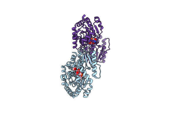
Structure Of The Campylobacter Concisus Glycosyltransferase Pgla R282V Variant
Organism: Campylobacter concisus
Method: X-RAY DIFFRACTION Resolution:2.27 Å Release Date: 2024-01-17 Classification: MEMBRANE PROTEIN Ligands: UD2 |
Organism: Campylobacter concisus
Method: X-RAY DIFFRACTION
Release Date: 2024-01-17
Ligands: UD2
 |
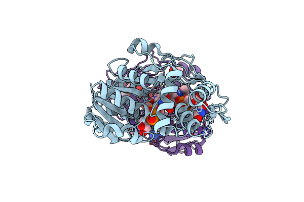
Crystal Structure Of P. Aeruginosa Waag In Complex With Udp-Galnac
Organism: Pseudomonas aeruginosa
Method: X-RAY DIFFRACTION Resolution:2.20 Å Release Date: 2023-10-04 Classification: TRANSFERASE Ligands: UD2, ACT, UDP |
Organism: Pseudomonas aeruginosa
Method: X-RAY DIFFRACTION
Release Date: 2023-10-04
Ligands: UD2, ACT, UDP
 |
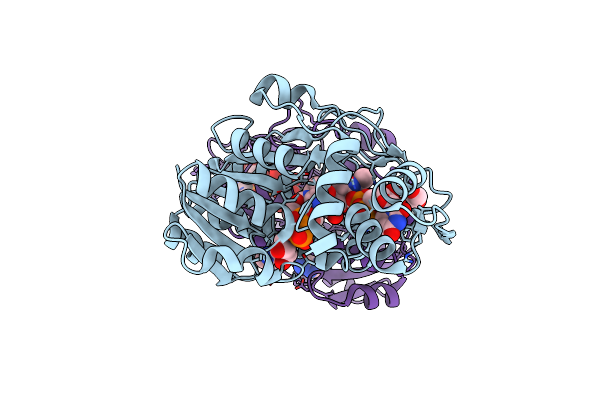
Crystal Structure Of Udp-Glucose 4-Epimerase (Rv3634C) Co-Crystallized With Udp-N-Acetylglucosamine From Mycobacterium Tuberculosis
Organism: Mycobacterium tuberculosis h37rv
Method: X-RAY DIFFRACTION Resolution:1.87 Å Release Date: 2023-08-16 Classification: ISOMERASE Ligands: NAD, UD2, ACT |
Organism: Mycobacterium tuberculosis h37rv
Method: X-RAY DIFFRACTION
Release Date: 2023-08-16
Ligands: NAD, UD2, ACT
 |
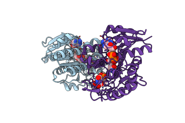
Crystal Structure Of Udp-Glucose 4-Epimerase (Rv3634C) Co-Crystallized With Udp-N-Acetylgalactosamine From Mycobacterium Tuberculosis
Organism: Mycobacterium tuberculosis h37rv
Method: X-RAY DIFFRACTION Resolution:2.16 Å Release Date: 2023-08-16 Classification: ISOMERASE Ligands: NAD, UD2, EDO |
Organism: Mycobacterium tuberculosis h37rv
Method: X-RAY DIFFRACTION
Release Date: 2023-08-16
Ligands: NAD, UD2, EDO
 |
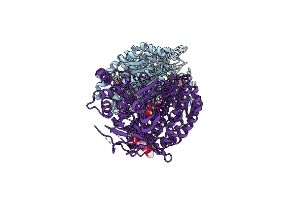
Udp-Glcnac C4-Epimerase Mutant S121A/Y146F From Pseudomonas Protegens In Complex With Udp-Galnac
Organism: Pseudomonas protegens
Method: X-RAY DIFFRACTION Resolution:2.09 Å Release Date: 2020-07-08 Classification: ISOMERASE Ligands: NAD, UD2 |
Organism: Pseudomonas protegens
Method: X-RAY DIFFRACTION
Release Date: 2020-07-08
Ligands: NAD, UD2
 |
Crystal Structure Of Human Gfat-1 In Complex With Glucose-6-Phosphate, L-Glu, And Udp-Galnac
Organism: Homo sapiens
Method: X-RAY DIFFRACTION Resolution:2.48 Å Release Date: 2020-01-15 Classification: TRANSFERASE Ligands: G6Q, GLU, UD2, MG |
Organism: Homo sapiens
Method: X-RAY DIFFRACTION
Release Date: 2020-01-15
Ligands: G6Q, GLU, UD2, MG
 |
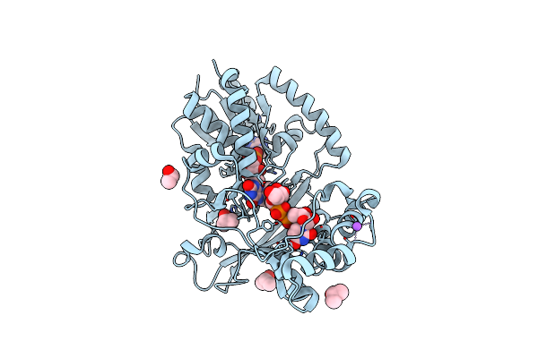
Blood Group Synthase Aaglyb In Complex With Udp-Galnac And Cryoprotected With Peg 3350
Organism: Homo sapiens
Method: X-RAY DIFFRACTION Resolution:1.60 Å Release Date: 2019-07-10 Classification: TRANSFERASE Ligands: MN, UD2, UDP, SO4, PEG |
Organism: Homo sapiens
Method: X-RAY DIFFRACTION
Release Date: 2019-07-10
Ligands: MN, UD2, UDP, SO4, PEG
 |
Crystal Structure Of Corynebacterium Diphtheriae Udp-Galactopyranose Mutase In Complex With Udp-Galnac (Open, Oxidized)
Organism: Corynebacterium diphtheriae
Method: X-RAY DIFFRACTION Resolution:2.15 Å Release Date: 2019-05-15 Classification: ISOMERASE Ligands: FAD, NA, UD2, IPA |
Organism: Corynebacterium diphtheriae
Method: X-RAY DIFFRACTION
Release Date: 2019-05-15
Ligands: FAD, NA, UD2, IPA
 |
Crystal Structure Of Galnac-T7 With Udp, Galnac And Mn2+
Organism: Homo sapiens
Method: X-RAY DIFFRACTION Resolution:2.60 Å Release Date: 2019-02-06 Classification: TRANSFERASE Ligands: MN, NAG, UDP, UD2 |
Organism: Homo sapiens
Method: X-RAY DIFFRACTION
Release Date: 2019-02-06
Ligands: MN, NAG, UDP, UD2
 |
Crystal Structure Of The Galnac-T2 F104S Mutant In Complex With Udp-Galnac
Organism: Homo sapiens
Method: X-RAY DIFFRACTION Resolution:2.70 Å Release Date: 2018-04-11 Classification: TRANSFERASE Ligands: MN, UD2 |
Organism: Homo sapiens
Method: X-RAY DIFFRACTION
Release Date: 2018-04-11
Ligands: MN, UD2
 |
Structure Of A Glycosyltransferase
Organism: Campylobacter jejuni
Method: X-RAY DIFFRACTION Resolution:2.30 Å Release Date: 2018-02-07 Classification: TRANSFERASE Ligands: UD2, K, GOL, NA, CL |
Organism: Campylobacter jejuni
Method: X-RAY DIFFRACTION
Release Date: 2018-02-07
Ligands: UD2, K, GOL, NA, CL
 |
Galnac-T2 Crystal Soaked With Udp-Galnac, Ea2 Peptide And Manganese
Organism: Homo sapiens, Synthetic construct
Method: X-RAY DIFFRACTION Resolution:2.45 Å Release Date: 2014-05-28 Classification: TRANSFERASE Ligands: MN, EDO, UD2, UDP, NGA |
Organism: Homo sapiens, Synthetic construct
Method: X-RAY DIFFRACTION
Release Date: 2014-05-28
Ligands: MN, EDO, UD2, UDP, NGA
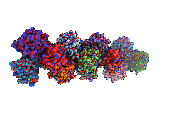 |
Monoclinic Crystal Form Of Bogt6A E192Q In Complex With Udp-Galnac, Udp And Galnac
Organism: Bacteroides ovatus
Method: X-RAY DIFFRACTION Resolution:3.50 Å Release Date: 2014-02-05 Classification: TRANSFERASE Ligands: GOL, UDP, A2G, 1PE, SO4, UD2, NGA |
Organism: Bacteroides ovatus
Method: X-RAY DIFFRACTION
Release Date: 2014-02-05
Ligands: GOL, UDP, A2G, 1PE, SO4, UD2, NGA
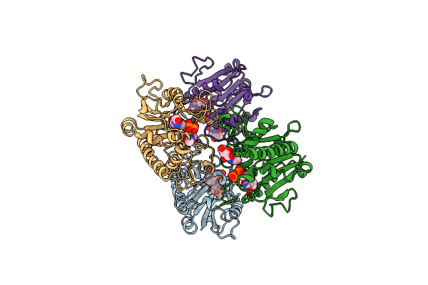 |
Orthorhombic Crystal Form Of Bogt6A E192Q In Complex With Udp-Galnac, Udp, Galnac
Organism: Bacteroides ovatus
Method: X-RAY DIFFRACTION Resolution:3.42 Å Release Date: 2014-02-05 Classification: TRANSFERASE Ligands: UD2, UDP, A2G |
Organism: Bacteroides ovatus
Method: X-RAY DIFFRACTION
Release Date: 2014-02-05
Ligands: UD2, UDP, A2G