

Search Count: 17
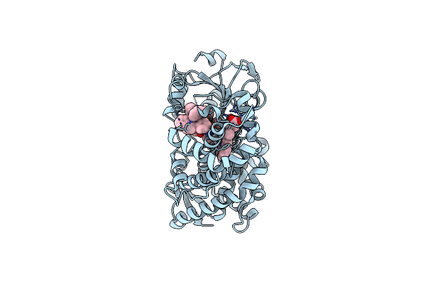 |
Crystal Structure Of Cyp3A4 Bound To An Inhibitor
Organism: Homo sapiens
Method: X-RAY DIFFRACTION Resolution:2.20 Å Release Date: 2023-02-01 Classification: OXIDOREDUCTASE/INHIBITOR Ligands: HEM, WZN |
Organism: Homo sapiens
Method: X-RAY DIFFRACTION
Release Date: 2023-02-01
Ligands: HEM, WZN
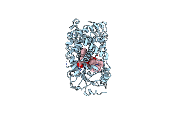 |
Crystal Structure Of Cyp3A4 Bound To An Inhibitor
Organism: Homo sapiens
Method: X-RAY DIFFRACTION Resolution:2.30 Å Release Date: 2023-02-01 Classification: OXIDOREDUCTASE/INHIBITOR Ligands: HEM, X0E |
Organism: Homo sapiens
Method: X-RAY DIFFRACTION
Release Date: 2023-02-01
Ligands: HEM, X0E
 |
Crystal Structure Of Cyp3A4 Bound To An Inhibitor
Organism: Homo sapiens
Method: X-RAY DIFFRACTION Resolution:2.35 Å Release Date: 2023-02-01 Classification: OXIDOREDUCTASE/INHIBITOR Ligands: HEM, X1C |
Organism: Homo sapiens
Method: X-RAY DIFFRACTION
Release Date: 2023-02-01
Ligands: HEM, X1C
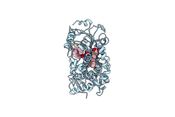 |
Crystal Structure Of Cyp3A4 Bound To An Inhibitor
Organism: Homo sapiens
Method: X-RAY DIFFRACTION Resolution:2.30 Å Release Date: 2023-02-01 Classification: OXIDOREDUCTASE/INHIBITOR Ligands: HEM, X1I |
Organism: Homo sapiens
Method: X-RAY DIFFRACTION
Release Date: 2023-02-01
Ligands: HEM, X1I
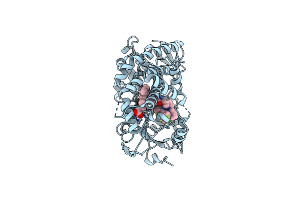 |
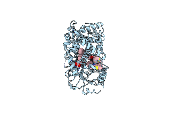
Crystal Structure Of Cyp3A4 Bound To An Inhibitor
Organism: Homo sapiens
Method: X-RAY DIFFRACTION Resolution:2.10 Å Release Date: 2023-02-01 Classification: OXIDOREDUCTASE/INHIBITOR Ligands: HEM, X1O |
Organism: Homo sapiens
Method: X-RAY DIFFRACTION
Release Date: 2023-02-01
Ligands: HEM, X1O
 |
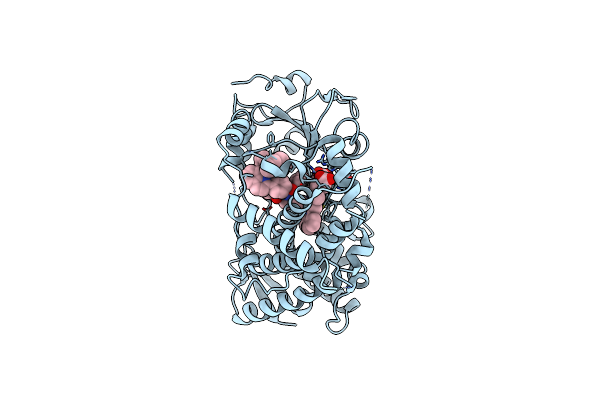
Crystal Structure Of Cyp3A4 Bound To An Inhibitor
Organism: Homo sapiens
Method: X-RAY DIFFRACTION Resolution:2.40 Å Release Date: 2023-02-01 Classification: OXIDOREDUCTASE/INHIBITOR Ligands: HEM, X2Q |
Organism: Homo sapiens
Method: X-RAY DIFFRACTION
Release Date: 2023-02-01
Ligands: HEM, X2Q
 |
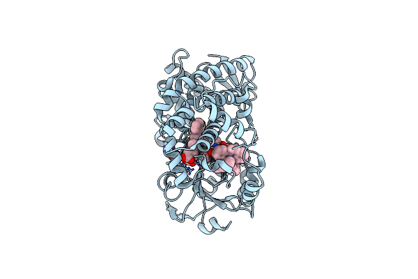
Crystal Structure Of Cyp3A4 Bound To An Inhibitor
Organism: Homo sapiens
Method: X-RAY DIFFRACTION Resolution:2.25 Å Release Date: 2023-02-01 Classification: OXIDOREDUCTASE/INHIBITOR Ligands: HEM, X2B |
Organism: Homo sapiens
Method: X-RAY DIFFRACTION
Release Date: 2023-02-01
Ligands: HEM, X2B
 |
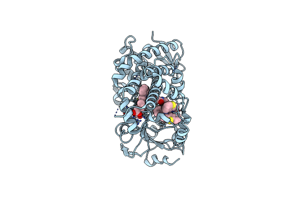
Crystal Structure Of Cyp3A4 Bound To An Inhibitor
Organism: Homo sapiens
Method: X-RAY DIFFRACTION Resolution:2.20 Å Release Date: 2023-02-01 Classification: OXIDOREDUCTASE/INHIBITOR Ligands: HEM, WZN |
Organism: Homo sapiens
Method: X-RAY DIFFRACTION
Release Date: 2023-02-01
Ligands: HEM, WZN
 |
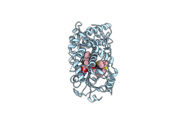
Crystal Structure Of Cyp3A4 Bound To An Inhibitor
Organism: Homo sapiens
Method: X-RAY DIFFRACTION Resolution:2.15 Å Release Date: 2023-02-01 Classification: OXIDOREDUCTASE/INHIBITOR Ligands: HEM, X2Q |
Organism: Homo sapiens
Method: X-RAY DIFFRACTION
Release Date: 2023-02-01
Ligands: HEM, X2Q
 |
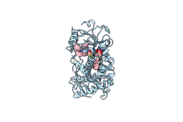
Crystal Structure Of Cyp3A4 Bound To An Inhibitor
Organism: Homo sapiens
Method: X-RAY DIFFRACTION Resolution:2.05 Å Release Date: 2023-02-01 Classification: OXIDOREDUCTASE/INHIBITOR Ligands: HEM, X4E |
Organism: Homo sapiens
Method: X-RAY DIFFRACTION
Release Date: 2023-02-01
Ligands: HEM, X4E
 |
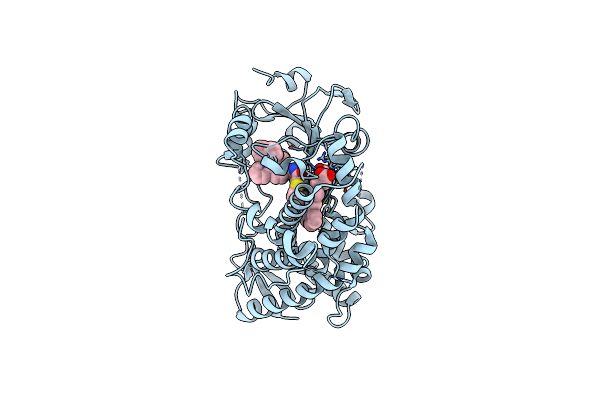
Crystal Structure Of Human Cyp3A4 With The Caged Inhibitor
Organism: Homo sapiens
Method: X-RAY DIFFRACTION Resolution:2.78 Å Release Date: 2022-08-31 Classification: OXIDOREDUCTASE/INHIBITOR Ligands: HEM, O39 |
Organism: Homo sapiens
Method: X-RAY DIFFRACTION
Release Date: 2022-08-31
Ligands: HEM, O39
 |
Crystal Structure Of Human Cyp3A4 With The Caged Inhibitor
Organism: Homo sapiens
Method: X-RAY DIFFRACTION Resolution:2.65 Å Release Date: 2022-08-31 Classification: OXIDOREDUCTASE/INHIBITOR Ligands: HEM, O3L |
Organism: Homo sapiens
Method: X-RAY DIFFRACTION
Release Date: 2022-08-31
Ligands: HEM, O3L
 |
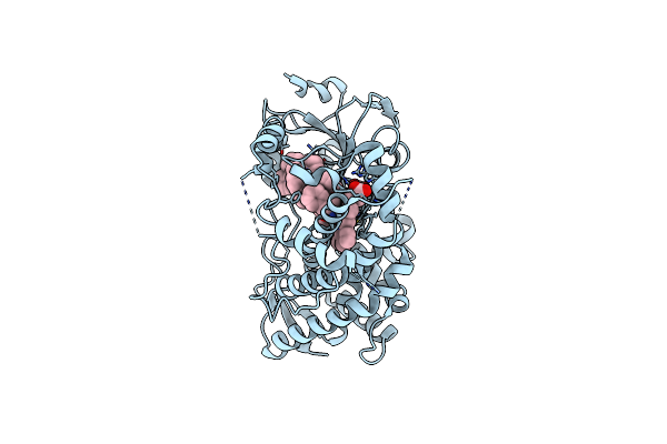
Crystal Structure Of Human Cyp3A4 With The Caged Inhibitor
Organism: Homo sapiens
Method: X-RAY DIFFRACTION Resolution:2.50 Å Release Date: 2021-06-02 Classification: OXIDOREDUCTASE/OXIDOREDUCTASE INHIBITOR Ligands: HEM, GOL, X8S |
Organism: Homo sapiens
Method: X-RAY DIFFRACTION
Release Date: 2021-06-02
Ligands: HEM, GOL, X8S
 |
Crystal Structure Of Human Cyp3A4 With The Caged Inhibitor
Organism: Homo sapiens
Method: X-RAY DIFFRACTION Resolution:2.50 Å Release Date: 2021-06-02 Classification: OXIDOREDUCTASE/OXIDOREDUCTASE INHIBITOR Ligands: HEM, X8P |
Organism: Homo sapiens
Method: X-RAY DIFFRACTION
Release Date: 2021-06-02
Ligands: HEM, X8P
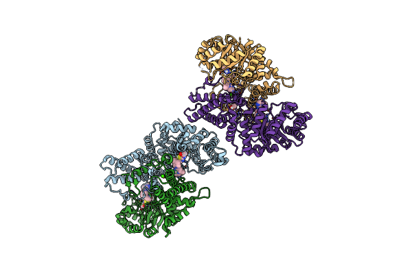 |
Ogoga In Complex With Ligand 55
Organism: Oceanicola granulosus
Method: X-RAY DIFFRACTION Resolution:1.78 Å Release Date: 2020-12-02 Classification: HYDROLASE/INHIBITOR Ligands: WG4, CA, EDO |
Organism: Oceanicola granulosus
Method: X-RAY DIFFRACTION
Release Date: 2020-12-02
Ligands: WG4, CA, EDO
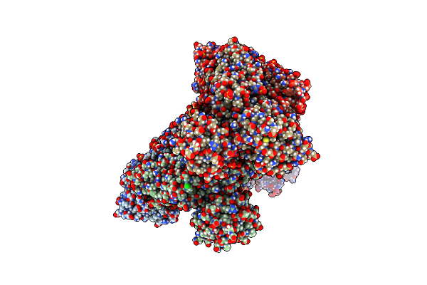 |
Cpoga In Complex With Ligand 54
Organism: Clostridium perfringens
Method: X-RAY DIFFRACTION Resolution:2.30 Å Release Date: 2020-12-02 Classification: HYDROLASE/INHIBITOR Ligands: X1A, CA, SO4, CL |
Organism: Clostridium perfringens
Method: X-RAY DIFFRACTION
Release Date: 2020-12-02
Ligands: X1A, CA, SO4, CL
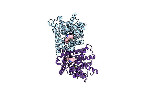 |
Discovery Of A Potent, Selective And Orally Active Pde10A Inhibitor For The Treatment Of Schizophrenia
Organism: Homo sapiens
Method: X-RAY DIFFRACTION Resolution:2.50 Å Release Date: 2013-10-16 Classification: HYDROLASE Ligands: LKF, ZN, MG |
Organism: Homo sapiens
Method: X-RAY DIFFRACTION
Release Date: 2013-10-16
Ligands: LKF, ZN, MG