

Search Count: 90
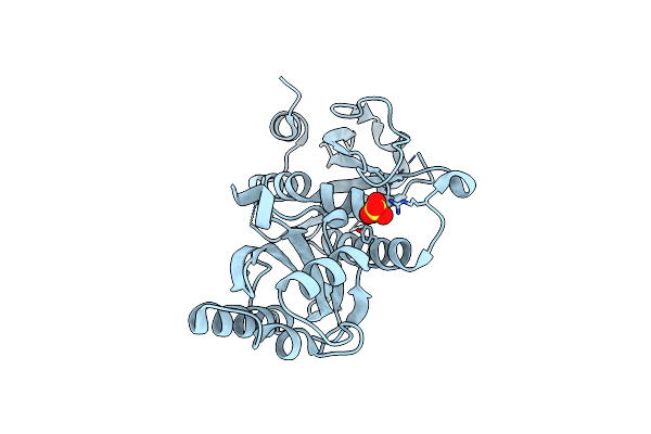 |
Sensor Region Of A Sensor Histidine Kinase
Organism: Mycobacterium tuberculosis
Method: X-RAY DIFFRACTION Resolution:2.00 Å Release Date: 2012-06-06 Classification: TRANSFERASE Ligands: SO4, BR |
Organism: Mycobacterium tuberculosis
Method: X-RAY DIFFRACTION
Release Date: 2012-06-06
Ligands: SO4, BR
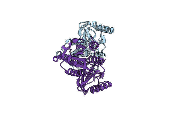 |
Sensor Region Of A Sensor Histidine Kinase
Organism: Mycobacterium tuberculosis
Method: X-RAY DIFFRACTION Resolution:2.78 Å Release Date: 2012-06-06 Classification: TRANSFERASE |
Organism: Mycobacterium tuberculosis
Method: X-RAY DIFFRACTION
Release Date: 2012-06-06
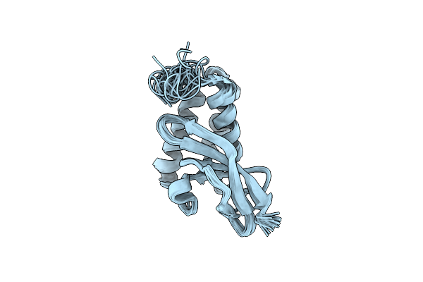 |
Nmr Structure Of The Arterivirus Nonstructural Protein 7 Alpha (Nsp7 Alpha)
Organism: Equine arteritis virus
Method: SOLUTION NMR Release Date: 2011-07-06 Classification: VIRAL PROTEIN |
Organism: Equine arteritis virus
Method: SOLUTION NMR
Release Date: 2011-07-06
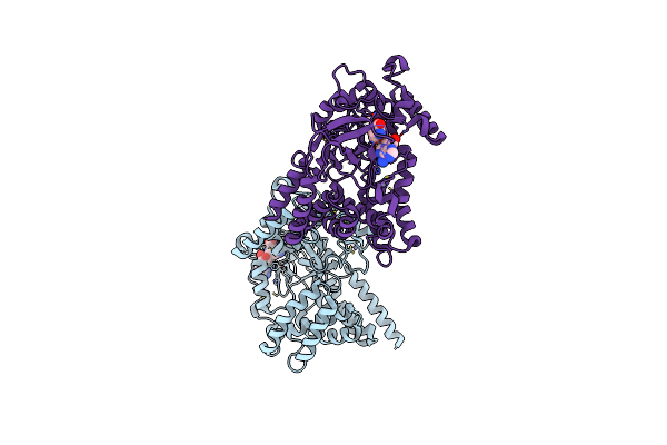 |
Cocrystal Structure Of Human Smyd3 With Inhibitor Sinefungin Bound
Organism: Homo sapiens
Method: X-RAY DIFFRACTION Resolution:1.85 Å Release Date: 2011-05-18 Classification: TRANSFERASE/TRANSFERASE INHIBITOR Ligands: SFG, ZN |
Organism: Homo sapiens
Method: X-RAY DIFFRACTION
Release Date: 2011-05-18
Ligands: SFG, ZN
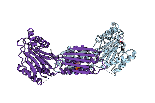 |
The Crystal Structure Of The Rsbu And Rsbw Domains Of Rv1364C From Mycobacterium Tuberculosis
Organism: Mycobacterium tuberculosis
Method: X-RAY DIFFRACTION Resolution:2.60 Å Release Date: 2010-11-10 Classification: UNKNOWN FUNCTION Ligands: SO4, GOL, MN |
Organism: Mycobacterium tuberculosis
Method: X-RAY DIFFRACTION
Release Date: 2010-11-10
Ligands: SO4, GOL, MN
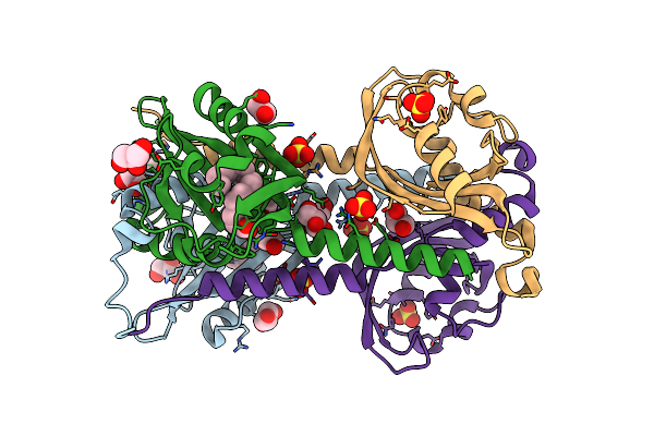 |
The N-Terminal Pas Domain Crystal Structure Of Rv1364C From Mycobacterium Tuberculosis At 1.62
Organism: Mycobacterium tuberculosis
Method: X-RAY DIFFRACTION Resolution:1.62 Å Release Date: 2010-09-29 Classification: SIGNALING PROTEIN Ligands: PLM, SO4, GOL |
Organism: Mycobacterium tuberculosis
Method: X-RAY DIFFRACTION
Release Date: 2010-09-29
Ligands: PLM, SO4, GOL
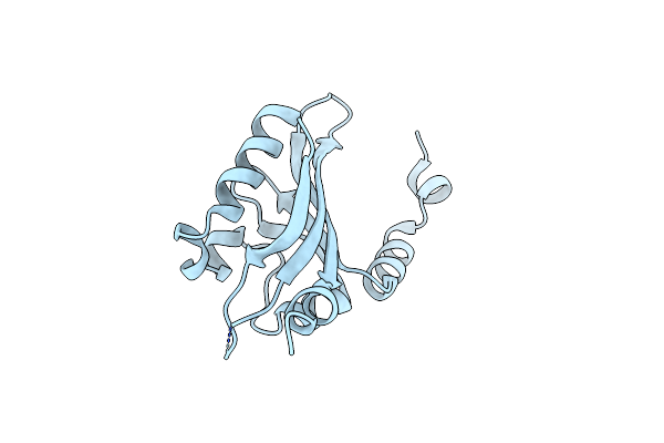 |
The N-Terminal Pas Domain Crystal Structure Of Rv1364C From Mycobacterium Tuberculosis At 2.3 Angstrom
Organism: Mycobacterium tuberculosis
Method: X-RAY DIFFRACTION Resolution:2.30 Å Release Date: 2010-09-29 Classification: SIGNALING PROTEIN |
Organism: Mycobacterium tuberculosis
Method: X-RAY DIFFRACTION
Release Date: 2010-09-29
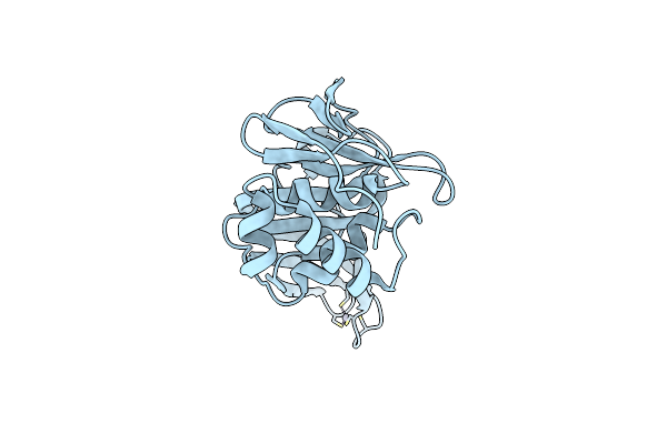 |
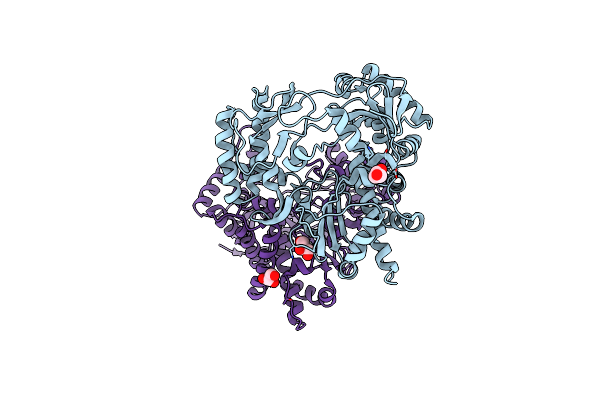
Crystal Structure Of Transmissible Gastroenteritis Virus Papain-Like Protease 1
Organism: Porcine transmissible gastroenteritis coronavirus
Method: X-RAY DIFFRACTION Resolution:2.50 Å Release Date: 2010-09-08 Classification: HYDROLASE Ligands: ZN |
Organism: Porcine transmissible gastroenteritis coronavirus
Method: X-RAY DIFFRACTION
Release Date: 2010-09-08
Ligands: ZN
 |
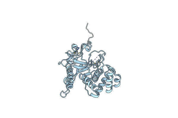
Dimeric Structure Of D347G D348G Mutant Of The Sapporovirus Rna Dependent Rna Polymerase
Organism: Sapporo virus
Method: X-RAY DIFFRACTION Resolution:2.98 Å Release Date: 2010-09-01 Classification: HYDROLASE Ligands: GOL |
Organism: Sapporo virus
Method: X-RAY DIFFRACTION
Release Date: 2010-09-01
Ligands: GOL
 |
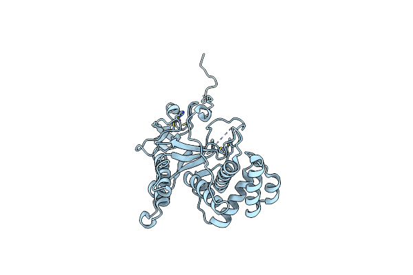
N512P Mutant Of The Dna Binding Domain Of The Adenovirus 5 Ssdna Binding Protein
Organism: Human adenovirus 5
Method: X-RAY DIFFRACTION Resolution:2.30 Å Release Date: 2010-03-31 Classification: DNA BINDING PROTEIN Ligands: ZN |
Organism: Human adenovirus 5
Method: X-RAY DIFFRACTION
Release Date: 2010-03-31
Ligands: ZN
 |
2.1 Resolution Structure Of The C-Terminal Domain Of The Human Adenovirus 5 Ssdna Binding Protein
Organism: Human adenovirus 5
Method: X-RAY DIFFRACTION Resolution:1.95 Å Release Date: 2010-03-31 Classification: DNA BINDING PROTEIN Ligands: ZN |
Organism: Human adenovirus 5
Method: X-RAY DIFFRACTION
Release Date: 2010-03-31
Ligands: ZN
 |
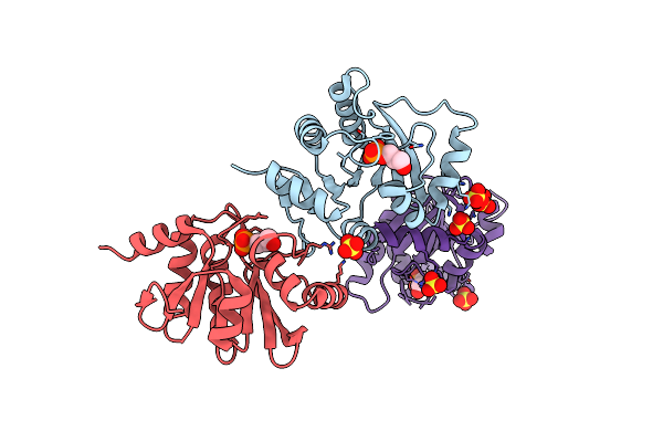
Structure Of A Cubic Crystal Form Of X (Adrp) Domain From Fcov With Adp-Ribose
Organism: Feline coronavirus
Method: X-RAY DIFFRACTION Resolution:3.91 Å Release Date: 2010-01-12 Classification: RNA BINDING PROTEIN Ligands: CL, APR, NA |
Organism: Feline coronavirus
Method: X-RAY DIFFRACTION
Release Date: 2010-01-12
Ligands: CL, APR, NA
 |
Structure Of The Tetragonal Crystal Form Of X (Adrp) Domain From Fcov
Organism: Feline infectious peritonitis virus
Method: X-RAY DIFFRACTION Resolution:3.10 Å Release Date: 2009-10-27 Classification: RNA BINDING PROTEIN Ligands: CL, SO4, 1GP |
Organism: Feline infectious peritonitis virus
Method: X-RAY DIFFRACTION
Release Date: 2009-10-27
Ligands: CL, SO4, 1GP
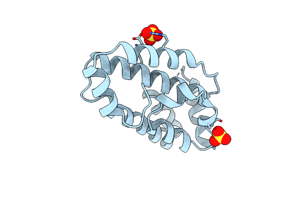 |
The Structure Of The Lipid Binding Protein Ce-Far-7 From Caenorhabditis Elegans
Organism: Caenorhabditis elegans
Method: X-RAY DIFFRACTION Resolution:1.80 Å Release Date: 2009-10-20 Classification: LIPID TRANSPORT Ligands: SO4 |
Organism: Caenorhabditis elegans
Method: X-RAY DIFFRACTION
Release Date: 2009-10-20
Ligands: SO4
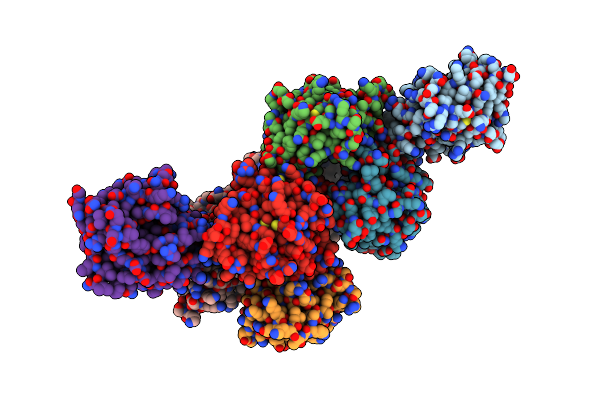 |
Structure Of A Cubic Crystal Form Of X (Adrp) Domain From Fcov
Organism: Feline infectious peritonitis virus
Method: X-RAY DIFFRACTION Resolution:2.20 Å Release Date: 2009-10-13 Classification: RNA BINDING PROTEIN |
Organism: Feline infectious peritonitis virus
Method: X-RAY DIFFRACTION
Release Date: 2009-10-13
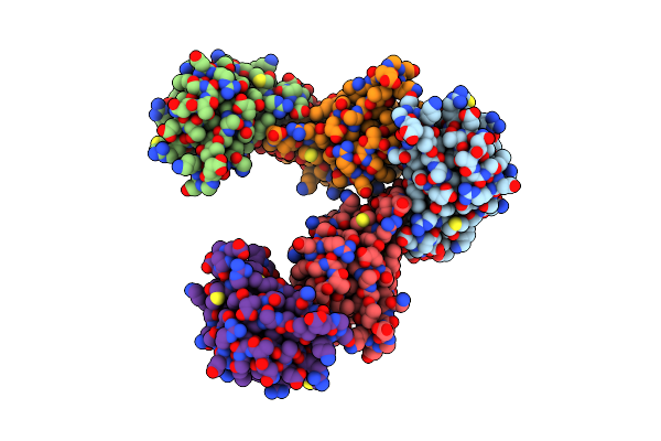 |
Structure Of The C-Terminal Domain Of Nsp4 From Feline Coronavirus
Organism: Feline coronavirus
Method: X-RAY DIFFRACTION Resolution:2.76 Å Release Date: 2009-08-18 Classification: VIRAL PROTEIN Ligands: SO4 |
Organism: Feline coronavirus
Method: X-RAY DIFFRACTION
Release Date: 2009-08-18
Ligands: SO4
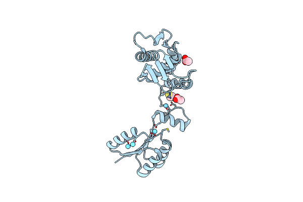 |
The Structure Of The Response Regulator Regx3 From Mycobacterium Tuberculosis
Organism: Mycobacterium tuberculosis h37rv
Method: X-RAY DIFFRACTION Resolution:2.03 Å Release Date: 2007-10-16 Classification: TRANSCRIPTION,SIGNALING PROTEIN Ligands: ACT, LA, BME |
Organism: Mycobacterium tuberculosis h37rv
Method: X-RAY DIFFRACTION
Release Date: 2007-10-16
Ligands: ACT, LA, BME
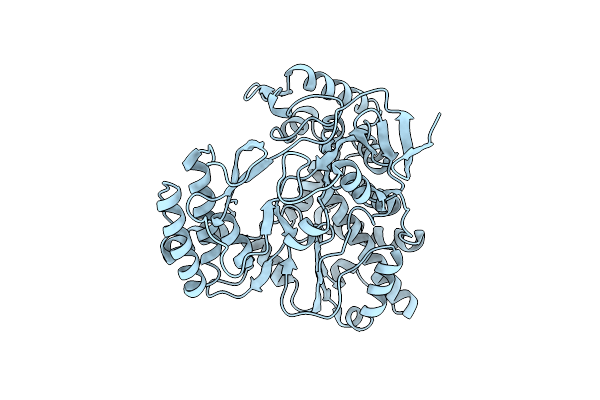 |
The 2.4 Angstrom Resolution Structure Of The D346G Mutant Of The Sapporo Virus Rdrp Polymerase
Organism: Sapporo virus
Method: X-RAY DIFFRACTION Resolution:2.40 Å Release Date: 2007-05-01 Classification: HYDROLASE |
Organism: Sapporo virus
Method: X-RAY DIFFRACTION
Release Date: 2007-05-01
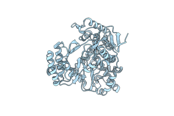 |
2.75 Angstrom Structure Of The D347G D348G Mutant Structure Of Sapporo Virus Rdrp Polymerase
Organism: Sapporo virus
Method: X-RAY DIFFRACTION Resolution:2.76 Å Release Date: 2007-05-01 Classification: HYDROLASE |
Organism: Sapporo virus
Method: X-RAY DIFFRACTION
Release Date: 2007-05-01
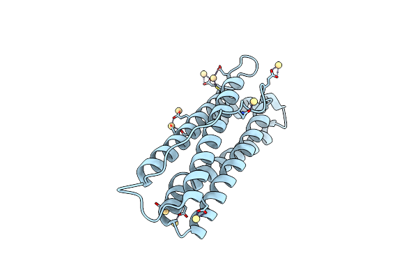 |
Anomalous Substructure Of Apoferritin
Organism: Equus caballus
Method: X-RAY DIFFRACTION Resolution:2.00 Å Release Date: 2007-03-06 Classification: METAL BINDING PROTEIN Ligands: CD, CL |
Organism: Equus caballus
Method: X-RAY DIFFRACTION
Release Date: 2007-03-06
Ligands: CD, CL