

Search Count: 7
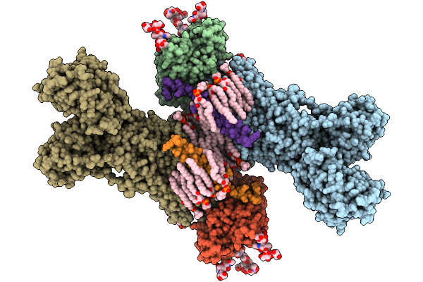 |
Cryo-Em Structure Of Palytoxin-Bound Na+,K+-Atpase In The E2P State
Organism: Squalus acanthias
Method: ELECTRON MICROSCOPY Release Date: 2025-10-01 Classification: MEMBRANE PROTEIN Ligands: CLR, PCW, MG, A1MA6 |
Organism: Squalus acanthias
Method: ELECTRON MICROSCOPY
Release Date: 2025-10-01
Ligands: CLR, PCW, MG, A1MA6
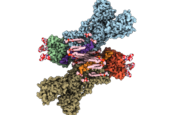 |
Cryo-Em Structure Of Palytoxin-Bound Na+,K+-Atpase In The Transient State Of Dephosphorylation (E2~P)
Organism: Squalus acanthias
Method: ELECTRON MICROSCOPY Release Date: 2025-10-01 Classification: MEMBRANE PROTEIN Ligands: CLR, PCW, ALF, MG, NA, A1MA6 |
Organism: Squalus acanthias
Method: ELECTRON MICROSCOPY
Release Date: 2025-10-01
Ligands: CLR, PCW, ALF, MG, NA, A1MA6
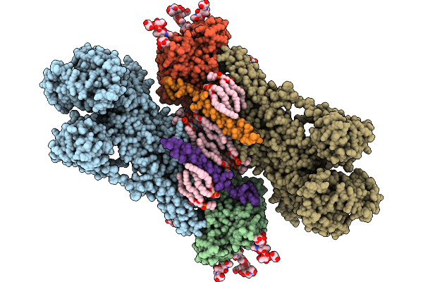 |
Cryo-Em Structure Of Na+,K+-Atpase That Forms A Cation Channel With Palytoxin (Atp Form)
Organism: Squalus acanthias
Method: ELECTRON MICROSCOPY Release Date: 2025-10-01 Classification: MEMBRANE PROTEIN Ligands: CLR, PCW, MG, ATP, NA, A1MA6 |
Organism: Squalus acanthias
Method: ELECTRON MICROSCOPY
Release Date: 2025-10-01
Ligands: CLR, PCW, MG, ATP, NA, A1MA6
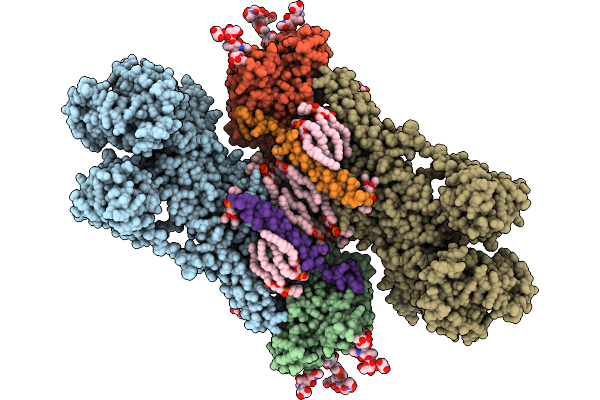 |
Cryo-Em Structure Of Na+,K+-Atpase That Forms A Cation Channel With Palytoxin (Adp Form)
Organism: Squalus acanthias
Method: ELECTRON MICROSCOPY Release Date: 2025-10-01 Classification: MEMBRANE PROTEIN Ligands: CLR, PCW, MG, ADP, NA, A1MA6 |
Organism: Squalus acanthias
Method: ELECTRON MICROSCOPY
Release Date: 2025-10-01
Ligands: CLR, PCW, MG, ADP, NA, A1MA6
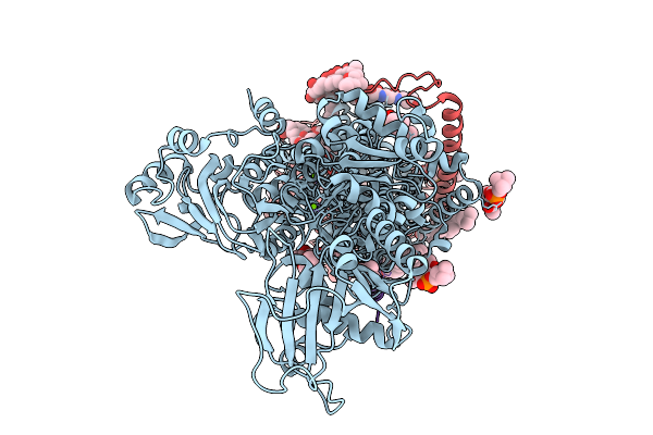 |
Crystal Structure Of Palytoxin-Bound Na+,K+-Atpase In The E2P State
Organism: Sus scrofa
Method: X-RAY DIFFRACTION Resolution:3.79 Å Release Date: 2025-10-01 Classification: MEMBRANE PROTEIN Ligands: MG, CLR, PCW, A1MA6, BEF, NAG |
Organism: Sus scrofa
Method: X-RAY DIFFRACTION
Release Date: 2025-10-01
Ligands: MG, CLR, PCW, A1MA6, BEF, NAG
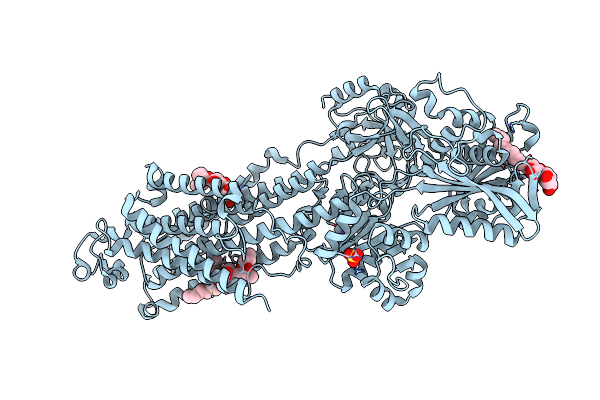 |
Crystal Structure Of The E309Q Mutant Of Sr Ca2+-Atpase In E2(Tg)
Organism: Oryctolagus cuniculus
Method: X-RAY DIFFRACTION Resolution:2.50 Å Release Date: 2019-04-03 Classification: HYDROLASE |
Organism: Oryctolagus cuniculus
Method: X-RAY DIFFRACTION
Release Date: 2019-04-03
 |
Crystal Structure Of The E309A Mutant Of Sr Ca2+-Atpase In E2(Tg)
Organism: Oryctolagus cuniculus
Method: X-RAY DIFFRACTION Resolution:3.30 Å Release Date: 2018-12-12 Classification: HYDROLASE Ligands: SO4, NA, TG1, PTY |
Organism: Oryctolagus cuniculus
Method: X-RAY DIFFRACTION
Release Date: 2018-12-12
Ligands: SO4, NA, TG1, PTY