

Search Count: 28
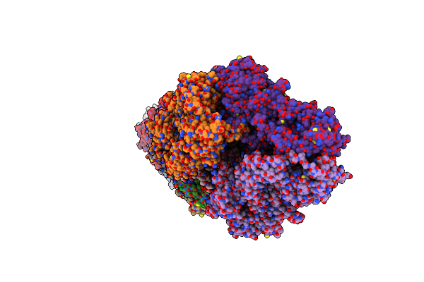 |
E. Coli (Lac Z) Beta-Galactosidase (Ncs Constrained Monomer-Monoclinic)
Organism: Escherichia coli
Method: X-RAY DIFFRACTION Resolution:2.50 Å Release Date: 2014-07-09 Classification: HYDROLASE Ligands: MG |
Organism: Escherichia coli
Method: X-RAY DIFFRACTION
Release Date: 2014-07-09
Ligands: MG
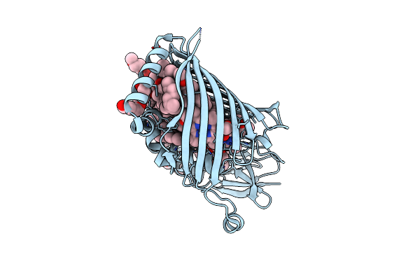 |
Structure Of The Fmo Protein From Pelodictyon Phaeum
Organism: Pelodictyon phaeum
Method: X-RAY DIFFRACTION Resolution:1.99 Å Release Date: 2012-01-25 Classification: PHOTOSYNTHESIS Ligands: BCL, PG4 |
Organism: Pelodictyon phaeum
Method: X-RAY DIFFRACTION
Release Date: 2012-01-25
Ligands: BCL, PG4
 |
3.0 A Model Of Iron Containing Urease Urea2B2 From Helicobacter Mustelae
Organism: Helicobacter mustelae
Method: X-RAY DIFFRACTION Resolution:3.00 Å Release Date: 2011-08-10 Classification: HYDROLASE Ligands: FE |
Organism: Helicobacter mustelae
Method: X-RAY DIFFRACTION
Release Date: 2011-08-10
Ligands: FE
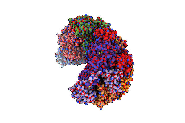 |
3.0 A Model Of Iron Containing Urease Urea2B2 From Helicobacter Mustelae (Refined W/ No Ordered Solvent)
Organism: Helicobacter mustelae
Method: X-RAY DIFFRACTION Resolution:3.00 Å Release Date: 2011-08-10 Classification: HYDROLASE Ligands: FE |
Organism: Helicobacter mustelae
Method: X-RAY DIFFRACTION
Release Date: 2011-08-10
Ligands: FE
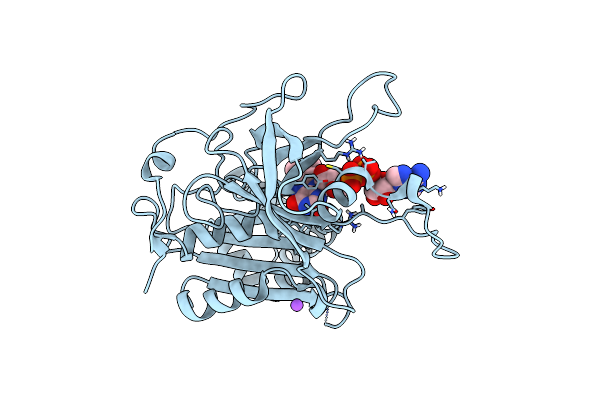 |
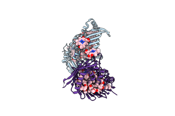
Crystal Structure Of The Oxidized Form Of Ferredoxin:Nadp+ Reductase From Maize Root At 1.05 Angstroms
Organism: Zea mays
Method: X-RAY DIFFRACTION Resolution:1.05 Å Release Date: 2010-03-16 Classification: OXIDOREDUCTASE Ligands: FAD, NA |
Organism: Zea mays
Method: X-RAY DIFFRACTION
Release Date: 2010-03-16
Ligands: FAD, NA
 |
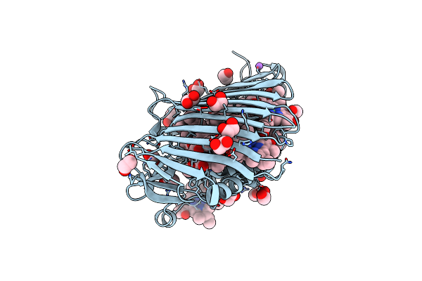
Crystal Structure Of The Fenna-Matthews-Olson Protein From Chlorobaculum Tepidum
Organism: Chlorobaculum tepidum
Method: X-RAY DIFFRACTION Resolution:2.20 Å Release Date: 2009-05-12 Classification: PHOTOSYNTHESIS Ligands: BCL |
Organism: Chlorobaculum tepidum
Method: X-RAY DIFFRACTION
Release Date: 2009-05-12
Ligands: BCL
 |
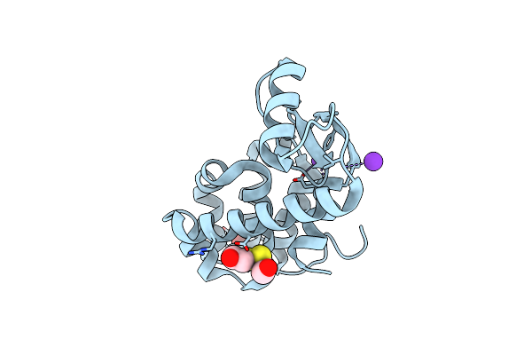
Fmo Protein From Prosthecochloris Aestuarii 2K At 1.3A Resolution
Organism: Prosthecochloris aestuarii 2k
Method: X-RAY DIFFRACTION Resolution:1.30 Å Release Date: 2009-05-12 Classification: PHOTOSYNTHESIS Ligands: BCL, EDO, NA, NH4 |
Organism: Prosthecochloris aestuarii 2k
Method: X-RAY DIFFRACTION
Release Date: 2009-05-12
Ligands: BCL, EDO, NA, NH4
 |
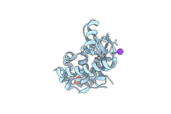
Evaulaution At Atomic Resolution Of The Role Of Strain In Destabilizing The Temperature Sensitive T4 Lysozyme Mutant Arg96-->His
Organism: Enterobacteria phage t4
Method: X-RAY DIFFRACTION Resolution:1.08 Å Release Date: 2009-02-17 Classification: HYDROLASE Ligands: HED, PO4, CL, NA, K |
Organism: Enterobacteria phage t4
Method: X-RAY DIFFRACTION
Release Date: 2009-02-17
Ligands: HED, PO4, CL, NA, K
 |
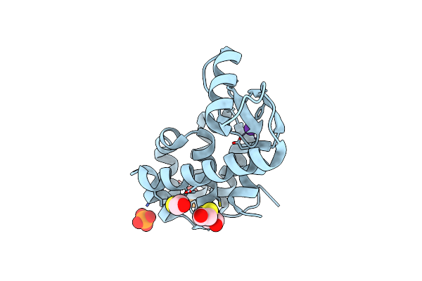
Evaulaution At Atomic Resolution Of The Role Of Strain In Destabilizing The Temperature Sensitive T4 Lysozyme Mutant Arg96-->His
Organism: Enterobacteria phage t4
Method: X-RAY DIFFRACTION Resolution:1.19 Å Release Date: 2009-02-17 Classification: HYDROLASE Ligands: PO4, NA, CL, K |
Organism: Enterobacteria phage t4
Method: X-RAY DIFFRACTION
Release Date: 2009-02-17
Ligands: PO4, NA, CL, K
 |
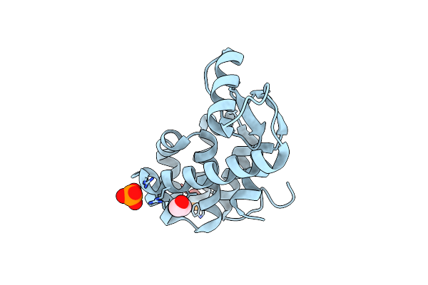
Evaulaution At Atomic Resolution Of The Role Of Strain In Destabilizing The Temperature Sensitive T4 Lysozyme Mutant Arg96-->His
Organism: Enterobacteria phage t4
Method: X-RAY DIFFRACTION Resolution:1.09 Å Release Date: 2009-02-17 Classification: HYDROLASE Ligands: HED, BME, PO4, CL, K |
Organism: Enterobacteria phage t4
Method: X-RAY DIFFRACTION
Release Date: 2009-02-17
Ligands: HED, BME, PO4, CL, K
 |
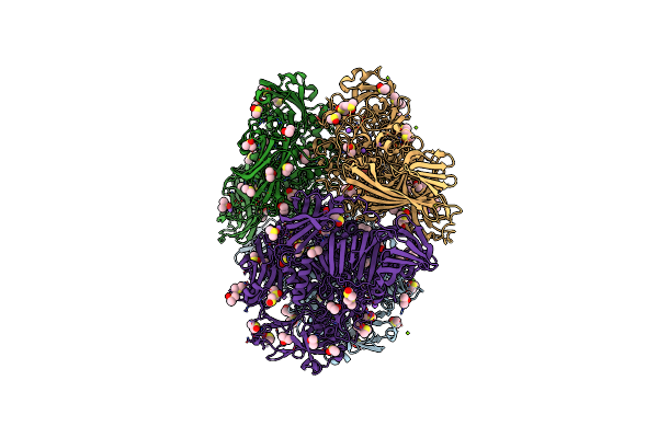
Evaulaution At Atomic Resolution Of The Role Of Strain In Destabilizing The Temperature Sensitive T4 Lysozyme Mutant Arg96-->His
Organism: Enterobacteria phage t4
Method: X-RAY DIFFRACTION Resolution:1.20 Å Release Date: 2009-02-17 Classification: HYDROLASE Ligands: PO4, BME |
Organism: Enterobacteria phage t4
Method: X-RAY DIFFRACTION
Release Date: 2009-02-17
Ligands: PO4, BME
 |
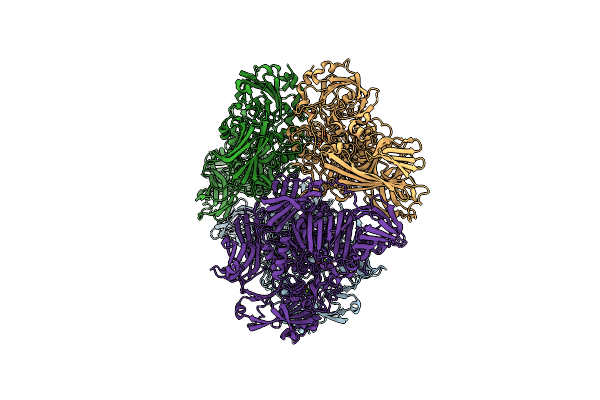
E. Coli Beta-Galactosidase At 1.7 Angstrom
Organism: Escherichia coli
Method: X-RAY DIFFRACTION Resolution:1.70 Å Release Date: 2001-02-21 Classification: HYDROLASE Ligands: MG, NA, DMS |
Organism: Escherichia coli
Method: X-RAY DIFFRACTION
Release Date: 2001-02-21
Ligands: MG, NA, DMS
 |
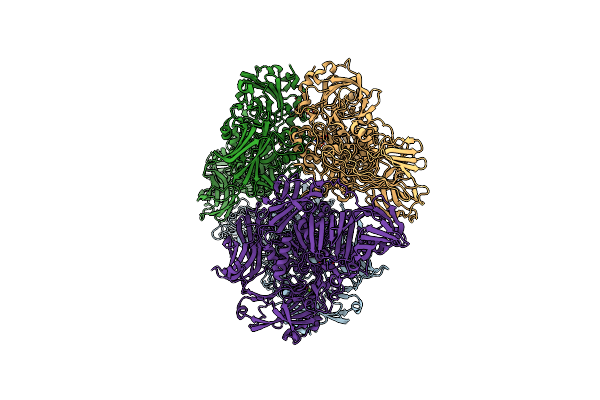
E. Coli (Lacz) Beta-Galactosidase (Ncs Constrained Monomer-Orthorhombic)
Organism: Escherichia coli
Method: X-RAY DIFFRACTION Resolution:2.80 Å Release Date: 2001-02-21 Classification: HYDROLASE Ligands: MG |
Organism: Escherichia coli
Method: X-RAY DIFFRACTION
Release Date: 2001-02-21
Ligands: MG
 |
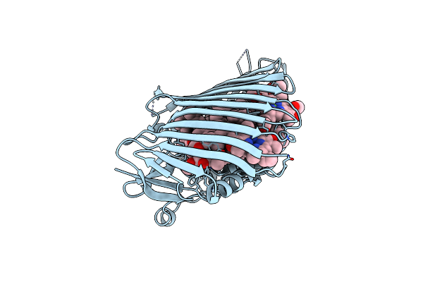
E. Coli (Lacz) Beta-Galactosidase (Orthorhombic)
Organism: Escherichia coli
Method: X-RAY DIFFRACTION Resolution:2.80 Å Release Date: 2001-02-21 Classification: HYDROLASE Ligands: MG |
Organism: Escherichia coli
Method: X-RAY DIFFRACTION
Release Date: 2001-02-21
Ligands: MG
 |
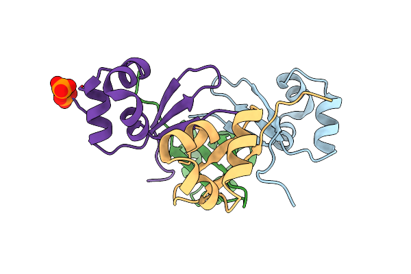
Fmo Protein From Prosthecochloris Aestuarii 2K At Room Temperature
Organism: Prosthecochloris aestuarii
Method: X-RAY DIFFRACTION Resolution:1.90 Å Release Date: 1998-09-16 Classification: ELECTRON TRANSPORT Ligands: BCL |
Organism: Prosthecochloris aestuarii
Method: X-RAY DIFFRACTION
Release Date: 1998-09-16
Ligands: BCL
 |
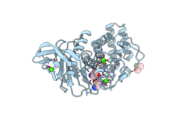
Refined Structure Of Cro Repressor Protein From Bacteriophage Lambda
Organism: Enterobacteria phage lambda
Method: X-RAY DIFFRACTION Resolution:2.30 Å Release Date: 1998-06-17 Classification: GENE REGULATING PROTEIN Ligands: PO4 |
Organism: Enterobacteria phage lambda
Method: X-RAY DIFFRACTION
Release Date: 1998-06-17
Ligands: PO4
 |
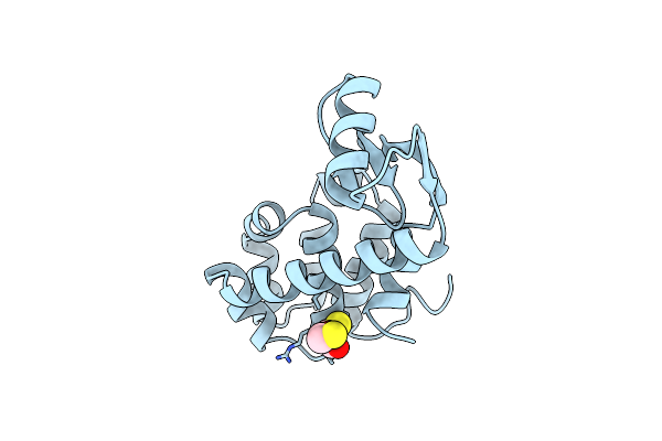
Structural Comparison Suggests That Thermolysin And Related Neutral Proteases Undergo Hinge-Bending Motion During Catalysis
Organism: Bacillus thermoproteolyticus
Method: X-RAY DIFFRACTION Resolution:1.60 Å Release Date: 1994-04-30 Classification: HYDROLASE(METALLOPROTEINASE) Ligands: VAL, LYS, CA, ZN, DMS |
Organism: Bacillus thermoproteolyticus
Method: X-RAY DIFFRACTION
Release Date: 1994-04-30
Ligands: VAL, LYS, CA, ZN, DMS
 |
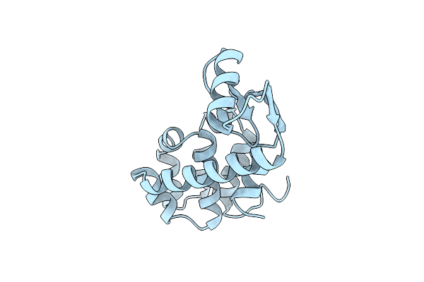
Analysis Of The Interaction Between Charged Side Chains And The Alpha-Helix Dipole Using Designed Thermostable Mutants Of Phage T4 Lysozyme
Organism: Enterobacteria phage t4
Method: X-RAY DIFFRACTION Resolution:1.80 Å Release Date: 1991-10-15 Classification: HYDROLASE (O-GLYCOSYL) Ligands: BME |
Organism: Enterobacteria phage t4
Method: X-RAY DIFFRACTION
Release Date: 1991-10-15
Ligands: BME
 |
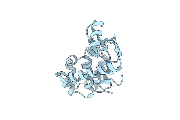
Analysis Of The Interaction Between Charged Side Chains And The Alpha-Helix Dipole Using Designed Thermostable Mutants Of Phage T4 Lysozyme
Organism: Enterobacteria phage t4
Method: X-RAY DIFFRACTION Resolution:1.70 Å Release Date: 1991-10-15 Classification: HYDROLASE (O-GLYCOSYL) |
Organism: Enterobacteria phage t4
Method: X-RAY DIFFRACTION
Release Date: 1991-10-15
 |
Contributions Of Left-Handed Helical Residues To The Structure And Stability Of Bacteriophage T4 Lysozyme
Organism: Enterobacteria phage t4
Method: X-RAY DIFFRACTION Resolution:1.85 Å Release Date: 1990-01-15 Classification: HYDROLASE (O-GLYCOSYL) |
Organism: Enterobacteria phage t4
Method: X-RAY DIFFRACTION
Release Date: 1990-01-15