

Search Count: 14
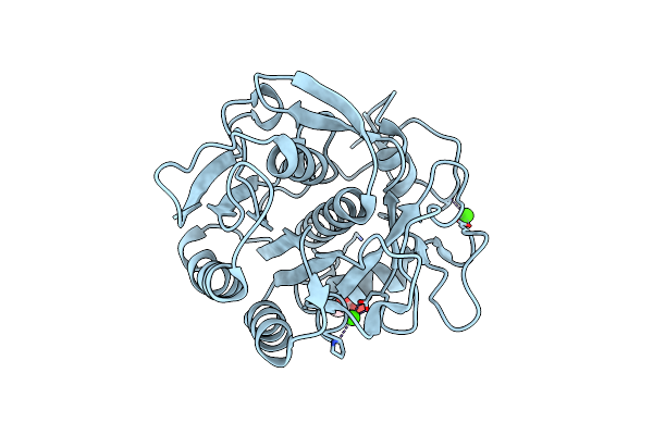 |
X-Ray Analysis Of Crystal Of Proteinase K Obtained From H2O Solution Using Peg 8000
Organism: Tritirachium album
Method: X-RAY DIFFRACTION Resolution:1.10 Å Release Date: 2011-06-22 Classification: HYDROLASE Ligands: CA, GOL |
Organism: Tritirachium album
Method: X-RAY DIFFRACTION
Release Date: 2011-06-22
Ligands: CA, GOL
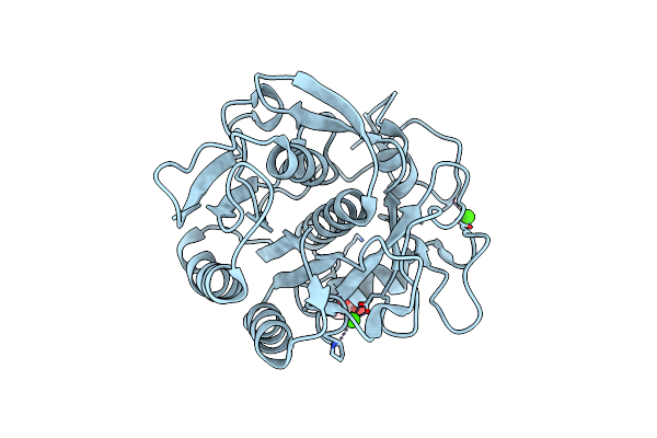 |
X-Ray Analysis Of Crystal Of Proteinase K Obtained From D2O Solution Using Peg 8000
Organism: Tritirachium album
Method: X-RAY DIFFRACTION Resolution:1.10 Å Release Date: 2011-06-22 Classification: HYDROLASE Ligands: CA, GOL |
Organism: Tritirachium album
Method: X-RAY DIFFRACTION
Release Date: 2011-06-22
Ligands: CA, GOL
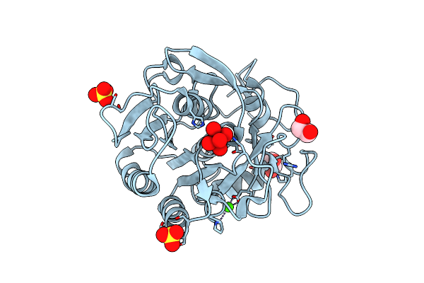 |
Sad Structure Solution Of Proteinase K Grown In Potassium Tellurate Solution
Organism: Tritirachium album
Method: X-RAY DIFFRACTION Resolution:1.55 Å Release Date: 2010-12-22 Classification: HYDROLASE Ligands: CA, TE6, SO4, EDO |
Organism: Tritirachium album
Method: X-RAY DIFFRACTION
Release Date: 2010-12-22
Ligands: CA, TE6, SO4, EDO
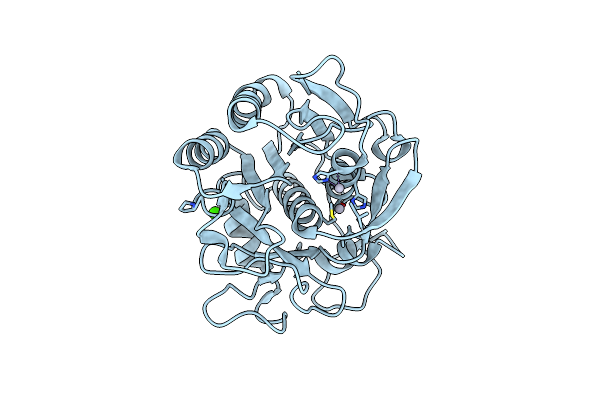 |
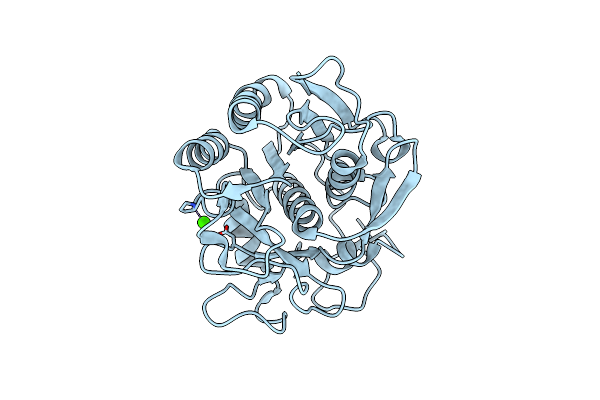
Proteinase K By Lb Nanotemplate Method After High X-Ray Dose On Id14-2 Beamline At Esrf
Organism: Tritirachium album
Method: X-RAY DIFFRACTION Resolution:1.00 Å Release Date: 2010-06-30 Classification: HYDROLASE Ligands: HG, CA |
Organism: Tritirachium album
Method: X-RAY DIFFRACTION
Release Date: 2010-06-30
Ligands: HG, CA
 |
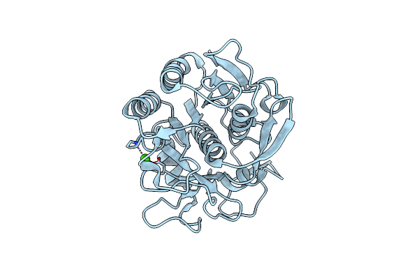
Proteinase K By Classical Hanging Drop Method Before High X-Ray Dose On Id14-2 Beamline At Esrf
Organism: Tritirachium album
Method: X-RAY DIFFRACTION Resolution:1.00 Å Release Date: 2010-06-09 Classification: HYDROLASE Ligands: CA |
Organism: Tritirachium album
Method: X-RAY DIFFRACTION
Release Date: 2010-06-09
Ligands: CA
 |
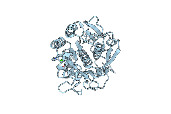
Proteinase K By Classical Hanging Drop Method After High X-Ray Dose On Id14-2 Beamline At Esrf
Organism: Tritirachium album
Method: X-RAY DIFFRACTION Resolution:0.99 Å Release Date: 2010-06-09 Classification: HYDROLASE Ligands: CA |
Organism: Tritirachium album
Method: X-RAY DIFFRACTION
Release Date: 2010-06-09
Ligands: CA
 |
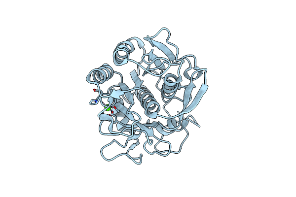
Proteinase K By Lb Nanotemplate Method Before High X-Ray Dose On Id14-2 Beamline At Esrf
Organism: Tritirachium album
Method: X-RAY DIFFRACTION Resolution:1.00 Å Release Date: 2010-06-09 Classification: HYDROLASE Ligands: CA |
Organism: Tritirachium album
Method: X-RAY DIFFRACTION
Release Date: 2010-06-09
Ligands: CA
 |
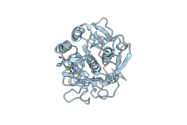
Proteinase K By Lb Nanotemplate Method Before High X-Ray Dose On Esrf Id14-2 Beamline
Organism: Tritirachium album
Method: X-RAY DIFFRACTION Resolution:1.02 Å Release Date: 2009-07-07 Classification: HYDROLASE Ligands: CA |
Organism: Tritirachium album
Method: X-RAY DIFFRACTION
Release Date: 2009-07-07
Ligands: CA
 |
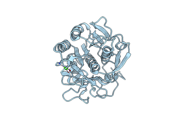
Proteinase K By Lb Nanotemplate Method After The First Step Of High X-Ray Dose On Esrf Id14-2 Beamline
Organism: Tritirachium album
Method: X-RAY DIFFRACTION Resolution:1.02 Å Release Date: 2009-07-07 Classification: HYDROLASE Ligands: CA |
Organism: Tritirachium album
Method: X-RAY DIFFRACTION
Release Date: 2009-07-07
Ligands: CA
 |
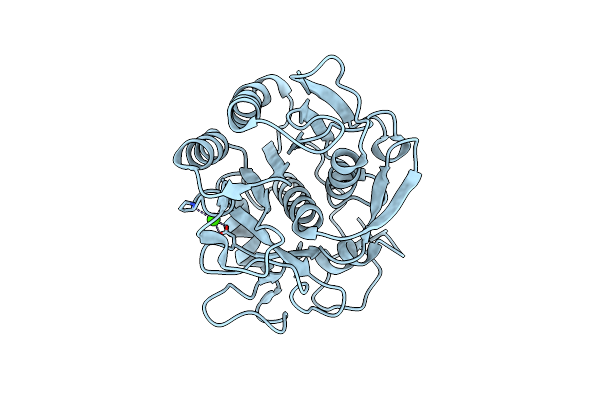
Proteinase K By Lb Nanotmplate Method After The Second Step Of High Dose On Esrf Id14-2 Beamline
Organism: Tritirachium album
Method: X-RAY DIFFRACTION Resolution:1.02 Å Release Date: 2009-07-07 Classification: HYDROLASE Ligands: CA |
Organism: Tritirachium album
Method: X-RAY DIFFRACTION
Release Date: 2009-07-07
Ligands: CA
 |
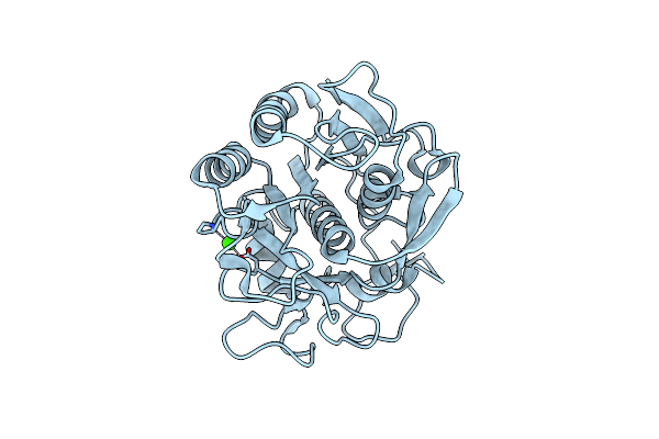
Proteinase K By Lb Nanotemplate Method After The Third Step High X-Ray Dose On Esrf Id14-2 Beamline
Organism: Tritirachium album
Method: X-RAY DIFFRACTION Resolution:1.03 Å Release Date: 2009-07-07 Classification: HYDROLASE Ligands: CA |
Organism: Tritirachium album
Method: X-RAY DIFFRACTION
Release Date: 2009-07-07
Ligands: CA
 |
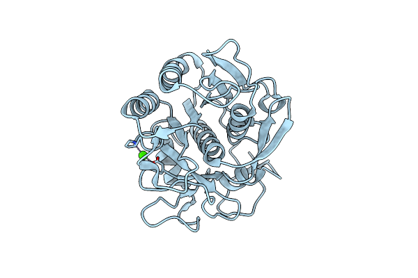
Proteinase K By Classical Hanging Drop Method Before High X Ray Dose On Esrf Id 14-2 Beamline
Organism: Tritirachium album
Method: X-RAY DIFFRACTION Resolution:0.99 Å Release Date: 2009-07-07 Classification: HYDROLASE Ligands: CA |
Organism: Tritirachium album
Method: X-RAY DIFFRACTION
Release Date: 2009-07-07
Ligands: CA
 |
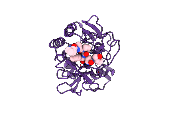
Proteinase K By Classical Hanging Drop Method After High X-Ray Dose On Esrf Id14-2 Beamline
Organism: Tritirachium album
Method: X-RAY DIFFRACTION Resolution:0.99 Å Release Date: 2009-07-07 Classification: HYDROLASE Ligands: CA |
Organism: Tritirachium album
Method: X-RAY DIFFRACTION
Release Date: 2009-07-07
Ligands: CA
 |
Structure Of The Complex Of Proteinase K With A Substrate-Analogue Hexa-Peptide Inhibitor At 2.2 Angstroms Resolution
Organism: Tritirachium album
Method: X-RAY DIFFRACTION Resolution:2.20 Å Release Date: 1994-01-31 Classification: HYDROLASE/HYDROLASE INHIBITOR |
Organism: Tritirachium album
Method: X-RAY DIFFRACTION
Release Date: 1994-01-31