
Search Count: 82
All
Selected
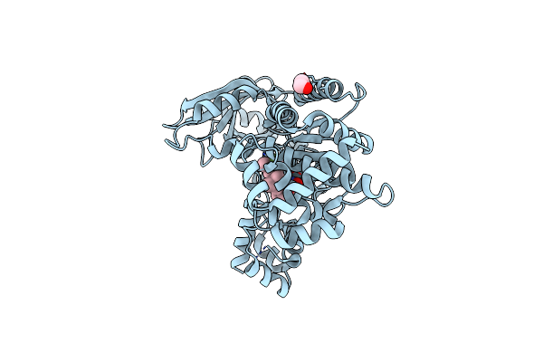 |
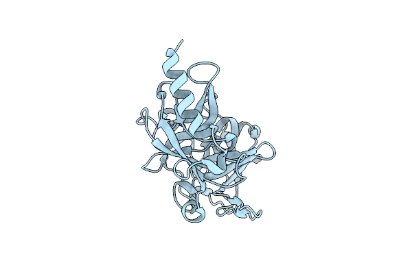
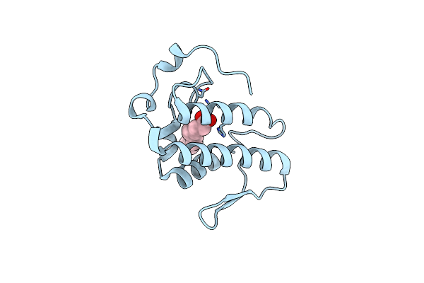
Organism: Bacillus anthracis
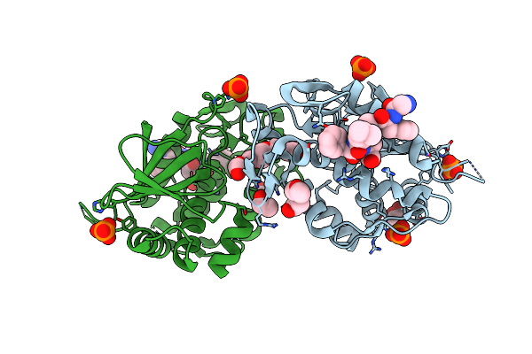
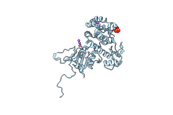
Method: X-RAY DIFFRACTION Resolution:2.35 Å Release Date: 2015-11-11 Classification: TOXIN,HYDROLASE Ligands: 41R, ZN, EDO |
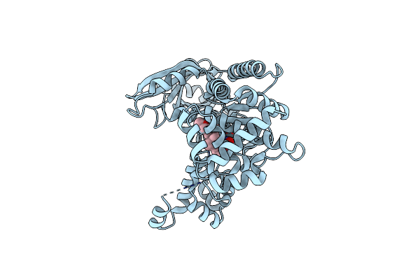 |
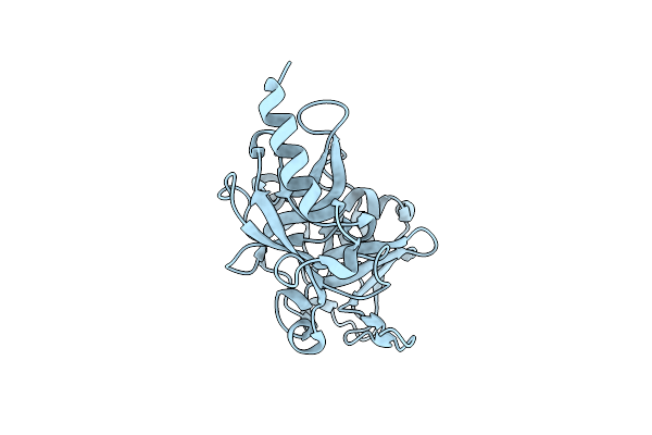
Organism: Bacillus anthracis
Method: X-RAY DIFFRACTION Resolution:2.70 Å Release Date: 2015-11-11 Classification: TOXIN,HYDROLASE Ligands: 41T, ZN |
 |
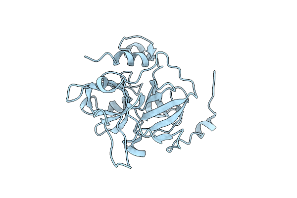
Organism: Bacillus anthracis
Method: X-RAY DIFFRACTION Resolution:2.70 Å Release Date: 2015-11-11 Classification: TOXIN,HYDROLASE Ligands: 41S, ZN, EDO |
 |
Human Acetylcholinesterase Complexed With Fasciculin-Ii, Glycosylated Protein
Organism: Homo sapiens, Dendroaspis angusticeps
Method: X-RAY DIFFRACTION Resolution:2.76 Å Release Date: 2001-01-17 Classification: HYDROLASE/TOXIN Ligands: NAG |
 |
Organism: Staphylococcus aureus
Method: X-RAY DIFFRACTION Resolution:2.00 Å Release Date: 2003-01-21 Classification: TOXIN, HYDROLASE |
 |
Organism: Staphylococcus aureus
Method: X-RAY DIFFRACTION Resolution:2.00 Å Release Date: 2003-01-21 Classification: TOXIN, HYDROLASE |
 |
Organism: Staphylococcus aureus
Method: X-RAY DIFFRACTION Resolution:2.80 Å Release Date: 2003-01-21 Classification: toxin, hydrolase |
 |
Crystal Structure Of Mutant E202Q Of Human Acetylcholinesterase Complexed With Green Mamba Venom Peptide Fasciculin-Ii
Organism: Homo sapiens, Dendroaspis angusticeps
Method: X-RAY DIFFRACTION Resolution:2.90 Å Release Date: 2001-01-17 Classification: HYDROLASE/TOXIN |
 |
Organism: Escherichia coli o104:h4, Escherichia coli
Method: X-RAY DIFFRACTION Resolution:2.01 Å Release Date: 2024-05-29 Classification: TOXIN, Hydrolase Ligands: 2PE, EDO, PO4, CL |
 |
Protein Serine/Threonine Phosphatase-1 (Alpha Isoform, Type 1) Complexed With Microcystin-Lr Toxin
Organism: Oryctolagus cuniculus, Microcystis aeruginosa
Method: X-RAY DIFFRACTION Resolution:2.10 Å Release Date: 1996-06-20 Classification: HYDROLASE/HYDROLASE INHIBITOR Ligands: MN, BME |
 |
Organism: Mus musculus, Dendroaspis angusticeps
Method: X-RAY DIFFRACTION Resolution:2.50 Å Release Date: 2003-12-23 Classification: HYDROLASE/TOXIN Ligands: NAG, EDO |
 |
Organism: Homo sapiens
Method: ELECTRON MICROSCOPY Release Date: 2021-03-10 Classification: HYDROLASE |
 |
The Crystal Structure Of Pesticin-T4 Lysozyme Hybrid Stabilized By Engineered Disulfide Bonds
Organism: Yersinia pestis, Enterobacteria phage t4
Method: X-RAY DIFFRACTION Resolution:1.74 Å Release Date: 2012-06-20 Classification: TOXIN, HYDROLASE Ligands: SO4, NA |
 |
Organism: Clostridioides difficile
Method: ELECTRON MICROSCOPY Release Date: 2022-03-02 Classification: HYDROLASE |
 |
Organism: Clostridioides difficile
Method: ELECTRON MICROSCOPY Release Date: 2022-03-02 Classification: HYDROLASE |
 |
The Crystal Structure Of An Engineered Phage Lysin Containing The Binding Domain Of Pesticin And The Killing Domain Of T4-Lysozyme
Organism: Yersinia pestis, Enterobacteria phage t4
Method: X-RAY DIFFRACTION Resolution:2.60 Å Release Date: 2012-06-20 Classification: TOXIN, HYDROLASE |
 |
Organism: Clostridium botulinum, Rattus norvegicus
Method: X-RAY DIFFRACTION Resolution:2.15 Å Release Date: 2006-12-19 Classification: TOXIN,HYDROLASE |
 |
Organism: Bothrops atrox
Method: X-RAY DIFFRACTION Resolution:1.95 Å Release Date: 2022-01-12 Classification: TOXIN, HYDROLASE Ligands: DAO |
 |
Organism: Clostridium botulinum
Method: X-RAY DIFFRACTION Resolution:2.00 Å Release Date: 2004-03-16 Classification: Toxin, hydrolase Ligands: CA |
 |
Structure Of Topi1 Inhibitor From Tityus Obscurus Scorpion Venom In Complex With Trypsin
Organism: Bos taurus, Tityus
Method: X-RAY DIFFRACTION Resolution:1.29 Å Release Date: 2020-07-01 Classification: hydrolase/hydrolase inhibitor Ligands: CA, SO4 |