

Search Count: 19
 |
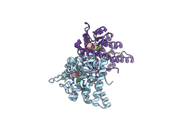
Progesterone Receptor Bound To An Alkylpyrrolidine Ligand.
Organism: Homo sapiens
Method: X-RAY DIFFRACTION Resolution:2.10 Å Release Date: 2010-03-02 Classification: hormone binding protein Ligands: SO4, GKK, GOL |
Organism: Homo sapiens
Method: X-RAY DIFFRACTION
Release Date: 2010-03-02
Ligands: SO4, GKK, GOL
 |
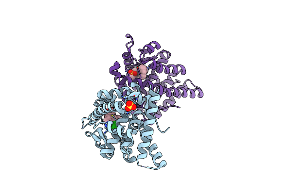
Progesterone Receptor With Bound Pyrrolidine 1
Organism: Homo sapiens
Method: X-RAY DIFFRACTION Resolution:1.90 Å Release Date: 2010-02-16 Classification: TRANSCRIPTION Ligands: 30X, SO4 |
Organism: Homo sapiens
Method: X-RAY DIFFRACTION
Release Date: 2010-02-16
Ligands: 30X, SO4
 |
Progesterone Receptor Bound To Sulfonamide Pyrrolidine Partial Agonist
Organism: Homo sapiens
Method: X-RAY DIFFRACTION Resolution:2.00 Å Release Date: 2009-12-08 Classification: TRANSCRIPTION Ligands: WOW, SO4 |
Organism: Homo sapiens
Method: X-RAY DIFFRACTION
Release Date: 2009-12-08
Ligands: WOW, SO4
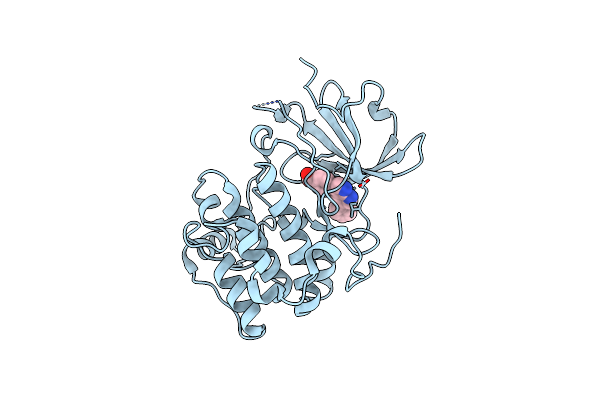 |
Crystal Structure Of Serum And Glucocorticoid-Regulated Kinase 1 In Complex With Compound 1
Organism: Homo sapiens
Method: X-RAY DIFFRACTION Resolution:2.60 Å Release Date: 2009-06-30 Classification: TRANSFERASE Ligands: MMG |
Organism: Homo sapiens
Method: X-RAY DIFFRACTION
Release Date: 2009-06-30
Ligands: MMG
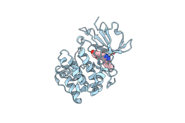 |
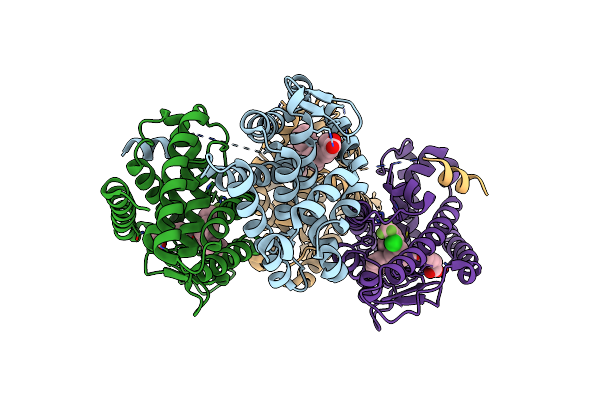
Crystal Structure Of Serum And Glucocorticoid-Regulated Kinase 1 In Complex With Compound 2
Organism: Homo sapiens
Method: X-RAY DIFFRACTION Resolution:3.10 Å Release Date: 2009-06-30 Classification: TRANSFERASE Ligands: GMG |
Organism: Homo sapiens
Method: X-RAY DIFFRACTION
Release Date: 2009-06-30
Ligands: GMG
 |
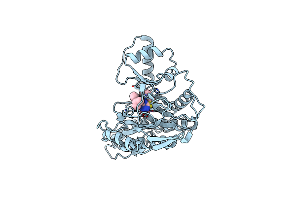
Hrxralpha & Mlxralpha With An Indole Pharmacophore, Sb786875
Organism: Homo sapiens, Mus musculus
Method: X-RAY DIFFRACTION Resolution:2.06 Å Release Date: 2009-02-10 Classification: TRANSCRIPTION Ligands: REA, LX2 |
Organism: Homo sapiens, Mus musculus
Method: X-RAY DIFFRACTION
Release Date: 2009-02-10
Ligands: REA, LX2
 |
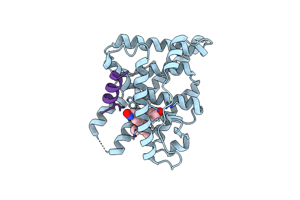
Human Methionine Aminopeptidase-2 Complexed With Sb-587094
Organism: Homo sapiens
Method: X-RAY DIFFRACTION Resolution:1.90 Å Release Date: 2007-06-19 Classification: HYDROLASE Ligands: CO, I96 |
Organism: Homo sapiens
Method: X-RAY DIFFRACTION
Release Date: 2007-06-19
Ligands: CO, I96
 |
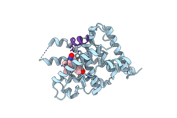
Progesterone Receptor With Bound Asoprisnil And A Peptide From The Co-Repressor Smrt
Organism: Homo sapiens
Method: X-RAY DIFFRACTION Resolution:2.00 Å Release Date: 2007-03-20 Classification: TRANSCRIPTION Ligands: AS0 |
Organism: Homo sapiens
Method: X-RAY DIFFRACTION
Release Date: 2007-03-20
Ligands: AS0
 |
Progesterone Receptor With Bound Asoprisnil And A Peptide From The Co-Repressor Ncor
Organism: Homo sapiens
Method: X-RAY DIFFRACTION Resolution:2.60 Å Release Date: 2007-03-20 Classification: TRANSCRIPTION Ligands: AS0 |
Organism: Homo sapiens
Method: X-RAY DIFFRACTION
Release Date: 2007-03-20
Ligands: AS0
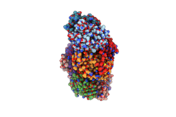 |
Liver X-Receptor Alpha Ligand Binding Domain With Sb313987
Organism: Homo sapiens, Mus musculus
Method: X-RAY DIFFRACTION Resolution:2.80 Å Release Date: 2005-09-20 Classification: TRANSCRIPTION Ligands: REA, L05 |
Organism: Homo sapiens, Mus musculus
Method: X-RAY DIFFRACTION
Release Date: 2005-09-20
Ligands: REA, L05
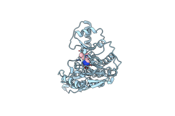 |
Human Methionine Aminopeptidase Complex With 4-Aryl-1,2,3-Triazole Inhibitor
Organism: Homo sapiens
Method: X-RAY DIFFRACTION Resolution:1.90 Å Release Date: 2005-09-13 Classification: HYDROLASE Ligands: CO, R20 |
Organism: Homo sapiens
Method: X-RAY DIFFRACTION
Release Date: 2005-09-13
Ligands: CO, R20
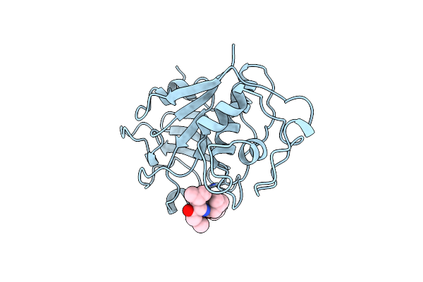 |
Use Of Papain As A Model For The Structure-Based Design Of Cathepsin K Inhibitors. Crystal Structures Of Two Papain Inhibitor Complexes Demonstrate Binding To S'-Subsites.
Organism: Carica papaya
Method: X-RAY DIFFRACTION Resolution:2.50 Å Release Date: 1999-08-16 Classification: HYDROLASE Ligands: SBA |
Organism: Carica papaya
Method: X-RAY DIFFRACTION
Release Date: 1999-08-16
Ligands: SBA
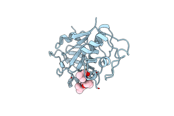 |
Use Of Papain As A Model For The Structure-Based Design Of Cathepsin K Inhibitors. Crystal Structures Of Two Papain Inhibitor Complexes Demonstrate Binding To S'-Subsites.
Organism: Carica papaya
Method: X-RAY DIFFRACTION Resolution:2.20 Å Release Date: 1999-08-12 Classification: HYDROLASE Ligands: ALD |
Organism: Carica papaya
Method: X-RAY DIFFRACTION
Release Date: 1999-08-12
Ligands: ALD
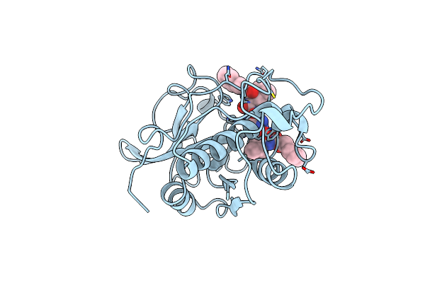 |
Crystal Structure Of Cysteine Protease Human Cathepsin K In Complex With A Covalent Symmetric Biscarbohydrazide Inhibitor
Organism: Homo sapiens
Method: X-RAY DIFFRACTION Resolution:2.20 Å Release Date: 1998-11-25 Classification: HYDROLASE Ligands: INA |
Organism: Homo sapiens
Method: X-RAY DIFFRACTION
Release Date: 1998-11-25
Ligands: INA
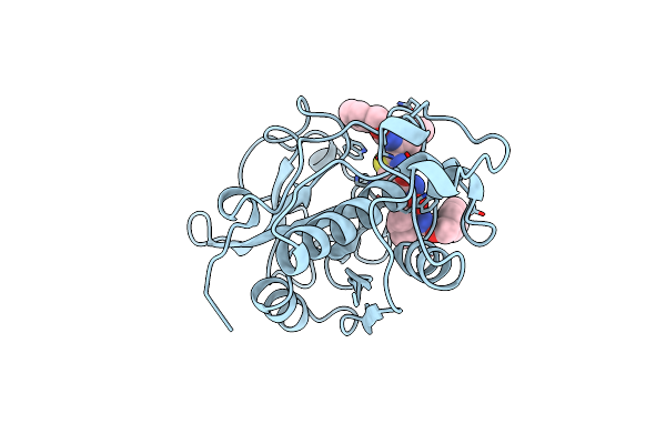 |
Crystal Structure Of Cysteine Protease Human Cathepsin K In Complex With A Covalent Thiazolhydrazide Inhibitor
Organism: Homo sapiens
Method: X-RAY DIFFRACTION Resolution:2.30 Å Release Date: 1998-11-25 Classification: HYDROLASE Ligands: IN6 |
Organism: Homo sapiens
Method: X-RAY DIFFRACTION
Release Date: 1998-11-25
Ligands: IN6
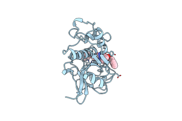 |
Crystal Structure Of Cysteine Protease Human Cathepsin K In Complex With A Covalent Benzyloxybenzoylcarbohydrazide Inhibitor
Organism: Homo sapiens
Method: X-RAY DIFFRACTION Resolution:2.40 Å Release Date: 1998-11-25 Classification: HYDROLASE Ligands: IN3 |
Organism: Homo sapiens
Method: X-RAY DIFFRACTION
Release Date: 1998-11-25
Ligands: IN3
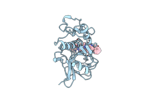 |
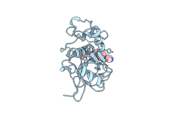
Crystal Structure Of The Cysteine Protease Human Cathepsin K In Complex With A Covalent Symmetric Diacylaminomethyl Ketone Inhibitor
Organism: Homo sapiens
Method: X-RAY DIFFRACTION Resolution:2.60 Å Release Date: 1998-10-14 Classification: HYDROLASE Ligands: SDK |
Organism: Homo sapiens
Method: X-RAY DIFFRACTION
Release Date: 1998-10-14
Ligands: SDK
 |
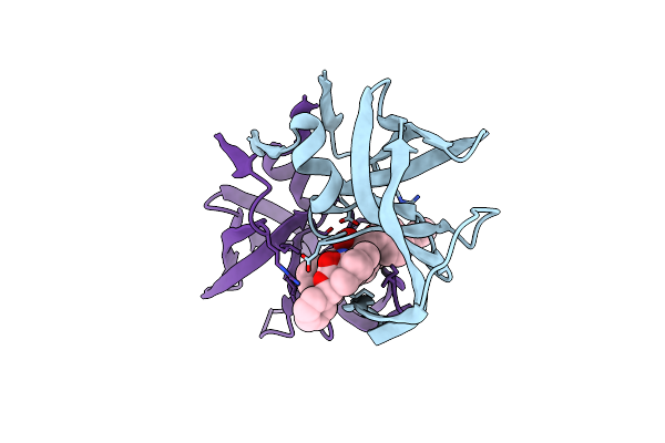
Crystal Structure Of The Cysteine Protease Human Cathepsin K In Complex With A Covalent Propanone Inhibitor
Organism: Homo sapiens
Method: X-RAY DIFFRACTION Resolution:2.60 Å Release Date: 1998-10-14 Classification: HYDROLASE Ligands: POS |
Organism: Homo sapiens
Method: X-RAY DIFFRACTION
Release Date: 1998-10-14
Ligands: POS
 |
Rational Design, Synthesis And Crystallographic Analysis Of A Hydroxyethylene-Based Hiv-1 Protease Inhibitor Containing A Heterocyclic P1'-P2' Amide Bond Isostere
Organism: Human immunodeficiency virus 1
Method: X-RAY DIFFRACTION Resolution:2.30 Å Release Date: 1994-08-31 Classification: HYDROLASE(ACID PROTEINASE) Ligands: RUN |
Organism: Human immunodeficiency virus 1
Method: X-RAY DIFFRACTION
Release Date: 1994-08-31
Ligands: RUN