

Search Count: 22
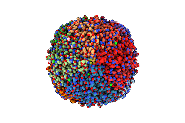 |
Crystal Structure Of Frog M Ferritin Mutant D80K
Organism: Rana catesbeiana
Method: X-RAY DIFFRACTION Resolution:1.91 Å Release Date: 2014-10-01 Classification: OXIDOREDUCTASE Ligands: SO4, EDO, ACT, CL |
Organism: Rana catesbeiana
Method: X-RAY DIFFRACTION
Release Date: 2014-10-01
Ligands: SO4, EDO, ACT, CL
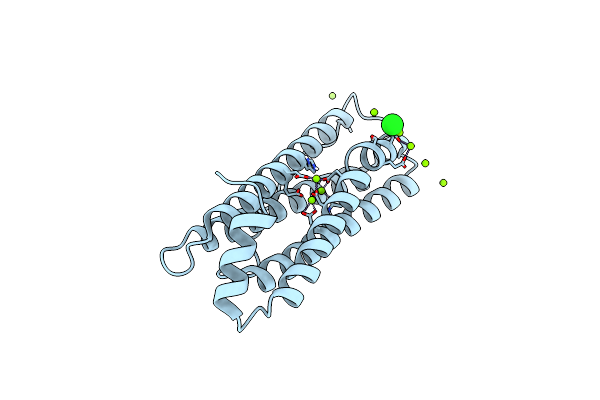 |
Frog M-Ferritin With Magnesium, D127E Mutant
Organism: Rana catesbeiana
Method: X-RAY DIFFRACTION Resolution:1.65 Å Release Date: 2014-07-16 Classification: OXIDOREDUCTASE Ligands: CL, MG |
Organism: Rana catesbeiana
Method: X-RAY DIFFRACTION
Release Date: 2014-07-16
Ligands: CL, MG
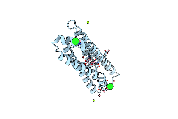 |
Frog M-Ferritin With Cobalt, D127E Mutant
Organism: Rana catesbeiana
Method: X-RAY DIFFRACTION Resolution:1.66 Å Release Date: 2014-07-16 Classification: OXIDOREDUCTASE Ligands: CO, CL, MG |
Organism: Rana catesbeiana
Method: X-RAY DIFFRACTION
Release Date: 2014-07-16
Ligands: CO, CL, MG
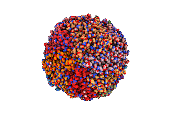 |
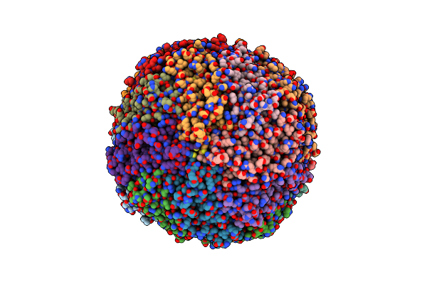
Crystal Structure Of Bullfrog M Ferritin
Organism: Rana catesbeiana
Method: X-RAY DIFFRACTION Resolution:2.56 Å Release Date: 2013-01-16 Classification: OXIDOREDUCTASE Ligands: PGE, EDO |
Organism: Rana catesbeiana
Method: X-RAY DIFFRACTION
Release Date: 2013-01-16
Ligands: PGE, EDO
 |
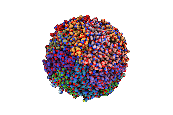
Iron Loaded Frog M Ferritin. Short Soaking Time
Organism: Rana catesbeiana
Method: X-RAY DIFFRACTION Resolution:2.89 Å Release Date: 2012-04-11 Classification: OXIDOREDUCTASE Ligands: FE |
Organism: Rana catesbeiana
Method: X-RAY DIFFRACTION
Release Date: 2012-04-11
Ligands: FE
 |
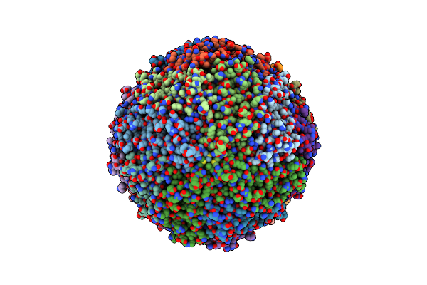
Bullfrog M Ferritin With Iron(Iii) Bound To The Ferroxidase Site
Organism: Rana catesbeiana
Method: X-RAY DIFFRACTION Resolution:2.70 Å Release Date: 2012-04-04 Classification: OXIDOREDUCTASE Ligands: FE |
Organism: Rana catesbeiana
Method: X-RAY DIFFRACTION
Release Date: 2012-04-04
Ligands: FE
 |
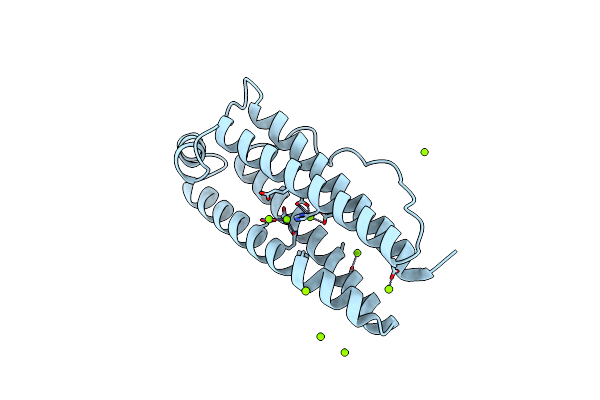
Copper (Ii) Loaded Bullfrog Ferritin M Chain
Organism: Rana catesbeiana
Method: X-RAY DIFFRACTION Resolution:2.82 Å Release Date: 2012-04-04 Classification: OXIDOREDUCTASE Ligands: CU |
Organism: Rana catesbeiana
Method: X-RAY DIFFRACTION
Release Date: 2012-04-04
Ligands: CU
 |
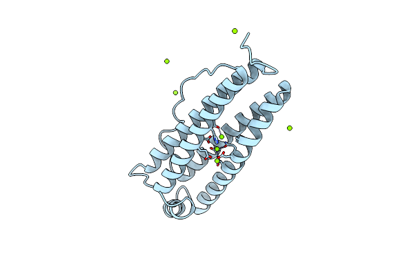
Frog M-Ferritin With Magnesium, R72D Mutant
Organism: Rana catesbeiana
Method: X-RAY DIFFRACTION Resolution:1.65 Å Release Date: 2012-02-29 Classification: OXIDOREDUCTASE Ligands: MG, CL |
Organism: Rana catesbeiana
Method: X-RAY DIFFRACTION
Release Date: 2012-02-29
Ligands: MG, CL
 |
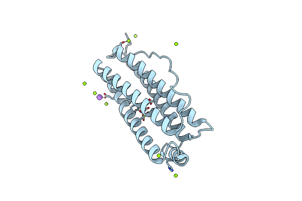
Frog M-Ferritin, D122R Mutant, With Magnesium
Organism: Rana catesbeiana
Method: X-RAY DIFFRACTION Resolution:1.40 Å Release Date: 2012-02-29 Classification: OXIDOREDUCTASE Ligands: MG, CL |
Organism: Rana catesbeiana
Method: X-RAY DIFFRACTION
Release Date: 2012-02-29
Ligands: MG, CL
 |
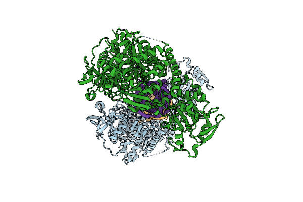
Frog M-Ferritin With Magnesium, L134P Mutant
Organism: Rana catesbeiana
Method: X-RAY DIFFRACTION Resolution:1.35 Å Release Date: 2012-02-29 Classification: OXIDOREDUCTASE Ligands: MG, CL, NA |
Organism: Rana catesbeiana
Method: X-RAY DIFFRACTION
Release Date: 2012-02-29
Ligands: MG, CL, NA
 |
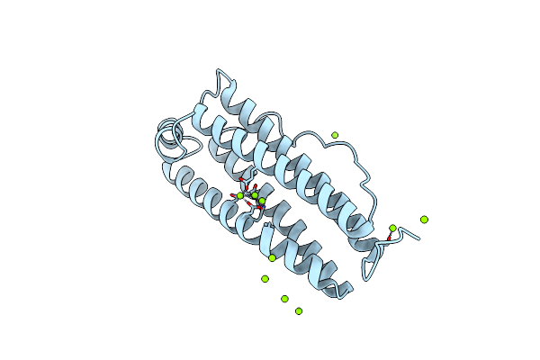
Crystal Structure Analysis Of Iron Regulatory Protein 1 In Complex With Ferritin H Ire Rna
Organism: Oryctolagus cuniculus
Method: X-RAY DIFFRACTION Resolution:2.80 Å Release Date: 2011-07-27 Classification: LYASE/RNA |
Organism: Oryctolagus cuniculus
Method: X-RAY DIFFRACTION
Release Date: 2011-07-27
 |
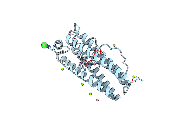
Frog M-Ferritin With Magnesium
Organism: Rana catesbeiana
Method: X-RAY DIFFRACTION Resolution:1.40 Å Release Date: 2010-10-06 Classification: OXIDOREDUCTASE Ligands: MG, CL |
Organism: Rana catesbeiana
Method: X-RAY DIFFRACTION
Release Date: 2010-10-06
Ligands: MG, CL
 |
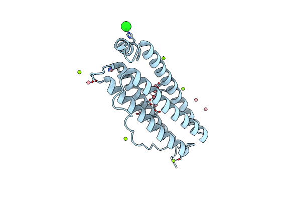
Frog M-Ferritin With Cobalt
Organism: Rana catesbeiana
Method: X-RAY DIFFRACTION Resolution:1.40 Å Release Date: 2010-10-06 Classification: OXIDOREDUCTASE Ligands: CO, CL, MG |
Organism: Rana catesbeiana
Method: X-RAY DIFFRACTION
Release Date: 2010-10-06
Ligands: CO, CL, MG
 |
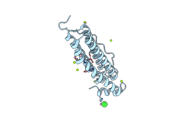
Frog M-Ferritin, Eed Mutant, With Cobalt
Organism: Rana catesbeiana
Method: X-RAY DIFFRACTION Resolution:1.40 Å Release Date: 2010-10-06 Classification: OXIDOREDUCTASE Ligands: CO, MG, CL |
Organism: Rana catesbeiana
Method: X-RAY DIFFRACTION
Release Date: 2010-10-06
Ligands: CO, MG, CL
 |
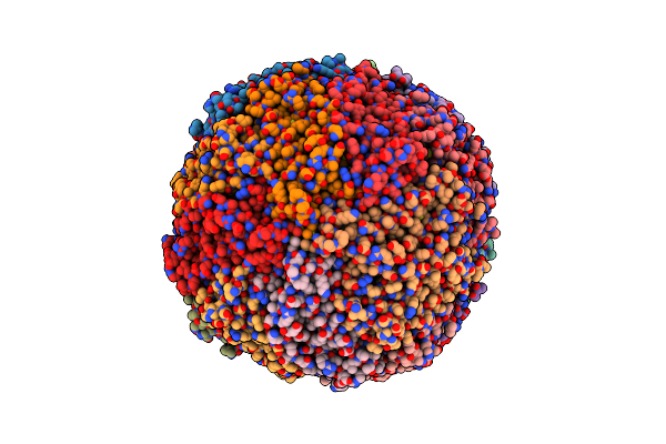
Frog M-Ferritin, Eqh Mutant, With Cobalt
Organism: Rana catesbeiana
Method: X-RAY DIFFRACTION Resolution:1.35 Å Release Date: 2010-10-06 Classification: OXIDOREDUCTASE Ligands: CO, MG, CL |
Organism: Rana catesbeiana
Method: X-RAY DIFFRACTION
Release Date: 2010-10-06
Ligands: CO, MG, CL
 |
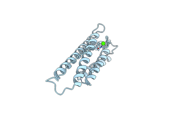
Crystal Structure Of M Ferritin
Organism: Rana catesbeiana
Method: X-RAY DIFFRACTION Resolution:2.80 Å Release Date: 1999-06-22 Classification: IRON STORAGE Ligands: MG |
Organism: Rana catesbeiana
Method: X-RAY DIFFRACTION
Release Date: 1999-06-22
Ligands: MG
 |
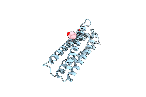
Localized Unfolding At The Junction Of Three Ferritin Subunits. A Mechanism For Iron Release?
Organism: Rana catesbeiana
Method: X-RAY DIFFRACTION Resolution:1.85 Å Release Date: 1999-01-13 Classification: IRON STORAGE Ligands: CA |
Organism: Rana catesbeiana
Method: X-RAY DIFFRACTION
Release Date: 1999-01-13
Ligands: CA
 |
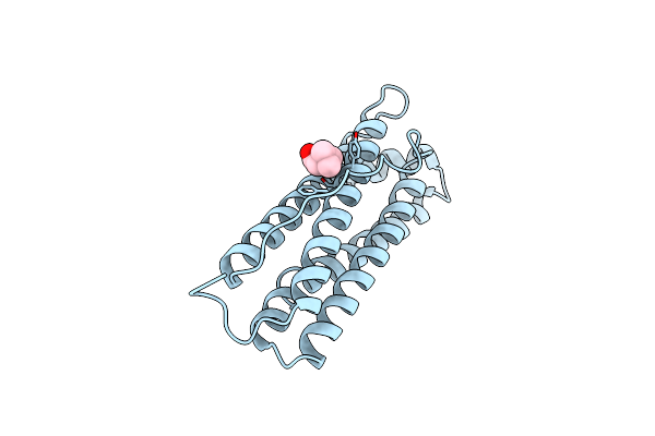
Bullfrog Red Cell L Ferritin Tartrate/Mg/Ph 5.5
Organism: Rana catesbeiana
Method: X-RAY DIFFRACTION Resolution:2.40 Å Release Date: 1995-11-14 Classification: IRON STORAGE Ligands: BET |
Organism: Rana catesbeiana
Method: X-RAY DIFFRACTION
Release Date: 1995-11-14
Ligands: BET
 |
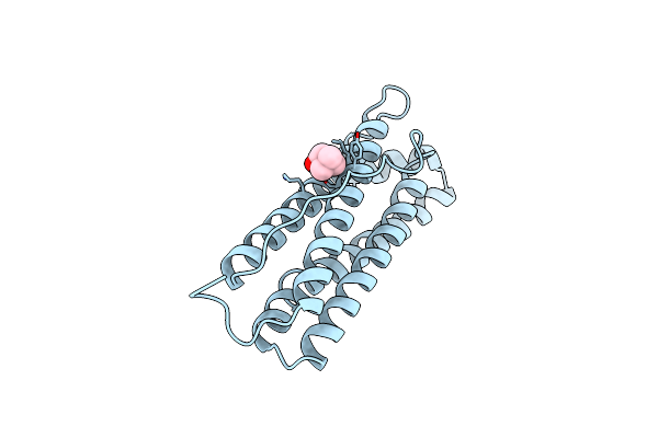
Bullfrog Red Cell L Ferritin Tartrate/Mg/Ph 5.5
Organism: Rana catesbeiana
Method: X-RAY DIFFRACTION Resolution:2.00 Å Release Date: 1995-11-14 Classification: IRON STORAGE Ligands: BET |
Organism: Rana catesbeiana
Method: X-RAY DIFFRACTION
Release Date: 1995-11-14
Ligands: BET
 |
Bullfrog Red Cell L Ferritin Sulfate/Mn/Ph 6.3
Organism: Rana catesbeiana
Method: X-RAY DIFFRACTION Resolution:2.40 Å Release Date: 1995-11-14 Classification: IRON STORAGE Ligands: BET |
Organism: Rana catesbeiana
Method: X-RAY DIFFRACTION
Release Date: 1995-11-14
Ligands: BET