

Search Count: 31
 |
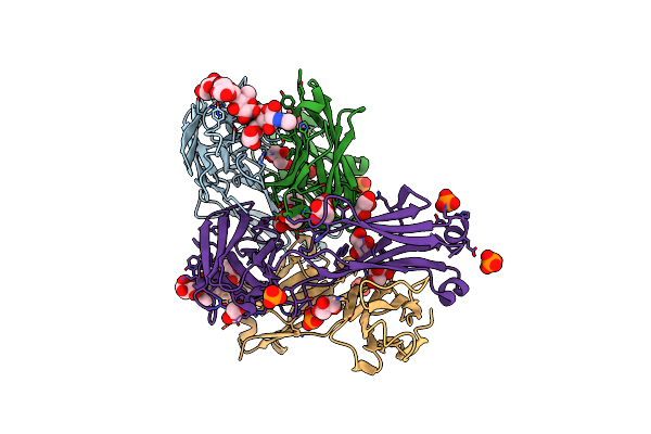
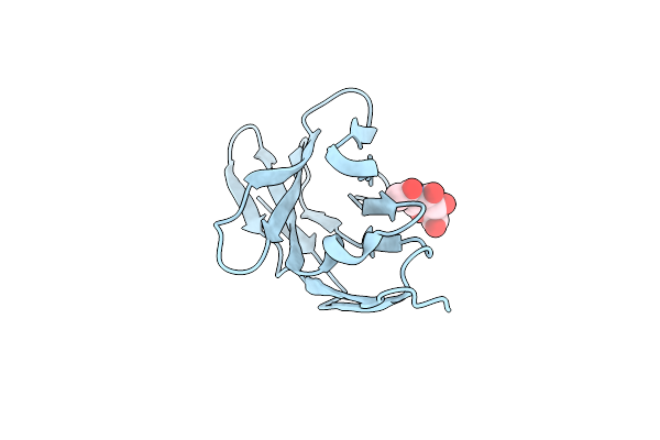
Crystal Structure Of Human Pancreatic Phospholipase A2
Organism: Homo sapiens
Method: X-RAY DIFFRACTION Resolution:1.90 Å Release Date: 2020-04-15 Classification: HYDROLASE Ligands: CL |
Organism: Homo sapiens
Method: X-RAY DIFFRACTION
Release Date: 2020-04-15
Ligands: CL
 |
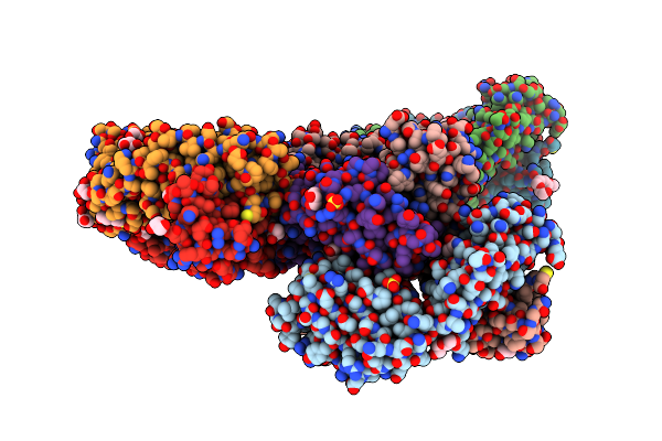
Crystal Structure Of Anti-Glycan Antibody Fab14.22 In Complex With Streptococcus Pneumoniae Serotype 14 Tetrasaccharide At 1.75 A
Organism: Mus musculus
Method: X-RAY DIFFRACTION Resolution:1.75 Å Release Date: 2017-03-22 Classification: IMMUNE SYSTEM Ligands: PO4, GOL |
Organism: Mus musculus
Method: X-RAY DIFFRACTION
Release Date: 2017-03-22
Ligands: PO4, GOL
 |
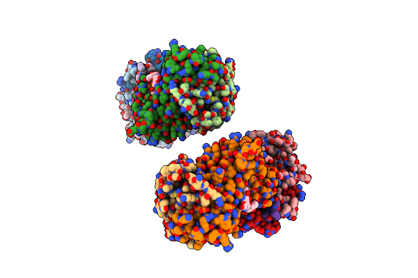
Crystal Structure Of Unbound Anti-Glycan Antibody Fab14.22 At 2.2 A
Organism: Mus musculus
Method: X-RAY DIFFRACTION Resolution:2.21 Å Release Date: 2017-03-22 Classification: IMMUNE SYSTEM Ligands: GOL, SO4, 1PE |
Organism: Mus musculus
Method: X-RAY DIFFRACTION
Release Date: 2017-03-22
Ligands: GOL, SO4, 1PE
 |
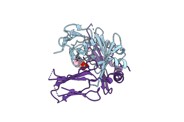
Tcr 21.30 In Complex With Mhc Class Ii I-Ag7Hel(11-27)
Organism: Mus musculus, Gallus gallus
Method: X-RAY DIFFRACTION Resolution:2.89 Å Release Date: 2010-05-12 Classification: IMMUNE SYSTEM Ligands: NAG |
Organism: Mus musculus, Gallus gallus
Method: X-RAY DIFFRACTION
Release Date: 2010-05-12
Ligands: NAG
 |
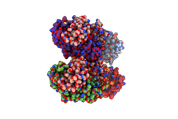
Crystal Structure Of The Mhc Class Ii Molecule I-Ag7 In Complex With The Peptide Gad221-235
Organism: Mus musculus
Method: X-RAY DIFFRACTION Resolution:3.09 Å Release Date: 2009-04-21 Classification: IMMUNE SYSTEM Ligands: NAG, EPE |
Organism: Mus musculus
Method: X-RAY DIFFRACTION
Release Date: 2009-04-21
Ligands: NAG, EPE
 |
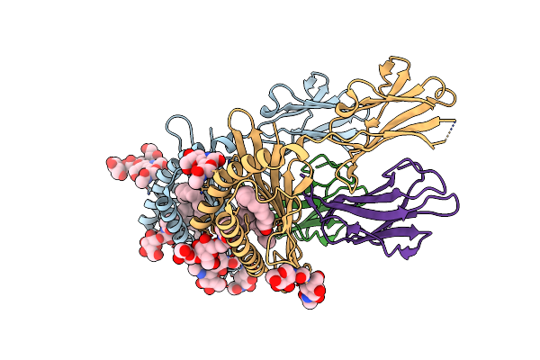
Crystal Structure Of Cd8Alpha-Beta In Complex With Yts 156.7 Fab
Organism: Mus musculus, Rattus rattus
Method: X-RAY DIFFRACTION Resolution:2.70 Å Release Date: 2008-11-11 Classification: IMMUNE SYSTEM Ligands: NAG |
Organism: Mus musculus, Rattus rattus
Method: X-RAY DIFFRACTION
Release Date: 2008-11-11
Ligands: NAG
 |
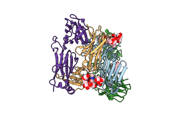
Structure Of The Endogenous Inkt Cell Ligand Igb3 Bound To Mcd1D
Organism: Mus musculus
Method: X-RAY DIFFRACTION Resolution:1.95 Å Release Date: 2008-04-01 Classification: IMMUNE SYSTEM Ligands: NAG, IGC, PLM |
Organism: Mus musculus
Method: X-RAY DIFFRACTION
Release Date: 2008-04-01
Ligands: NAG, IGC, PLM
 |
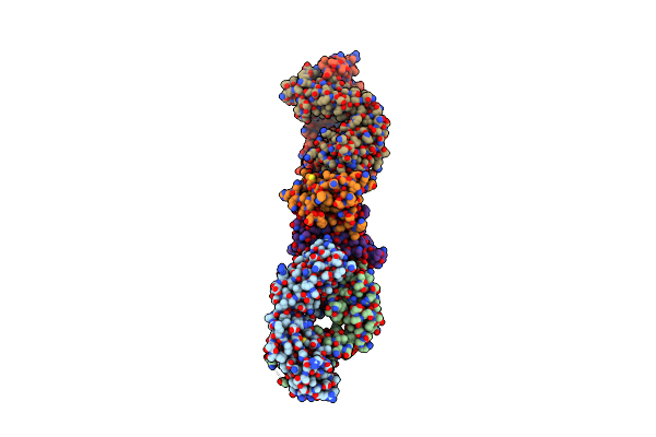
Structure Of The Mouse Invariant Nkt Cell Receptor Valpha14
Organism: Mus musculus
Method: X-RAY DIFFRACTION Resolution:1.85 Å Release Date: 2008-04-01 Classification: IMMUNE SYSTEM Ligands: NAG |
Organism: Mus musculus
Method: X-RAY DIFFRACTION
Release Date: 2008-04-01
Ligands: NAG
 |
Cd8Alpha-Alpha In Complex With Yts 105.18 Fab
Organism: Mus musculus, Rattus norvegicus
Method: X-RAY DIFFRACTION Resolution:2.88 Å Release Date: 2006-05-30 Classification: IMMUNE SYSTEM |
Organism: Mus musculus, Rattus norvegicus
Method: X-RAY DIFFRACTION
Release Date: 2006-05-30
 |
Structure Of A Highly Potent Short-Chain Galactosyl Ceramide Agonist Bound To Cd1D
Organism: Mus musculus
Method: X-RAY DIFFRACTION Resolution:2.20 Å Release Date: 2005-07-19 Classification: IMMUNE SYSTEM Ligands: NAG, PBS, R16 |
Organism: Mus musculus
Method: X-RAY DIFFRACTION
Release Date: 2005-07-19
Ligands: NAG, PBS, R16
 |
Crystal Structure Of Human Triggering Receptor Expressed On Myeloid Cells 1 (Trem-1) At 1.47 .
Organism: Homo sapiens
Method: X-RAY DIFFRACTION Resolution:1.47 Å Release Date: 2004-09-21 Classification: IMMUNE SYSTEM Ligands: TLA |
Organism: Homo sapiens
Method: X-RAY DIFFRACTION
Release Date: 2004-09-21
Ligands: TLA
 |
Drosophila Peptidoglycan Recognition Protein (Pgrp)-Sa
Organism: Drosophila melanogaster
Method: X-RAY DIFFRACTION Resolution:1.56 Å Release Date: 2004-06-29 Classification: IMMUNE SYSTEM Ligands: SO4, EDO |
Organism: Drosophila melanogaster
Method: X-RAY DIFFRACTION
Release Date: 2004-06-29
Ligands: SO4, EDO
 |
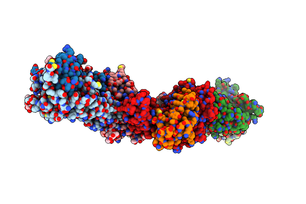
Crystal Structure Of Cd1A In Complex With A Sulfatide
Organism: Homo sapiens
Method: X-RAY DIFFRACTION Resolution:2.15 Å Release Date: 2003-08-05 Classification: IMMUNE SYSTEM Ligands: FUC, SLF, NAG |
Organism: Homo sapiens
Method: X-RAY DIFFRACTION
Release Date: 2003-08-05
Ligands: FUC, SLF, NAG
 |
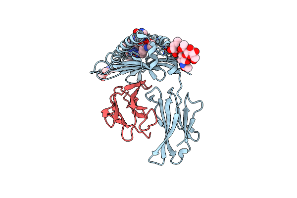
2C/H-2Kbm3/Dev8 Allogeneic Complex
Organism: Mus musculus
Method: X-RAY DIFFRACTION Resolution:2.40 Å Release Date: 2002-11-27 Classification: IMMUNE SYSTEM Ligands: GOL, NAG, ACY |
Organism: Mus musculus
Method: X-RAY DIFFRACTION
Release Date: 2002-11-27
Ligands: GOL, NAG, ACY
 |
Crystal Structure Of Mhc Class I H-2Kb Heavy Chain Complexed With Beta-2 Microglobulin And Yeast Alpha-Glucosidase
Organism: Mus musculus
Method: X-RAY DIFFRACTION Resolution:1.50 Å Release Date: 2002-07-17 Classification: IMMUNE SYSTEM Ligands: NAG |
Organism: Mus musculus
Method: X-RAY DIFFRACTION
Release Date: 2002-07-17
Ligands: NAG
 |
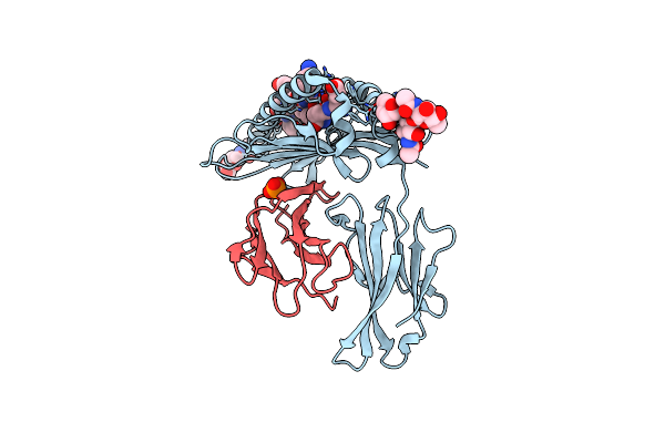
Crystal Structure Of Mhc Class I H-2Kb Heavy Chain Complexed With Beta-2 Microglobulin And Muc1 Vntr Peptide Sapdtrpa
Organism: Mus musculus
Method: X-RAY DIFFRACTION Resolution:1.60 Å Release Date: 2002-07-17 Classification: IMMUNE SYSTEM Ligands: NAG |
Organism: Mus musculus
Method: X-RAY DIFFRACTION
Release Date: 2002-07-17
Ligands: NAG
 |
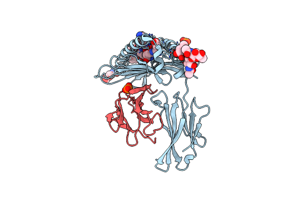
Crystal Structure Of H-2Kbm3 Bound To Dev8
Organism: Mus musculus
Method: X-RAY DIFFRACTION Resolution:2.15 Å Release Date: 2002-06-26 Classification: IMMUNE SYSTEM Ligands: NAG, PO4 |
Organism: Mus musculus
Method: X-RAY DIFFRACTION
Release Date: 2002-06-26
Ligands: NAG, PO4
 |
Crystal Structure Of H-2Kb Bound To The Dev8 Peptide
Organism: Mus musculus
Method: X-RAY DIFFRACTION Resolution:1.75 Å Release Date: 2002-06-19 Classification: IMMUNE SYSTEM Ligands: NAG, PO4 |
Organism: Mus musculus
Method: X-RAY DIFFRACTION
Release Date: 2002-06-19
Ligands: NAG, PO4
 |
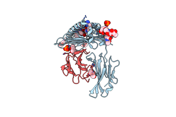
Tcr Domain
Organism: Mus musculus
Method: X-RAY DIFFRACTION Resolution:2.50 Å Release Date: 2001-12-05 Classification: IMMUNE SYSTEM Ligands: NAG |
Organism: Mus musculus
Method: X-RAY DIFFRACTION
Release Date: 2001-12-05
Ligands: NAG
 |
Mhc Class I Natural Mutant H-2Kbm1 Heavy Chain Complexed With Beta-2 Microglobulin And Vesicular Stomatitis Virus Nucleoprotein
Organism: Mus musculus
Method: X-RAY DIFFRACTION Resolution:1.90 Å Release Date: 2001-03-28 Classification: IMMUNE SYSTEM Ligands: NAG, PO4, MPD |
Organism: Mus musculus
Method: X-RAY DIFFRACTION
Release Date: 2001-03-28
Ligands: NAG, PO4, MPD