

Search Count: 30
 |
Crystal Structure Of L-Threonate 3-Dehydrogenase From Paracoccus Litorisediminis (Ligand-Free Form)
Organism: Paracoccus litorisediminis
Method: X-RAY DIFFRACTION Resolution:1.90 Å Release Date: 2026-03-18 Classification: OXIDOREDUCTASE Ligands: SO4, GOL, PEG |
Organism: Paracoccus litorisediminis
Method: X-RAY DIFFRACTION
Release Date: 2026-03-18
Ligands: SO4, GOL, PEG
 |
Crystal Structure Of L-Threonate 3-Dehydrogenase From Paracoccus Litorisediminis (Nadp+ And Tartronate Bound Form)
Organism: Paracoccus litorisediminis
Method: X-RAY DIFFRACTION Resolution:2.08 Å Release Date: 2026-03-18 Classification: OXIDOREDUCTASE Ligands: NAP, TTN |
Organism: Paracoccus litorisediminis
Method: X-RAY DIFFRACTION
Release Date: 2026-03-18
Ligands: NAP, TTN
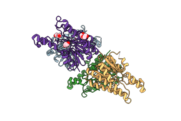 |
Crystal Structure Of L-2-Keto-3-Deoxyfuconate 4-Dehydrogenase From Herbaspillum Huttiense (Apo Form)
Organism: Herbaspirillum huttiense
Method: X-RAY DIFFRACTION Resolution:1.27 Å Release Date: 2024-07-10 Classification: OXIDOREDUCTASE Ligands: PEG |
Organism: Herbaspirillum huttiense
Method: X-RAY DIFFRACTION
Release Date: 2024-07-10
Ligands: PEG
 |
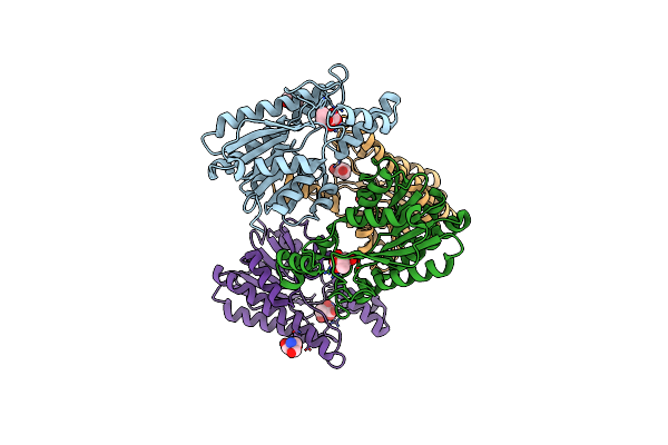
Crystal Structure Of L-2-Keto-3-Deoxyfuconate 4-Dehydrogenase Bound To Nad(H) And Sulfate Ion
Organism: Herbaspirillum huttiense
Method: X-RAY DIFFRACTION Resolution:1.77 Å Release Date: 2024-07-10 Classification: OXIDOREDUCTASE Ligands: NAD, SO4, PEG, TRS, GOL |
Organism: Herbaspirillum huttiense
Method: X-RAY DIFFRACTION
Release Date: 2024-07-10
Ligands: NAD, SO4, PEG, TRS, GOL
 |
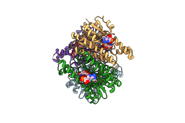
Crystal Structure Of L-2-Keto-3-Deoxyfuconate 4-Dehydrogenase Bound To L-Kdf Or L-2,4-Dkdf
Organism: Herbaspirillum huttiense
Method: X-RAY DIFFRACTION Resolution:1.29 Å Release Date: 2024-07-10 Classification: OXIDOREDUCTASE Ligands: TRS, A1LXA, A1LXB, PEG |
Organism: Herbaspirillum huttiense
Method: X-RAY DIFFRACTION
Release Date: 2024-07-10
Ligands: TRS, A1LXA, A1LXB, PEG
 |
Crystal Structure Of L-2-Keto-3-Deoxyfuconate 4-Dehydrogenase Bound To L-2,4-Dkdf And Nadh
Organism: Herbaspirillum huttiense
Method: X-RAY DIFFRACTION Resolution:1.67 Å Release Date: 2024-07-10 Classification: OXIDOREDUCTASE Ligands: NAD, A1LXB |
Organism: Herbaspirillum huttiense
Method: X-RAY DIFFRACTION
Release Date: 2024-07-10
Ligands: NAD, A1LXB
 |
Crystal Structure Of L-2-Keto-3-Deoxyfuconate 4-Dehydrogenase Bound To D-Kdp
Organism: Herbaspirillum huttiense
Method: X-RAY DIFFRACTION Resolution:1.51 Å Release Date: 2024-07-10 Classification: OXIDOREDUCTASE Ligands: TRS, A1LXD, PEG |
Organism: Herbaspirillum huttiense
Method: X-RAY DIFFRACTION
Release Date: 2024-07-10
Ligands: TRS, A1LXD, PEG
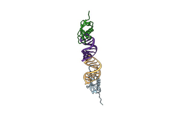 |
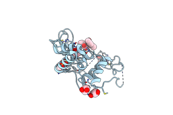
Crystal Structure Of The Ubiquitin-Like Domain In The Sf3A1 Subunit Of Human U2 Snrnp Complexed With The Stem-Loop 4 Of U1 Snrna
Organism: Homo sapiens
Method: X-RAY DIFFRACTION Resolution:1.80 Å Release Date: 2023-05-17 Classification: RNA BINDING PROTEIN/RNA |
Organism: Homo sapiens
Method: X-RAY DIFFRACTION
Release Date: 2023-05-17
 |
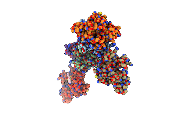
Hoip-Hoipin1 Complex
Organism: Homo sapiens
Method: X-RAY DIFFRACTION Resolution:1.54 Å Release Date: 2020-04-15 Classification: LIGASE Ligands: ZN, D5U, GOL, EPE |
Organism: Homo sapiens
Method: X-RAY DIFFRACTION
Release Date: 2020-04-15
Ligands: ZN, D5U, GOL, EPE
 |
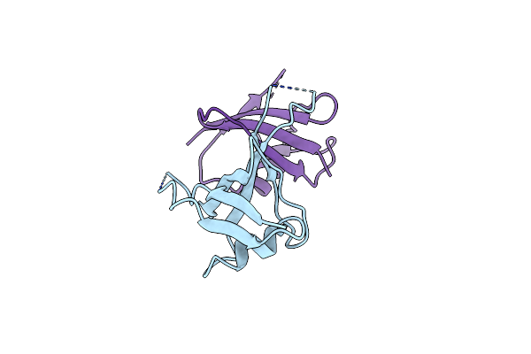
Hoip-Hoipin8 Complex
Organism: Homo sapiens
Method: X-RAY DIFFRACTION Resolution:2.12 Å Release Date: 2020-04-15 Classification: LIGASE Ligands: ZN, D60, CL, GOL |
Organism: Homo sapiens
Method: X-RAY DIFFRACTION
Release Date: 2020-04-15
Ligands: ZN, D60, CL, GOL
 |
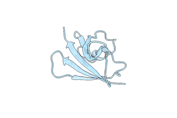
Complex Structure Of Axin-Dix And Dvl2-Dix
Organism: Homo sapiens
Method: X-RAY DIFFRACTION Resolution:3.09 Å Release Date: 2020-01-15 Classification: SIGNALING PROTEIN |
Organism: Homo sapiens
Method: X-RAY DIFFRACTION
Release Date: 2020-01-15
 |
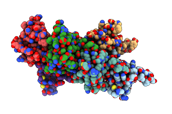
High Resolution Structure Of Dvl2-Dix Y27W/C80S Mutant
Organism: Homo sapiens
Method: X-RAY DIFFRACTION Resolution:1.64 Å Release Date: 2019-02-20 Classification: SIGNALING PROTEIN |
Organism: Homo sapiens
Method: X-RAY DIFFRACTION
Release Date: 2019-02-20
 |
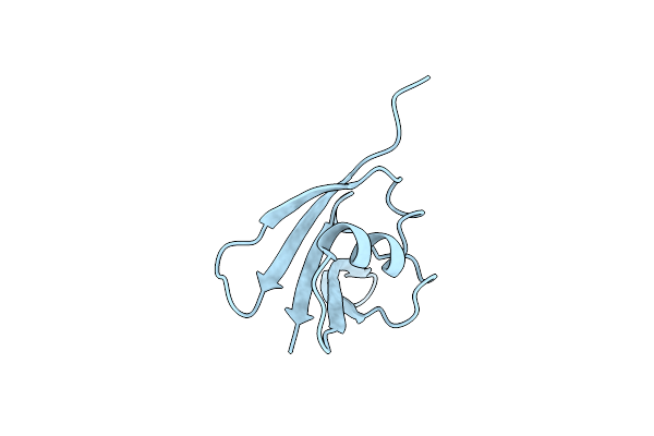
Crystal Structure Of Mouse Ccd1 Dix Domain
Organism: Mus musculus
Method: X-RAY DIFFRACTION Resolution:3.00 Å Release Date: 2017-09-06 Classification: SIGNALING PROTEIN |
Organism: Mus musculus
Method: X-RAY DIFFRACTION
Release Date: 2017-09-06
 |
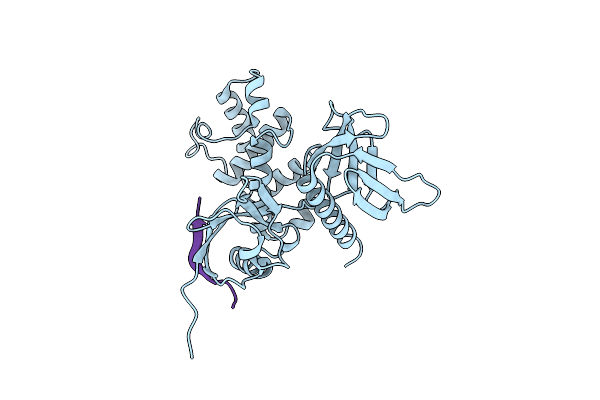
Crystal Structure Of Zebrafish Ccd1 Dix Domain
Organism: Danio rerio
Method: X-RAY DIFFRACTION Resolution:1.96 Å Release Date: 2017-09-06 Classification: SIGNALING PROTEIN |
Organism: Danio rerio
Method: X-RAY DIFFRACTION
Release Date: 2017-09-06
 |
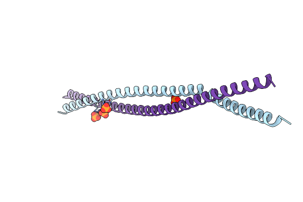
Radixin Complex
Organism: Mus musculus, Homo sapiens
Method: X-RAY DIFFRACTION Resolution:2.40 Å Release Date: 2015-10-21 Classification: CELL INVASION |
Organism: Mus musculus, Homo sapiens
Method: X-RAY DIFFRACTION
Release Date: 2015-10-21
 |
Crystal Structure Of The C-Terminal Coiled Coil Of Mouse Bicaudal D1
Organism: Mus musculus
Method: X-RAY DIFFRACTION Resolution:1.50 Å Release Date: 2015-04-08 Classification: TRANSPORT PROTEIN Ligands: PO4 |
Organism: Mus musculus
Method: X-RAY DIFFRACTION
Release Date: 2015-04-08
Ligands: PO4
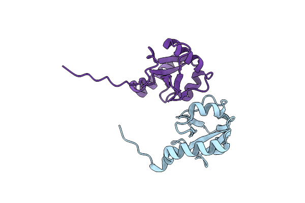 |
Crystal Structure Of Plekhm1 Lir-Fused Human Lc3B_2-119
Organism: Homo sapiens
Method: X-RAY DIFFRACTION Resolution:2.71 Å Release Date: 2015-01-14 Classification: PROTEIN BINDING |
Organism: Homo sapiens
Method: X-RAY DIFFRACTION
Release Date: 2015-01-14
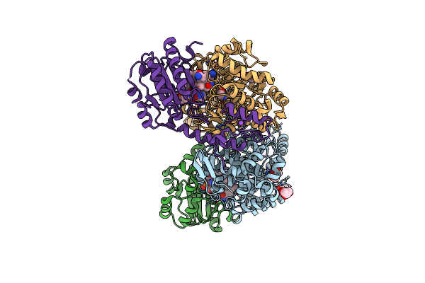 |
Crystal Structure Of Ethanolamine Ammonia-Lyase From Escherichia Coli Complexed With Cn-Cbl And Ethanolamine
Organism: Escherichia coli
Method: X-RAY DIFFRACTION Resolution:2.10 Å Release Date: 2010-06-02 Classification: LYASE Ligands: ETA, GOL, B12, NA |
Organism: Escherichia coli
Method: X-RAY DIFFRACTION
Release Date: 2010-06-02
Ligands: ETA, GOL, B12, NA
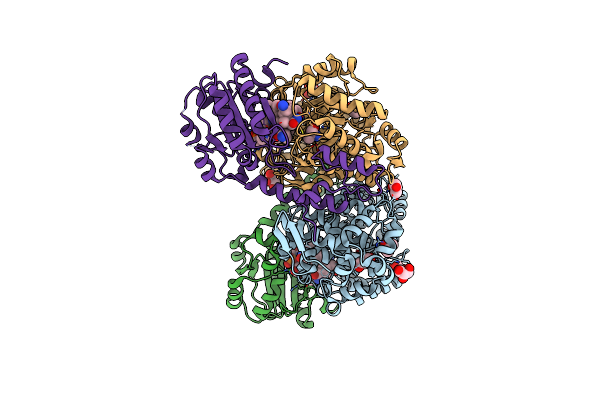 |
Crystal Structure Of Ethanolamine Ammonia-Lyase From Escherichia Coli Complexed With Cn-Cbl And 2-Amino-1-Propanol
Organism: Escherichia coli
Method: X-RAY DIFFRACTION Resolution:2.05 Å Release Date: 2010-06-02 Classification: LYASE Ligands: 2A1, GOL, B12 |
Organism: Escherichia coli
Method: X-RAY DIFFRACTION
Release Date: 2010-06-02
Ligands: 2A1, GOL, B12
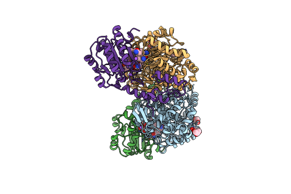 |
Crystal Structure Of Ethanolamine Ammonia-Lyase From Escherichia Coli Complexed With Cn-Cbl (Substrate-Free Form)
Organism: Escherichia coli
Method: X-RAY DIFFRACTION Resolution:2.10 Å Release Date: 2010-06-02 Classification: LYASE Ligands: GOL, NA, B12 |
Organism: Escherichia coli
Method: X-RAY DIFFRACTION
Release Date: 2010-06-02
Ligands: GOL, NA, B12