

Search Count: 26
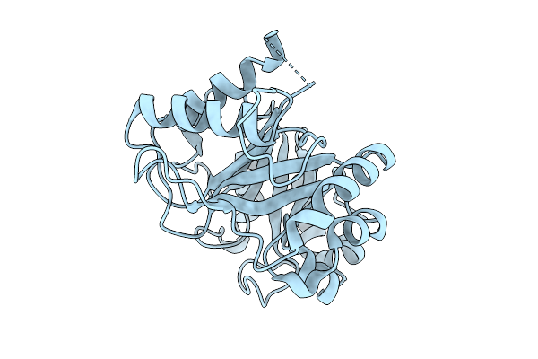 |
Crystal Structure Of Xylanase Inhibitor Protein (Osxip) From Rice
Organism: Oryza sativa subsp. japonica
Method: X-RAY DIFFRACTION Resolution:1.30 Å Release Date: 2025-09-03 Classification: PLANT PROTEIN |
Organism: Oryza sativa subsp. japonica
Method: X-RAY DIFFRACTION
Release Date: 2025-09-03
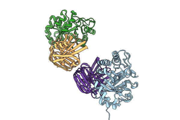 |
Crystal Structure Of Family 11 Xylanase In Complex With Inhibitor (Osxip)
Organism: Rhizopus arrhizus, Oryza sativa subsp. japonica
Method: X-RAY DIFFRACTION Resolution:2.21 Å Release Date: 2025-09-03 Classification: PROTEIN BINDING |
Organism: Rhizopus arrhizus, Oryza sativa subsp. japonica
Method: X-RAY DIFFRACTION
Release Date: 2025-09-03
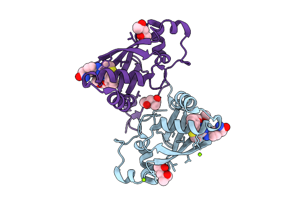 |
Crystal Structure Of P110Alpha-Ras Binding Domain (Rbd) In Complex With Molecular Glue D927
Organism: Homo sapiens
Method: X-RAY DIFFRACTION Resolution:1.75 Å Release Date: 2025-07-23 Classification: SIGNALING PROTEIN Ligands: A1ATF, MG, MRD, MPD |
Organism: Homo sapiens
Method: X-RAY DIFFRACTION
Release Date: 2025-07-23
Ligands: A1ATF, MG, MRD, MPD
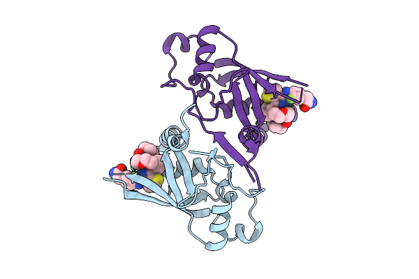 |
Crystal Structure Of P110Alpha-Ras Binding Domain (Rbd) In Complex With Molecular Glue D223
Organism: Homo sapiens
Method: X-RAY DIFFRACTION Resolution:2.01 Å Release Date: 2025-07-23 Classification: SIGNALING PROTEIN Ligands: A1AZD |
Organism: Homo sapiens
Method: X-RAY DIFFRACTION
Release Date: 2025-07-23
Ligands: A1AZD
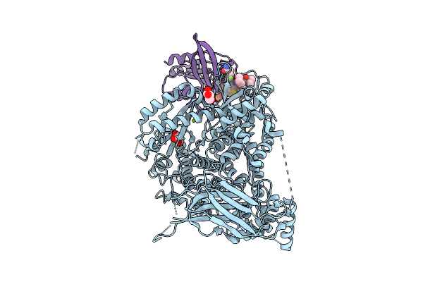 |
Crystal Structure Of The Kras-P110Alpha Complex In The Presence Of Molecular Glue D223
Organism: Homo sapiens
Method: X-RAY DIFFRACTION Resolution:3.01 Å Release Date: 2025-07-23 Classification: ONCOPROTEIN Ligands: A1AZD, GOL, MG, GNP |
Organism: Homo sapiens
Method: X-RAY DIFFRACTION
Release Date: 2025-07-23
Ligands: A1AZD, GOL, MG, GNP
 |
X-Ray Structure Of The Human Heart Fatty Acid-Binding Protein Complexed With R-Ibuprofen
Organism: Homo sapiens
Method: X-RAY DIFFRACTION Resolution:1.35 Å Release Date: 2025-04-30 Classification: LIPID BINDING PROTEIN Ligands: IZP, PG4, PEG, ACY, PGE, GOL |
Organism: Homo sapiens
Method: X-RAY DIFFRACTION
Release Date: 2025-04-30
Ligands: IZP, PG4, PEG, ACY, PGE, GOL
 |
Sfx Structure Of An Mn-Carbonyl Complex Immobilized In Hen Egg White Lysozyme Microcrystals, 100 Ns After Photoexcitation At Rt.
Organism: Gallus gallus
Method: X-RAY DIFFRACTION Resolution:1.60 Å Release Date: 2024-09-04 Classification: HYDROLASE Ligands: CL, NA, XTX |
Organism: Gallus gallus
Method: X-RAY DIFFRACTION
Release Date: 2024-09-04
Ligands: CL, NA, XTX
 |
Dark State Sfx Structure Of Hen Egg White Lysozyme Microcrystals Immobilized With A Mn Carbonyl Complex At Rt.
Organism: Gallus gallus
Method: X-RAY DIFFRACTION Resolution:1.60 Å Release Date: 2024-07-31 Classification: HYDROLASE Ligands: CL, NA, XTX |
Organism: Gallus gallus
Method: X-RAY DIFFRACTION
Release Date: 2024-07-31
Ligands: CL, NA, XTX
 |
Sfx Structure Of An Mn-Carbonyl Complex Immobilized In Hen Egg White Lysozyme Microcrystals, 10 Ns After Photoexcitation At Rt.
Organism: Gallus gallus
Method: X-RAY DIFFRACTION Resolution:1.60 Å Release Date: 2024-07-31 Classification: HYDROLASE Ligands: CL, NA, XTX |
Organism: Gallus gallus
Method: X-RAY DIFFRACTION
Release Date: 2024-07-31
Ligands: CL, NA, XTX
 |
Sfx Structure Of An Mn-Carbonyl Complex Immobilized In Hen Egg White Lysozyme Microcrystals, 1 Microsecond After Photoexcitation At Rt.
Organism: Gallus gallus
Method: X-RAY DIFFRACTION Resolution:1.60 Å Release Date: 2024-07-31 Classification: HYDROLASE Ligands: CL, NA, XTX |
Organism: Gallus gallus
Method: X-RAY DIFFRACTION
Release Date: 2024-07-31
Ligands: CL, NA, XTX
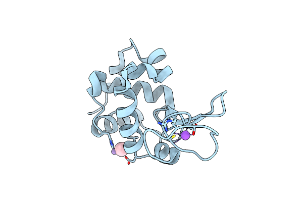 |
Sfx Structure Of An Mn-Carbonyl Complex Immobilized In Hen Egg White Lysozyme Microcrystals, 1 Microsecond After Photoexcitation With 40 Microjoules Laser Intensity At Rt.
Organism: Gallus gallus
Method: X-RAY DIFFRACTION Resolution:1.60 Å Release Date: 2024-07-31 Classification: HYDROLASE Ligands: CL, NA, XTX |
Organism: Gallus gallus
Method: X-RAY DIFFRACTION
Release Date: 2024-07-31
Ligands: CL, NA, XTX
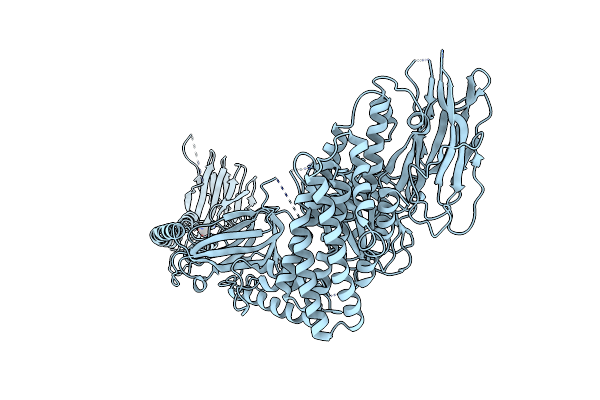 |
Crystal Structure Of The In-Cell Cry1Aa Purified From Bacillus Thuringiensis
Organism: Bacillus thuringiensis
Method: X-RAY DIFFRACTION Resolution:3.60 Å Release Date: 2024-03-06 Classification: TOXIN Ligands: UNX |
Organism: Bacillus thuringiensis
Method: X-RAY DIFFRACTION
Release Date: 2024-03-06
Ligands: UNX
 |
Crystal Structure Of A Rice Class Iiib Chitinase, Oschib2
Organism: Oryza sativa
Method: X-RAY DIFFRACTION Resolution:1.18 Å Release Date: 2023-05-03 Classification: HYDROLASE Ligands: EDO, ACT |
Organism: Oryza sativa
Method: X-RAY DIFFRACTION
Release Date: 2023-05-03
Ligands: EDO, ACT
 |
Crystal Structure Of Wild Type Cypovirus Polyhedra Produced By Cell-Free Protein Synthesis
Organism: Bombyx mori cypovirus 1
Method: X-RAY DIFFRACTION Resolution:1.80 Å Release Date: 2023-02-01 Classification: VIRAL PROTEIN Ligands: ACE, CL |
Organism: Bombyx mori cypovirus 1
Method: X-RAY DIFFRACTION
Release Date: 2023-02-01
Ligands: ACE, CL
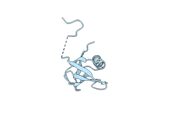 |
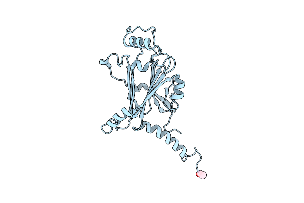
Crystal Structure Of Cipa Crystal Produced By Cell-Free Protein Synthesis
Organism: Photorhabdus
Method: X-RAY DIFFRACTION Resolution:2.11 Å Release Date: 2023-02-01 Classification: Crystalline inclusion protein |
Organism: Photorhabdus
Method: X-RAY DIFFRACTION
Release Date: 2023-02-01
 |
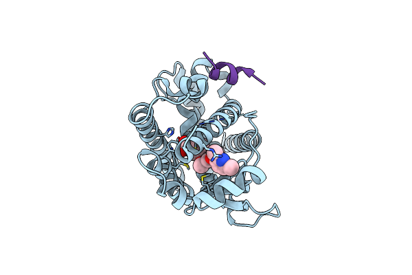
Crystal Structure Of Wild Type Cypovirus Polyhedra Produced By Cell-Free Protein Synthesis With Small Volume
Organism: Bombyx mori cypovirus 1
Method: X-RAY DIFFRACTION Resolution:1.95 Å Release Date: 2023-02-01 Classification: VIRAL PROTEIN Ligands: ACE, CL |
Organism: Bombyx mori cypovirus 1
Method: X-RAY DIFFRACTION
Release Date: 2023-02-01
Ligands: ACE, CL
 |
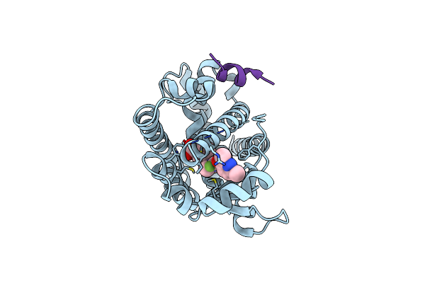
Crystal Structure Of The Ppargamma-Lbd Complexed With Compound 1L
Organism: Homo sapiens
Method: X-RAY DIFFRACTION Resolution:1.80 Å Release Date: 2019-03-27 Classification: TRANSCRIPTION Ligands: B0X |
Organism: Homo sapiens
Method: X-RAY DIFFRACTION
Release Date: 2019-03-27
Ligands: B0X
 |
Crystal Structure Of The Ppargamma-Lbd Complexed With Compound 3G
Organism: Homo sapiens
Method: X-RAY DIFFRACTION Resolution:1.75 Å Release Date: 2019-03-27 Classification: TRANSCRIPTION Ligands: B1O |
Organism: Homo sapiens
Method: X-RAY DIFFRACTION
Release Date: 2019-03-27
Ligands: B1O
 |
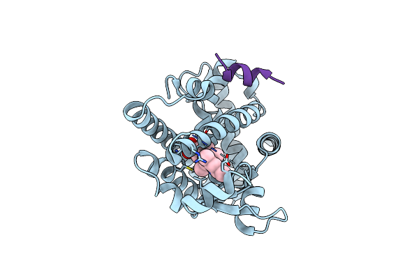
Crystal Structure Of The Ppargamma-Lbd Complexed With Compound 13Ab
Organism: Homo sapiens
Method: X-RAY DIFFRACTION Resolution:1.80 Å Release Date: 2018-10-17 Classification: TRANSCRIPTION Ligands: RTE, CL |
Organism: Homo sapiens
Method: X-RAY DIFFRACTION
Release Date: 2018-10-17
Ligands: RTE, CL
 |
Crystal Structure Of The Ppargamma-Lbd Complexed With Compound Ds-6930
Organism: Homo sapiens
Method: X-RAY DIFFRACTION Resolution:1.80 Å Release Date: 2018-10-17 Classification: TRANSCRIPTION Ligands: RTF, CL |
Organism: Homo sapiens
Method: X-RAY DIFFRACTION
Release Date: 2018-10-17
Ligands: RTF, CL