
Search Count: 37
All
Selected
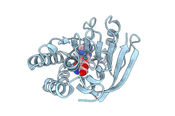 |
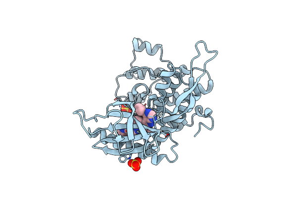
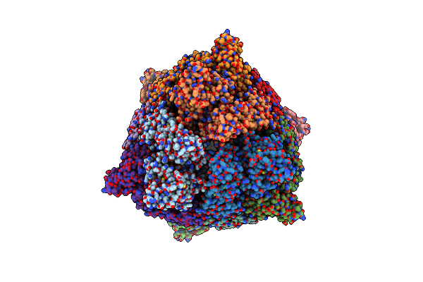
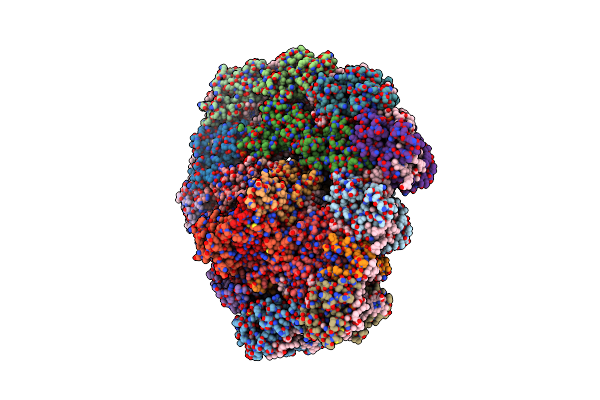
Crystal Structure Of A Bacterial Dot1L Methyl-Transferase In Complex With S-Adenosyl Homocysteine
Organism: Legionella pneumophila
Method: X-RAY DIFFRACTION Resolution:2.11 Å Release Date: 2026-02-25 Classification: TRANSFERASE Ligands: SAH |
 |
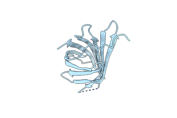
Organism: Phage metagenome
Method: X-RAY DIFFRACTION Resolution:1.56 Å Release Date: 2025-09-17 Classification: VIRAL PROTEIN Ligands: OJC |
 |
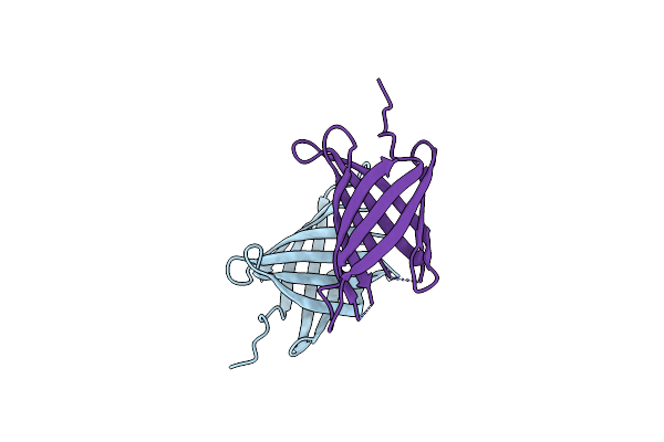
Organism: Phage metagenome
Method: X-RAY DIFFRACTION Resolution:1.62 Å Release Date: 2025-07-30 Classification: VIRAL PROTEIN Ligands: OJC |
 |
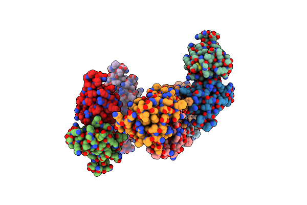
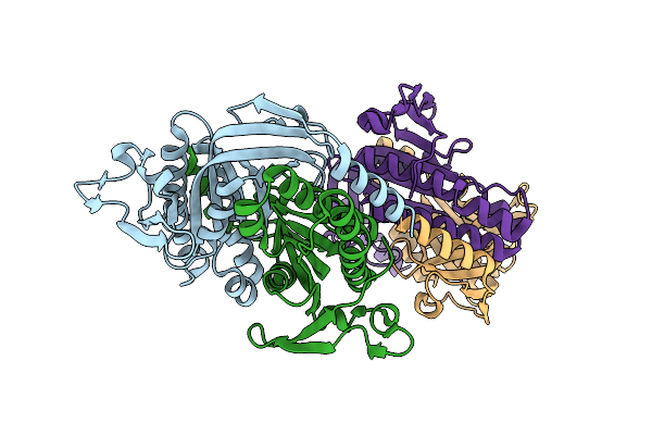
Structure Of The C-Terminal Half Of Lrrk2 Bound To Rn277 (Type-Ii Inhibitor)
Organism: Homo sapiens, Synthetic construct
Method: ELECTRON MICROSCOPY Resolution:3.35 Å Release Date: 2025-06-18 Classification: PROTEIN BINDING Ligands: A1A7Q |
 |
Organism: Homo sapiens
Method: X-RAY DIFFRACTION Resolution:2.70 Å Release Date: 2024-09-11 Classification: TRANSFERASE Ligands: SO4, A1H74 |
 |
Organism: Mus musculus, Saccharomyces cerevisiae
Method: ELECTRON MICROSCOPY Release Date: 2024-06-26 Classification: RIBOSOME Ligands: MG, CL, SPM, SPD, ZN |
 |
Organism: Saccharomyces cerevisiae
Method: ELECTRON MICROSCOPY Release Date: 2024-06-12 Classification: RIBOSOME Ligands: MG, SPM, SPD, ZN |
 |
Organism: Mus musculus, Saccharomyces cerevisiae
Method: ELECTRON MICROSCOPY Release Date: 2024-06-12 Classification: RIBOSOME Ligands: MG, CL, SPD, SPM, ZN |
 |
Organism: Saccharomyces cerevisiae
Method: ELECTRON MICROSCOPY Release Date: 2024-06-12 Classification: RIBOSOME Ligands: MG, CL, SPM, SPD, ZN |
 |
Organism: Escherichia coli
Method: ELECTRON MICROSCOPY Release Date: 2023-02-01 Classification: IMMUNE SYSTEM |
 |
Organism: Streptococcus suis
Method: ELECTRON MICROSCOPY Release Date: 2023-02-01 Classification: IMMUNE SYSTEM |
 |
Organism: Escherichia coli
Method: ELECTRON MICROSCOPY Release Date: 2023-02-01 Classification: IMMUNE SYSTEM Ligands: ZN |
 |
Organism: Escherichia coli
Method: ELECTRON MICROSCOPY Release Date: 2023-02-01 Classification: IMMUNE SYSTEM Ligands: ZN |
 |
Organism: Escherichia coli k-12
Method: X-RAY DIFFRACTION Resolution:1.88 Å Release Date: 2022-03-09 Classification: UNKNOWN FUNCTION |
 |
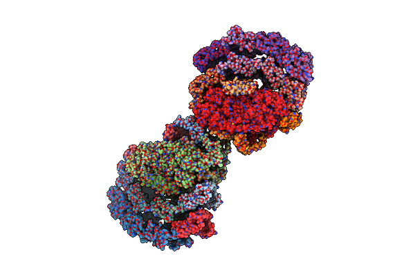 |
Dimeric Photosystem I Of A Temperature Sensitive Mutant Chlamydomonas Reinhardtii
Organism: Chlamydomonas reinhardtii
Method: ELECTRON MICROSCOPY Release Date: 2021-12-15 Classification: PHOTOSYNTHESIS |
 |
 |
Structure Of Yersinia Aleksiciae Cap15 Cyclic Dinucleotide Receptor, Crystal Form 1
Organism: Yersinia aleksiciae
Method: X-RAY DIFFRACTION Resolution:1.90 Å Release Date: 2021-11-17 Classification: IMMUNE SYSTEM |
 |
Structure Of Yersinia Aleksiciae Cap15 Cyclic Dinucleotide Receptor, Crystal Form 2
Organism: Yersinia aleksiciae
Method: X-RAY DIFFRACTION Resolution:2.60 Å Release Date: 2021-11-17 Classification: IMMUNE SYSTEM |
 |
Crystal Structure Of A Bacterial Cyclic Ump Synthase From Burkholderia Cepacia Lk29
Organism: Burkholderia cepacia
Method: X-RAY DIFFRACTION Resolution:1.45 Å Release Date: 2021-10-13 Classification: TRANSFERASE |