

Search Count: 20
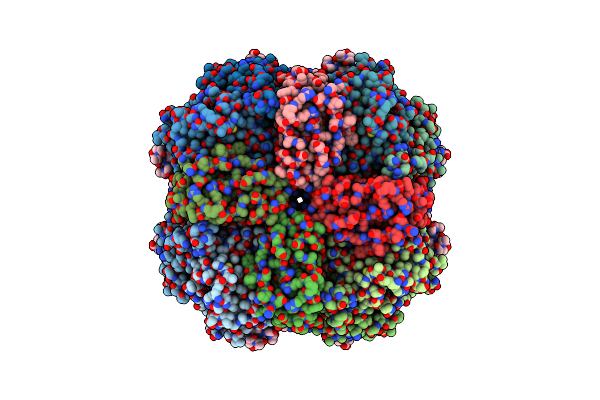 |
Rubisco From Phaeodactylum Tricornutum Bound To Pyco1(452-592)
Organism: Phaeodactylum tricornutum
Method: ELECTRON MICROSCOPY Release Date: 2023-06-21 Classification: PHOTOSYNTHESIS Ligands: CAP |
Organism: Phaeodactylum tricornutum
Method: ELECTRON MICROSCOPY
Release Date: 2023-06-21
Ligands: CAP
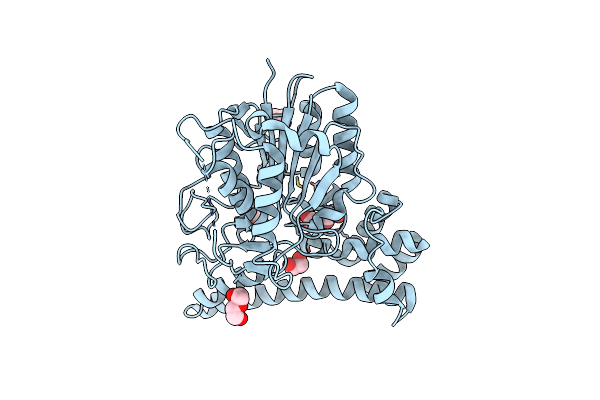 |
Turning An Asparaginyl Endopeptidase Into A Peptide Ligase
Organism: Clitoria ternatea
Method: X-RAY DIFFRACTION Resolution:1.35 Å Release Date: 2020-10-07 Classification: HYDROLASE Ligands: EDO, PEG |
Organism: Clitoria ternatea
Method: X-RAY DIFFRACTION
Release Date: 2020-10-07
Ligands: EDO, PEG
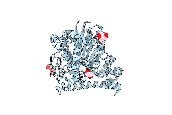 |
Turning An Asparaginyl Endopeptidase Into A Peptide Ligase
Organism: Clitoria ternatea
Method: X-RAY DIFFRACTION Resolution:1.66 Å Release Date: 2020-10-07 Classification: HYDROLASE Ligands: NAG, EDO, PEG |
Organism: Clitoria ternatea
Method: X-RAY DIFFRACTION
Release Date: 2020-10-07
Ligands: NAG, EDO, PEG
 |
Turning An Asparaginyl Endopeptidase Into A Peptide Ligase
Organism: Clitoria ternatea
Method: X-RAY DIFFRACTION Resolution:2.64 Å Release Date: 2020-10-07 Classification: HYDROLASE |
Organism: Clitoria ternatea
Method: X-RAY DIFFRACTION
Release Date: 2020-10-07
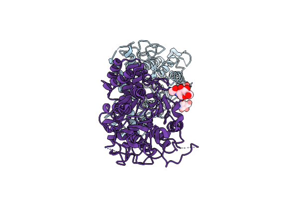 |
Turning An Asparaginyl Endopeptidase Into A Peptide Ligase
Organism: Clitoria ternatea
Method: X-RAY DIFFRACTION Resolution:1.50 Å Release Date: 2020-10-07 Classification: HYDROLASE Ligands: NAG, EDO, PEG |
Organism: Clitoria ternatea
Method: X-RAY DIFFRACTION
Release Date: 2020-10-07
Ligands: NAG, EDO, PEG
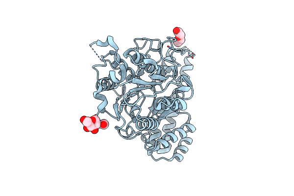 |
Turning An Asparaginyl Endopeptidase Into A Peptide Ligase
Organism: Clitoria ternatea
Method: X-RAY DIFFRACTION Resolution:2.00 Å Release Date: 2020-10-07 Classification: HYDROLASE Ligands: NAG, PEG |
Organism: Clitoria ternatea
Method: X-RAY DIFFRACTION
Release Date: 2020-10-07
Ligands: NAG, PEG
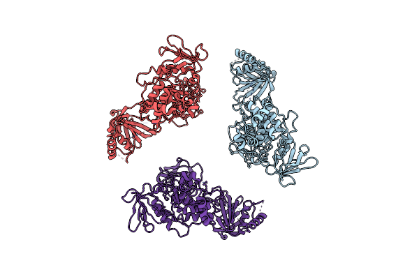 |
Structure Of Ferm Protein
Organism: Homo sapiens
Method: X-RAY DIFFRACTION Resolution:3.60 Å Release Date: 2020-06-03 Classification: SIGNALING PROTEIN |
Organism: Homo sapiens
Method: X-RAY DIFFRACTION
Release Date: 2020-06-03
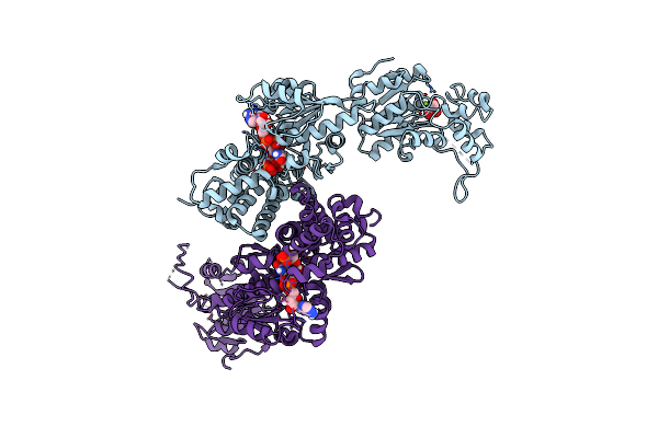 |
Structure Of Dsgpdh Of Dunaliella Salina
Organism: Dunaliella salina
Method: X-RAY DIFFRACTION Resolution:2.20 Å Release Date: 2019-12-04 Classification: OXIDOREDUCTASE Ligands: NAD, 13P, GOL, MG |
Organism: Dunaliella salina
Method: X-RAY DIFFRACTION
Release Date: 2019-12-04
Ligands: NAD, 13P, GOL, MG
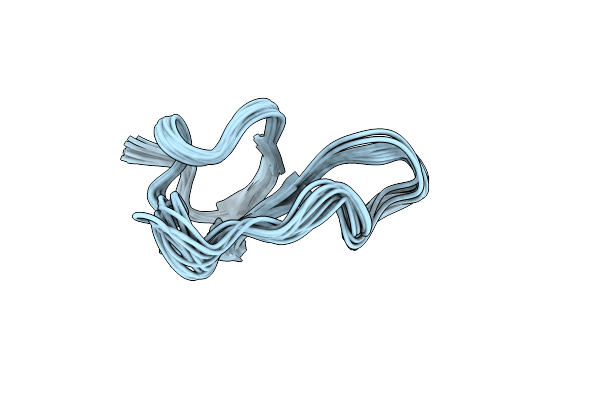 |
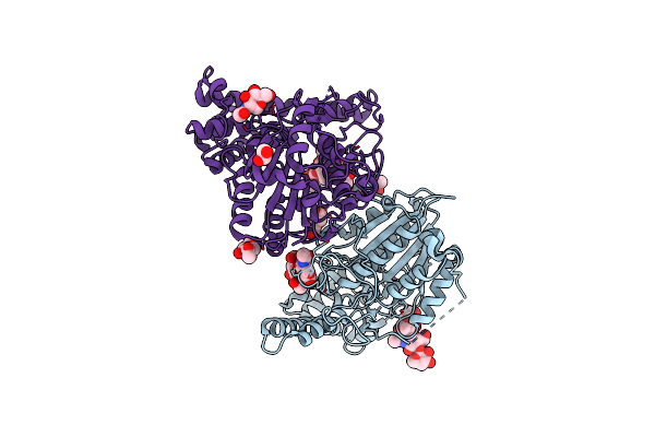
Nmr Solution Structure Of Roseltide Rt7
Organism: Hibiscus sabdariffa
Method: SOLUTION NMR Release Date: 2019-11-20 Classification: PLANT PROTEIN |
Organism: Hibiscus sabdariffa
Method: SOLUTION NMR
Release Date: 2019-11-20
 |
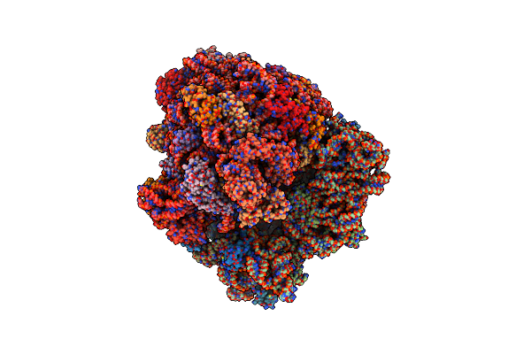
Peptide Asparaginyl Ligases From Viola Yedoensis
Organism: Viola philippica
Method: X-RAY DIFFRACTION Resolution:2.40 Å Release Date: 2019-05-15 Classification: LIGASE Ligands: NAG, EDO, PEG |
Organism: Viola philippica
Method: X-RAY DIFFRACTION
Release Date: 2019-05-15
Ligands: NAG, EDO, PEG
 |
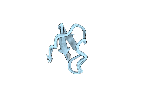
Ribosome Structure Bound To Abc-F Protein.
Organism: Pseudomonas aeruginosa, Thermus thermophilus hb8, Thermus thermophilus (strain hb8 / atcc 27634 / dsm 579)
Method: ELECTRON MICROSCOPY Resolution:3.60 Å Release Date: 2018-08-01 Classification: RIBOSOME Ligands: MG, ANP |
Organism: Pseudomonas aeruginosa, Thermus thermophilus hb8, Thermus thermophilus (strain hb8 / atcc 27634 / dsm 579)
Method: ELECTRON MICROSCOPY
Release Date: 2018-08-01
Ligands: MG, ANP
 |
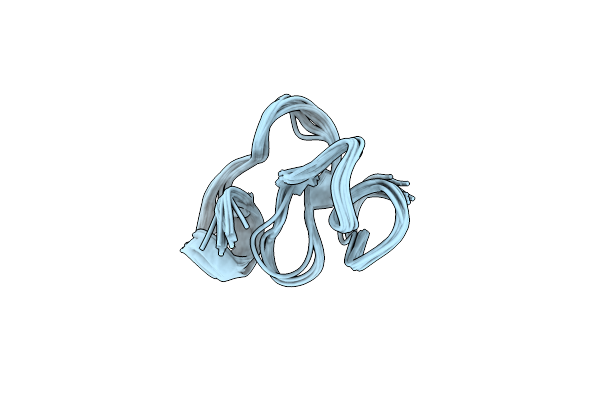
Highly Disulfide-Constrained Antifeedant Jasmintides From Jasminum Sambac Flowers
Organism: Jasminum sambac
Method: SOLUTION NMR Release Date: 2018-01-17 Classification: UNKNOWN FUNCTION |
Organism: Jasminum sambac
Method: SOLUTION NMR
Release Date: 2018-01-17
 |
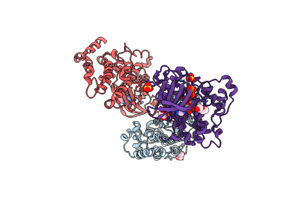
Vaccatide: Antifungal Glutamine-Rich 8C-Hevein-Like Peptide, Vh1
Organism: Vaccaria hispanica
Method: SOLUTION NMR Release Date: 2017-07-19 Classification: ANTIFUNGAL PROTEIN |
Organism: Vaccaria hispanica
Method: SOLUTION NMR
Release Date: 2017-07-19
 |
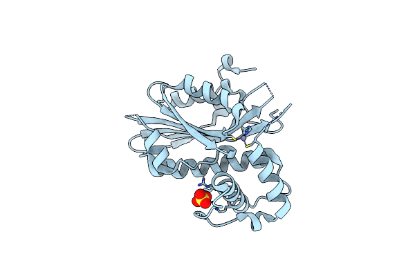
Crystal Structure Of The Protein Kinase Ck2 Catalytic Domain From Plasmodium Falciparum Bound To Atp
Organism: Plasmodium falciparum (isolate 3d7)
Method: X-RAY DIFFRACTION Resolution:3.00 Å Release Date: 2017-07-12 Classification: TRANSFERASE Ligands: SO4, CL, ATP, EDO, PEG |
Organism: Plasmodium falciparum (isolate 3d7)
Method: X-RAY DIFFRACTION
Release Date: 2017-07-12
Ligands: SO4, CL, ATP, EDO, PEG
 |
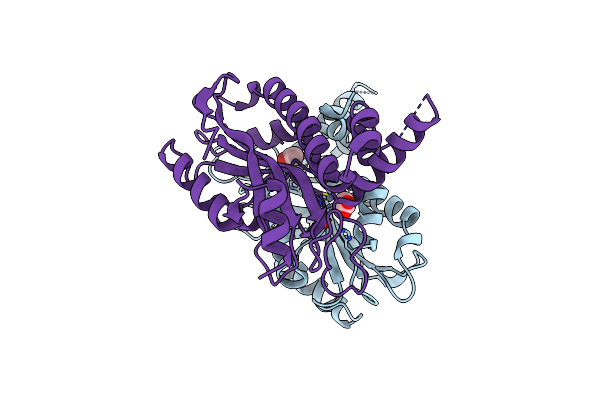
Crystal Structure Of Limiting Co2-Inducible Protein Lcic
Organism: Chlamydomonas reinhardtii
Method: X-RAY DIFFRACTION Resolution:2.51 Å Release Date: 2016-12-07 Classification: METAL BINDING PROTEIN Ligands: ZN, SO4 |
Organism: Chlamydomonas reinhardtii
Method: X-RAY DIFFRACTION
Release Date: 2016-12-07
Ligands: ZN, SO4
 |
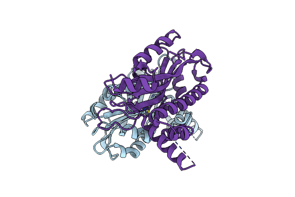
Crystal Structure Of Ptlcib4, A Homolog Of The Limiting Co2-Inducible Protein Lcib
Organism: Phaeodactylum tricornutum
Method: X-RAY DIFFRACTION Resolution:1.75 Å Release Date: 2016-12-07 Classification: METAL BINDING PROTEIN Ligands: ZN, ACT |
Organism: Phaeodactylum tricornutum
Method: X-RAY DIFFRACTION
Release Date: 2016-12-07
Ligands: ZN, ACT
 |
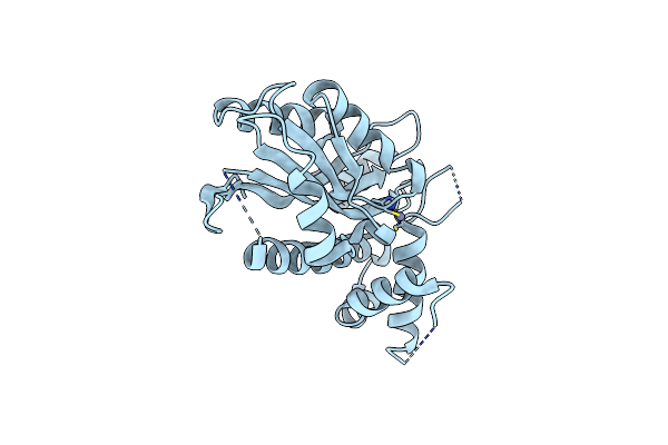
Crystal Structure Of Ptlcib4 H88A Mutant, A Homolog Of The Limiting Co2-Inducible Protein Lcib
Organism: Phaeodactylum tricornutum
Method: X-RAY DIFFRACTION Resolution:1.60 Å Release Date: 2016-12-07 Classification: METAL BINDING PROTEIN Ligands: ZN |
Organism: Phaeodactylum tricornutum
Method: X-RAY DIFFRACTION
Release Date: 2016-12-07
Ligands: ZN
 |
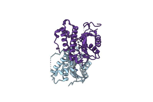
Crystal Structure Of Ptlcib4 S47R Mutant, A Homolog Of The Limiting Co2-Inducible Protein Lcib
Organism: Phaeodactylum tricornutum
Method: X-RAY DIFFRACTION Resolution:2.20 Å Release Date: 2016-12-07 Classification: METAL BINDING PROTEIN Ligands: ZN, CL |
Organism: Phaeodactylum tricornutum
Method: X-RAY DIFFRACTION
Release Date: 2016-12-07
Ligands: ZN, CL
 |
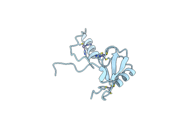
Crystal Structure Of Limiting Co2-Inducible Protein Lcib
Organism: Chlamydomonas reinhardtii
Method: X-RAY DIFFRACTION Resolution:2.59 Å Release Date: 2016-12-07 Classification: METAL BINDING PROTEIN Ligands: ZN |
Organism: Chlamydomonas reinhardtii
Method: X-RAY DIFFRACTION
Release Date: 2016-12-07
Ligands: ZN
 |
Crystal Structure Of A Phosphotyrosine Binding Domain
Organism: Mus musculus
Method: X-RAY DIFFRACTION Resolution:1.90 Å Release Date: 2012-01-25 Classification: LIGASE Ligands: ZN |
Organism: Mus musculus
Method: X-RAY DIFFRACTION
Release Date: 2012-01-25
Ligands: ZN