

Search Count: 16
 |
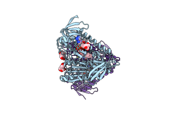
Crystal Structure Of The I318T Pathogenic Variant Of The Human Dihydrolipoamide Dehydrogenase
Organism: Homo sapiens
Method: X-RAY DIFFRACTION Resolution:2.89 Å Release Date: 2023-06-14 Classification: OXIDOREDUCTASE Ligands: FAD, BTB, EDO |
Organism: Homo sapiens
Method: X-RAY DIFFRACTION
Release Date: 2023-06-14
Ligands: FAD, BTB, EDO
 |
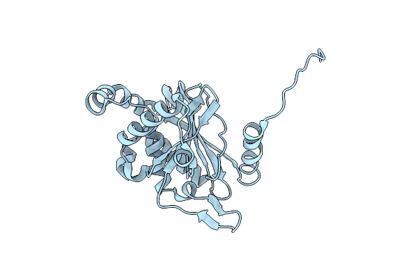
Crystal Structure Of The Disease-Causing I358T Mutant Of The Human Dihydrolipoamide Dehydrogenase
Organism: Homo sapiens
Method: X-RAY DIFFRACTION Resolution:2.44 Å Release Date: 2023-04-05 Classification: OXIDOREDUCTASE Ligands: FAD, BTB |
Organism: Homo sapiens
Method: X-RAY DIFFRACTION
Release Date: 2023-04-05
Ligands: FAD, BTB
 |
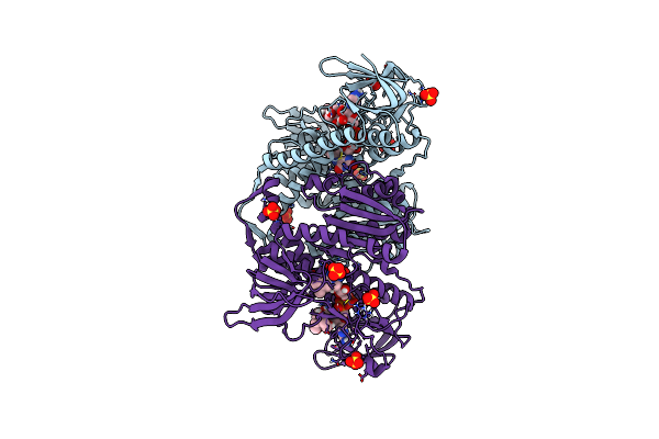
Cryo-Electron Microscopic Structure Of The Dihydrolipoamide Succinyltransferase (E2) Component Of The Human Alpha-Ketoglutarate (2-Oxoglutarate) Dehydrogenase Complex [Residues 218-453]
Organism: Homo sapiens
Method: ELECTRON MICROSCOPY Release Date: 2020-01-22 Classification: TRANSFERASE |
Organism: Homo sapiens
Method: ELECTRON MICROSCOPY
Release Date: 2020-01-22
 |
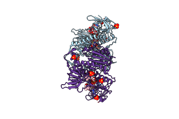
Crystal Structure Of The Disease-Causing G194C Mutant Of The Human Dihydrolipoamide Dehydrogenase
Organism: Homo sapiens
Method: X-RAY DIFFRACTION Resolution:1.60 Å Release Date: 2019-11-20 Classification: OXIDOREDUCTASE Ligands: FAD, SO4, BTB |
Organism: Homo sapiens
Method: X-RAY DIFFRACTION
Release Date: 2019-11-20
Ligands: FAD, SO4, BTB
 |
Crystal Structure Of The Human Dihydrolipoamide Dehydrogenase At 1.75 Angstrom Resolution
Organism: Homo sapiens
Method: X-RAY DIFFRACTION Resolution:1.75 Å Release Date: 2019-11-20 Classification: OXIDOREDUCTASE Ligands: FAD, BTB, SO4 |
Organism: Homo sapiens
Method: X-RAY DIFFRACTION
Release Date: 2019-11-20
Ligands: FAD, BTB, SO4
 |
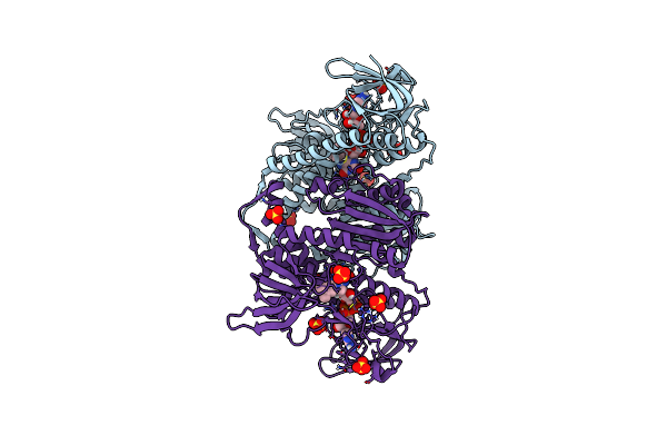
Crystal Structure Of The Disease-Causing R460G Mutant Of The Human Dihydrolipoamide Dehydrogenase At 1.44 Angstrom Resolution
Organism: Homo sapiens
Method: X-RAY DIFFRACTION Resolution:1.44 Å Release Date: 2019-11-20 Classification: OXIDOREDUCTASE Ligands: FAD, SO4, BTB |
Organism: Homo sapiens
Method: X-RAY DIFFRACTION
Release Date: 2019-11-20
Ligands: FAD, SO4, BTB
 |
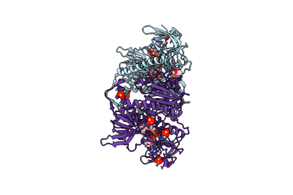
Crystal Structure Of The Disease-Causing R447G Mutant Of The Human Dihydrolipoamide Dehydrogenase
Organism: Homo sapiens
Method: X-RAY DIFFRACTION Resolution:1.75 Å Release Date: 2019-11-20 Classification: OXIDOREDUCTASE Ligands: FAD, SO4 |
Organism: Homo sapiens
Method: X-RAY DIFFRACTION
Release Date: 2019-11-20
Ligands: FAD, SO4
 |
Crystal Structure Of The Disease-Causing I445M Mutant Of The Human Dihydrolipoamide Dehydrogenase
Organism: Homo sapiens
Method: X-RAY DIFFRACTION Resolution:1.82 Å Release Date: 2019-11-20 Classification: OXIDOREDUCTASE Ligands: FAD, SO4, 1PE |
Organism: Homo sapiens
Method: X-RAY DIFFRACTION
Release Date: 2019-11-20
Ligands: FAD, SO4, 1PE
 |
Crystal Structure Of The Disease-Causing G426E Mutant Of The Human Dihydrolipoamide Dehydrogenase
Organism: Homo sapiens
Method: X-RAY DIFFRACTION Resolution:1.84 Å Release Date: 2019-11-20 Classification: OXIDOREDUCTASE Ligands: FAD, SO4, 1PE |
Organism: Homo sapiens
Method: X-RAY DIFFRACTION
Release Date: 2019-11-20
Ligands: FAD, SO4, 1PE
 |
Crystal Structure Of The Disease-Causing P453L Mutant Of The Human Dihydrolipoamide Dehydrogenase
Organism: Homo sapiens
Method: X-RAY DIFFRACTION Resolution:2.34 Å Release Date: 2019-11-20 Classification: OXIDOREDUCTASE Ligands: FAD, SO4 |
Organism: Homo sapiens
Method: X-RAY DIFFRACTION
Release Date: 2019-11-20
Ligands: FAD, SO4
 |
Crystal Structure Of The R460G Disease-Causing Mutant Of The Human Dihydrolipoamide Dehydrogenase.
Organism: Homo sapiens
Method: X-RAY DIFFRACTION Resolution:1.76 Å Release Date: 2019-09-04 Classification: OXIDOREDUCTASE Ligands: FAD, SO4, TRS |
Organism: Homo sapiens
Method: X-RAY DIFFRACTION
Release Date: 2019-09-04
Ligands: FAD, SO4, TRS
 |
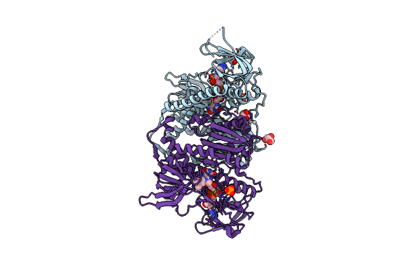
Crystal Structure Of The Human Dihydrolipoamide Dehydrogenase
Organism: Homo sapiens
Method: X-RAY DIFFRACTION Resolution:2.27 Å Release Date: 2018-05-16 Classification: OXIDOREDUCTASE Ligands: FAD, TRS, BTB |
Organism: Homo sapiens
Method: X-RAY DIFFRACTION
Release Date: 2018-05-16
Ligands: FAD, TRS, BTB
 |
Crystal Structure Of The D444V Disease-Causing Mutant Of The Human Dihydrolipoamide Dehydrogenase
Organism: Homo sapiens
Method: X-RAY DIFFRACTION Resolution:1.84 Å Release Date: 2017-11-15 Classification: OXIDOREDUCTASE Ligands: FAD, PO4, GOL |
Organism: Homo sapiens
Method: X-RAY DIFFRACTION
Release Date: 2017-11-15
Ligands: FAD, PO4, GOL
 |
Crystal Structure Analysis Of The Mutant S189D Rat Chymotrypsin
Organism: Rattus norvegicus
Method: X-RAY DIFFRACTION Resolution:2.55 Å Release Date: 2003-06-10 Classification: HYDROLASE Ligands: CA |
Organism: Rattus norvegicus
Method: X-RAY DIFFRACTION
Release Date: 2003-06-10
Ligands: CA
 |
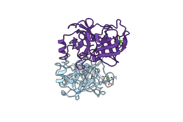
D254E, D256E Mutant Of D-Xylose Isomerase Complexed With Al3 And Xylitol
Organism: Arthrobacter sp. nrrl
Method: X-RAY DIFFRACTION Resolution:2.40 Å Release Date: 1998-01-28 Classification: ISOMERASE Ligands: XYL, AL |
Organism: Arthrobacter sp. nrrl
Method: X-RAY DIFFRACTION
Release Date: 1998-01-28
Ligands: XYL, AL
 |
Uncomplexed Rat Trypsin Mutant With Asp 189 Replaced With Ser (D189S)
Organism: Rattus rattus
Method: X-RAY DIFFRACTION Resolution:2.50 Å Release Date: 1997-12-24 Classification: HYDROLASE Ligands: CA |
Organism: Rattus rattus
Method: X-RAY DIFFRACTION
Release Date: 1997-12-24
Ligands: CA