

Search Count: 15
 |
Structure Of Hect E3 Trip12 Forming K29/K48-Branched Ubiquitin Chains
Organism: Homo sapiens
Method: ELECTRON MICROSCOPY Release Date: 2025-05-21 Classification: LIGASE Ligands: SY8 |
Organism: Homo sapiens
Method: ELECTRON MICROSCOPY
Release Date: 2025-05-21
Ligands: SY8
 |
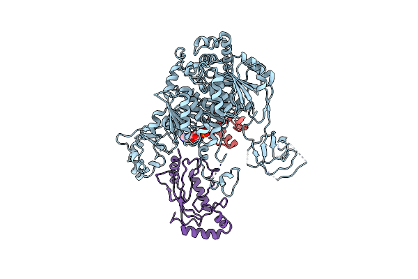
Structure Of Hect E3 Trip12 Forming K29-Linked Ubiquitin Chains
Organism: Homo sapiens
Method: ELECTRON MICROSCOPY Release Date: 2025-05-21 Classification: LIGASE Ligands: SY8 |
Organism: Homo sapiens
Method: ELECTRON MICROSCOPY
Release Date: 2025-05-21
Ligands: SY8
 |
Structure Of The Ube1L Activating Enzyme Bound To Isg15 And Ube2L6
Organism: Homo sapiens
Method: ELECTRON MICROSCOPY Release Date: 2023-12-13 Classification: ANTIVIRAL PROTEIN Ligands: AMP |
Organism: Homo sapiens
Method: ELECTRON MICROSCOPY
Release Date: 2023-12-13
Ligands: AMP
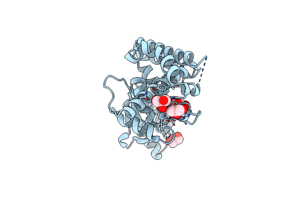 |
Structure Of The Otu Domain Of Otulin G281R Mutant
Organism: Homo sapiens
Method: X-RAY DIFFRACTION Resolution:1.77 Å Release Date: 2019-03-06 Classification: HYDROLASE Ligands: GOL, CL |
Organism: Homo sapiens
Method: X-RAY DIFFRACTION
Release Date: 2019-03-06
Ligands: GOL, CL
 |
Structure Of Chlamydia Trachomatis Effector Protein Chladub1 Bound To Ubiquitin
Organism: Chlamydia trachomatis serovar l2 (strain 434/bu / atcc vr-902b), Homo sapiens
Method: X-RAY DIFFRACTION Resolution:1.90 Å Release Date: 2018-11-14 Classification: HYDROLASE Ligands: GOL, SO4, AYE |
Organism: Chlamydia trachomatis serovar l2 (strain 434/bu / atcc vr-902b), Homo sapiens
Method: X-RAY DIFFRACTION
Release Date: 2018-11-14
Ligands: GOL, SO4, AYE
 |
Structure Of Chlamydia Trachomatis Effector Protein Chladub1 Bound To Coenzyme A
Organism: Chlamydia trachomatis
Method: X-RAY DIFFRACTION Resolution:2.10 Å Release Date: 2018-11-14 Classification: HYDROLASE Ligands: GOL, SO4, COA |
Organism: Chlamydia trachomatis
Method: X-RAY DIFFRACTION
Release Date: 2018-11-14
Ligands: GOL, SO4, COA
 |
Structure Of Chlamydia Abortus Effector Protein Chladub
Organism: Chlamydia abortus
Method: X-RAY DIFFRACTION Resolution:1.47 Å Release Date: 2018-11-14 Classification: TRANSFERASE Ligands: GOL, ZN |
Organism: Chlamydia abortus
Method: X-RAY DIFFRACTION
Release Date: 2018-11-14
Ligands: GOL, ZN
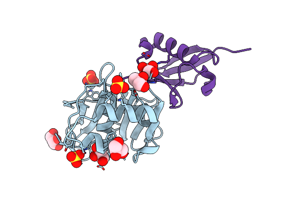 |
Fmdv Leader Protease Bound To Substrate Isg15
Organism: Foot-and-mouth disease virus, Homo sapiens
Method: X-RAY DIFFRACTION Resolution:1.50 Å Release Date: 2018-02-21 Classification: HYDROLASE Ligands: GOL, SO4 |
Organism: Foot-and-mouth disease virus, Homo sapiens
Method: X-RAY DIFFRACTION
Release Date: 2018-02-21
Ligands: GOL, SO4
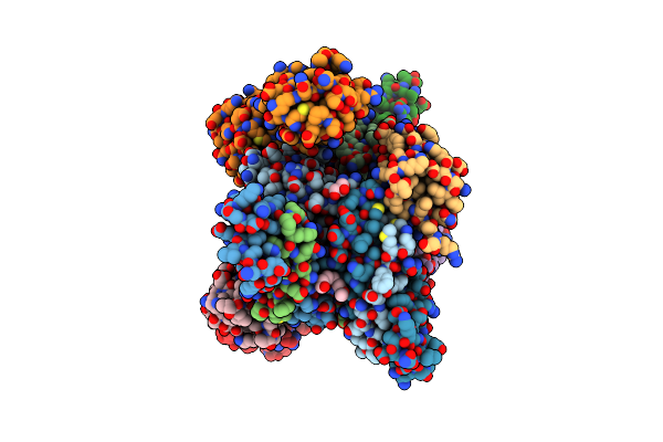 |
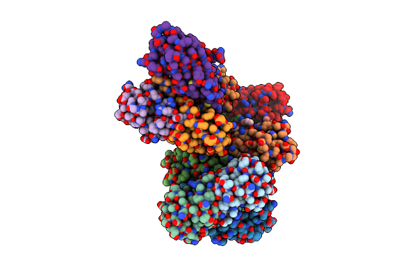
K6-Specific Affimer Bound To K6 Diub
Organism: Synthetic construct, Homo sapiens
Method: X-RAY DIFFRACTION Resolution:2.50 Å Release Date: 2017-10-04 Classification: SIGNALING PROTEIN Ligands: GOL, 15P |
Organism: Synthetic construct, Homo sapiens
Method: X-RAY DIFFRACTION
Release Date: 2017-10-04
Ligands: GOL, 15P
 |
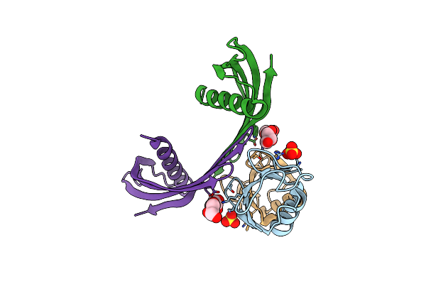
K33-Specific Affimer Bound To K33 Diub
Organism: Homo sapiens, Synthetic construct
Method: X-RAY DIFFRACTION Resolution:3.80 Å Release Date: 2017-10-04 Classification: SIGNALING PROTEIN Ligands: 15P |
Organism: Homo sapiens, Synthetic construct
Method: X-RAY DIFFRACTION
Release Date: 2017-10-04
Ligands: 15P
 |
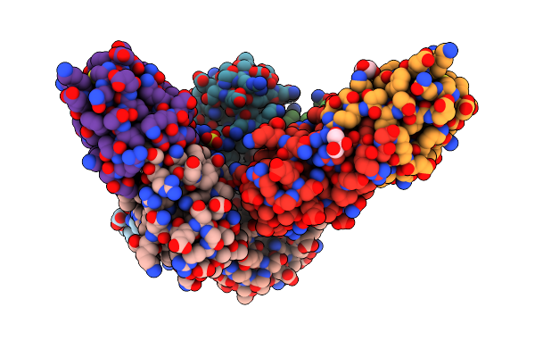
K33-Specific Affimer Bound To K33 Diub
Organism: Homo sapiens, Synthetic construct
Method: X-RAY DIFFRACTION Resolution:2.80 Å Release Date: 2017-10-04 Classification: SIGNALING PROTEIN Ligands: SO4, GOL |
Organism: Homo sapiens, Synthetic construct
Method: X-RAY DIFFRACTION
Release Date: 2017-10-04
Ligands: SO4, GOL
 |
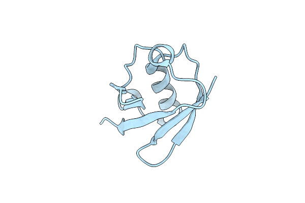
Structure Of Lys33-Linked Diub
Organism: Homo sapiens
Method: X-RAY DIFFRACTION Resolution:1.85 Å Release Date: 2015-03-25 Classification: SIGNALING PROTEIN Ligands: GOL, SO4 |
Organism: Homo sapiens
Method: X-RAY DIFFRACTION
Release Date: 2015-03-25
Ligands: GOL, SO4
 |
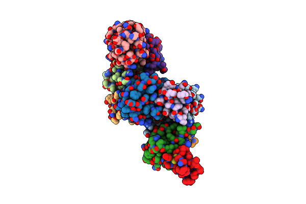
Structure Of Lys33-Linked Triub S.G. P 212121
Organism: Homo sapiens
Method: X-RAY DIFFRACTION Resolution:1.68 Å Release Date: 2015-03-25 Classification: SIGNALING PROTEIN |
Organism: Homo sapiens
Method: X-RAY DIFFRACTION
Release Date: 2015-03-25
 |
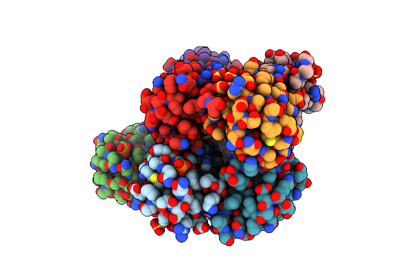
Structure Of Lys33-Linked Diub Bound To Trabid Nzf1
Organism: Homo sapiens
Method: X-RAY DIFFRACTION Resolution:3.40 Å Release Date: 2015-03-25 Classification: SIGNALING PROTEIN Ligands: ZN |
Organism: Homo sapiens
Method: X-RAY DIFFRACTION
Release Date: 2015-03-25
Ligands: ZN
 |
Ser65 Phosphorylated Ubiquitin, Major Conformation
Organism: Homo sapiens
Method: X-RAY DIFFRACTION Resolution:1.90 Å Release Date: 2015-01-14 Classification: SIGNALING PROTEIN Ligands: SO4 |
Organism: Homo sapiens
Method: X-RAY DIFFRACTION
Release Date: 2015-01-14
Ligands: SO4