

Search Count: 17
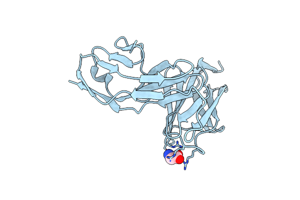 |
Crystal Structure Of The N-Terminal Ig1-2 Module Of Drosophila Receptor Protein Tyrosine Phosphatase Dlar
Organism: Drosophila melanogaster
Method: X-RAY DIFFRACTION Resolution:1.80 Å Release Date: 2011-04-13 Classification: HYDROLASE Ligands: GLY |
Organism: Drosophila melanogaster
Method: X-RAY DIFFRACTION
Release Date: 2011-04-13
Ligands: GLY
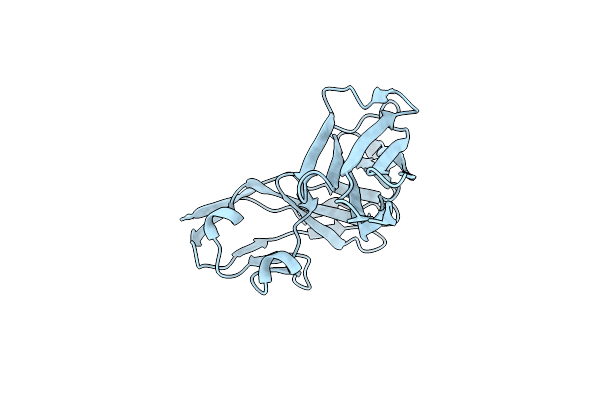 |
Crystal Structure Of The N-Terminal Ig1-2 Module Of Human Receptor Protein Tyrosine Phosphatase Sigma
Organism: Homo sapiens
Method: X-RAY DIFFRACTION Resolution:2.55 Å Release Date: 2011-04-13 Classification: HYDROLASE Ligands: CL, IOD |
Organism: Homo sapiens
Method: X-RAY DIFFRACTION
Release Date: 2011-04-13
Ligands: CL, IOD
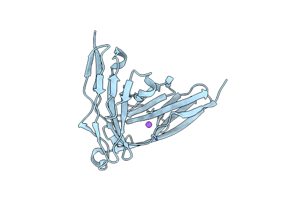 |
Crystal Structure Of The N-Terminal Ig1-2 Module Of Human Receptor Protein Tyrosine Phosphatase Sigma
Organism: Homo sapiens
Method: X-RAY DIFFRACTION Resolution:2.30 Å Release Date: 2011-04-13 Classification: HYDROLASE |
Organism: Homo sapiens
Method: X-RAY DIFFRACTION
Release Date: 2011-04-13
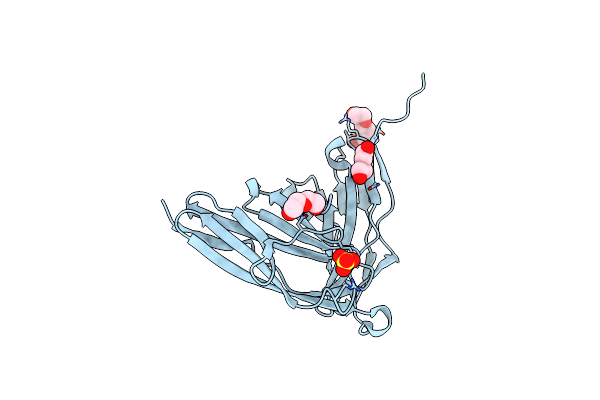 |
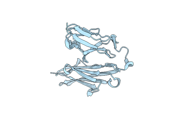
Crystal Structure Of The N-Terminal Ig1-2 Module Of Chicken Receptor Protein Tyrosine Phosphatase Sigma
Organism: Gallus gallus
Method: X-RAY DIFFRACTION Resolution:1.65 Å Release Date: 2011-04-13 Classification: HYDROLASE Ligands: CL, PGE, PEG, SO4 |
Organism: Gallus gallus
Method: X-RAY DIFFRACTION
Release Date: 2011-04-13
Ligands: CL, PGE, PEG, SO4
 |
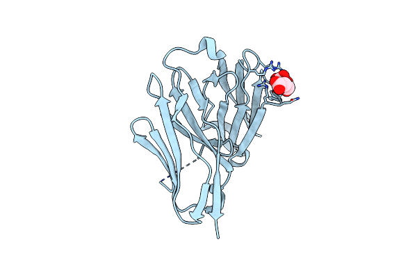
Crystal Structure Of The N-Terminal Ig1-2 Module Of Human Receptor Protein Tyrosine Phosphatase Lar
Organism: Homo sapiens
Method: X-RAY DIFFRACTION Resolution:2.20 Å Release Date: 2011-04-13 Classification: HYDROLASE |
Organism: Homo sapiens
Method: X-RAY DIFFRACTION
Release Date: 2011-04-13
 |
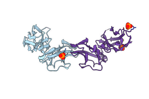
Crystal Structure Of The N-Terminal Ig1-2 Module Of Human Receptor Protein Tyrosine Phosphatase Delta
Organism: Homo sapiens
Method: X-RAY DIFFRACTION Resolution:1.35 Å Release Date: 2011-04-13 Classification: HYDROLASE Ligands: CL, FLC |
Organism: Homo sapiens
Method: X-RAY DIFFRACTION
Release Date: 2011-04-13
Ligands: CL, FLC
 |
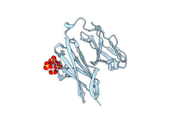
Crystal Structure Of The N-Terminal Ig1-2 Module Of Human Receptor Protein Tyrosine Phosphatase Delta
Organism: Homo sapiens
Method: X-RAY DIFFRACTION Resolution:1.98 Å Release Date: 2011-04-13 Classification: HYDROLASE Ligands: PO4 |
Organism: Homo sapiens
Method: X-RAY DIFFRACTION
Release Date: 2011-04-13
Ligands: PO4
 |
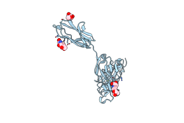
Crystal Structure Of The N-Terminal Ig1-2 Module Of Human Receptor Protein Tyrosine Phosphatase Lar In Complex With Sucrose Octasulphate
Organism: Homo sapiens
Method: X-RAY DIFFRACTION Resolution:2.05 Å Release Date: 2011-04-13 Classification: HYDROLASE Ligands: YYJ |
Organism: Homo sapiens
Method: X-RAY DIFFRACTION
Release Date: 2011-04-13
Ligands: YYJ
 |
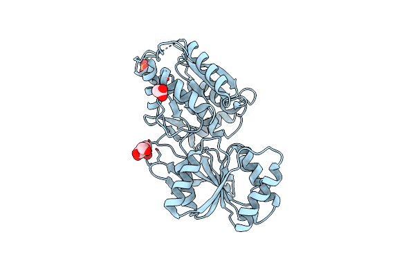
Crystal Structure Of The N-Terminal Ig1-3 Module Of Human Receptor Protein Tyrosine Phosphatase Sigma
Organism: Homo sapiens
Method: X-RAY DIFFRACTION Resolution:2.60 Å Release Date: 2011-04-13 Classification: HYDROLASE Ligands: CL, IOD, NAG, B3P |
Organism: Homo sapiens
Method: X-RAY DIFFRACTION
Release Date: 2011-04-13
Ligands: CL, IOD, NAG, B3P
 |
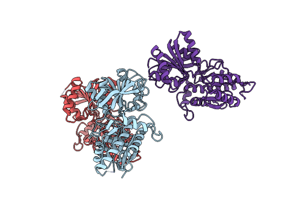
Crystal Structure Of The Human Ionotropic Glutamate Receptor Glur2 Atd Region At 1.8 A Resolution
Organism: Homo sapiens
Method: X-RAY DIFFRACTION Resolution:1.80 Å Release Date: 2009-08-18 Classification: TRANSPORT PROTEIN Ligands: NAG, CL, SO4, ACT |
Organism: Homo sapiens
Method: X-RAY DIFFRACTION
Release Date: 2009-08-18
Ligands: NAG, CL, SO4, ACT
 |
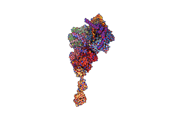
Crystal Structure Of The Human Ionotropic Glutamate Receptor Glur2 Atd Region At 4.1 A Resolution
Organism: Homo sapiens
Method: X-RAY DIFFRACTION Resolution:4.10 Å Release Date: 2009-08-18 Classification: TRANSPORT PROTEIN |
Organism: Homo sapiens
Method: X-RAY DIFFRACTION
Release Date: 2009-08-18
 |
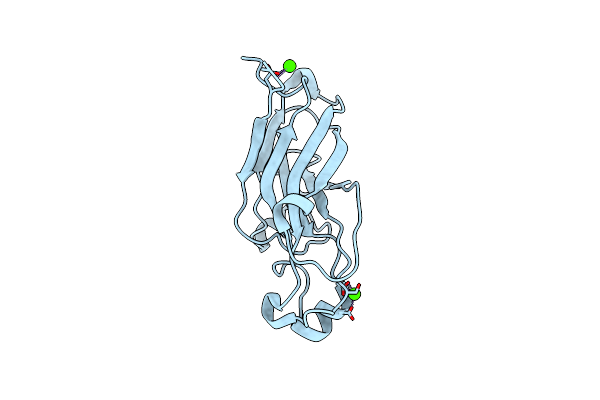
X-Ray Structure Of The Entire Lipid-Containing Bacteriophage Pm2
Organism: Pseudoalteromonas phage pm2
Method: X-RAY DIFFRACTION Resolution:7.00 Å Release Date: 2008-09-23 Classification: VIRUS Ligands: CA |
Organism: Pseudoalteromonas phage pm2
Method: X-RAY DIFFRACTION
Release Date: 2008-09-23
Ligands: CA
 |
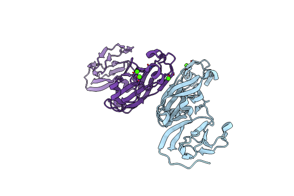
Crystal Structure Of The Receptor Binding Domain Of The Spike Protein P1 From Bacteriophage Pm2
Organism: Pseudoalteromonas phage pm2
Method: X-RAY DIFFRACTION Resolution:2.26 Å Release Date: 2008-09-16 Classification: VIRAL PROTEIN Ligands: CA |
Organism: Pseudoalteromonas phage pm2
Method: X-RAY DIFFRACTION
Release Date: 2008-09-16
Ligands: CA
 |
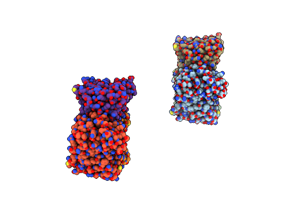
Crystal Structure Of The Stem And Receptor Binding Domain Of The Spike Protein P1 From Bacteriophage Pm2
Organism: Pseudoalteromonas phage pm2
Method: X-RAY DIFFRACTION Resolution:1.77 Å Release Date: 2008-09-16 Classification: VIRAL PROTEIN Ligands: CA, CL |
Organism: Pseudoalteromonas phage pm2
Method: X-RAY DIFFRACTION
Release Date: 2008-09-16
Ligands: CA, CL
 |
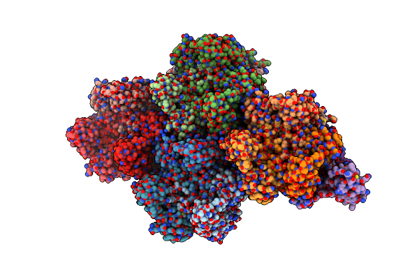
Crystal Structure Of The Major Capsid Protein P2 From Bacteriophage Pm2
Organism: Pseudoalteromonas phage pm2
Method: X-RAY DIFFRACTION Resolution:2.50 Å Release Date: 2008-09-16 Classification: VIRAL PROTEIN Ligands: CA |
Organism: Pseudoalteromonas phage pm2
Method: X-RAY DIFFRACTION
Release Date: 2008-09-16
Ligands: CA
 |
Structural Analysis Of Prd1
Organism: Enterobacteria phage prd1
Method: X-RAY DIFFRACTION Resolution:4.20 Å Release Date: 2004-11-11 Classification: VIRUS |
Organism: Enterobacteria phage prd1
Method: X-RAY DIFFRACTION
Release Date: 2004-11-11
 |
The Bluetongue Virus (Btv) Core Binds Dsrna
Organism: Bluetongue virus
Method: X-RAY DIFFRACTION Resolution:10.00 Å Release Date: 2002-09-26 Classification: VIRUS/VIRAL PROTEIN/RNA |
Organism: Bluetongue virus
Method: X-RAY DIFFRACTION
Release Date: 2002-09-26