

Search Count: 23
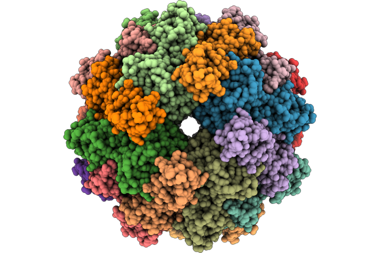 |
The Cryo-Em Structure Of Chum Salmon (Oncorhynchus Keta) 24-Mer Rhamnose-Binding Lectin Csl2
Organism: Oncorhynchus keta
Method: ELECTRON MICROSCOPY Resolution:1.82 Å Release Date: 2026-07-22 Classification: SUGAR BINDING PROTEIN |
Organism: Oncorhynchus keta
Method: ELECTRON MICROSCOPY
Release Date: 2026-07-22
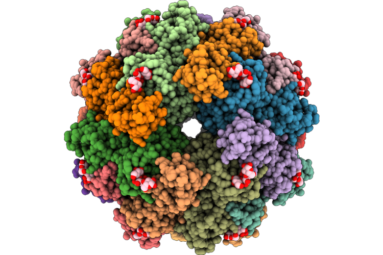 |
The Cryo-Em Structure Of Chum Salmon (Oncorhynchus Keta) 24-Mer Rhamnose-Binding Lectin Csl2 Bound With Gb3
Organism: Oncorhynchus keta
Method: ELECTRON MICROSCOPY Resolution:1.89 Å Release Date: 2026-07-22 Classification: SUGAR BINDING PROTEIN |
Organism: Oncorhynchus keta
Method: ELECTRON MICROSCOPY
Release Date: 2026-07-22
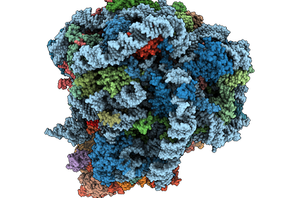 |
Cryo-Em Structure Of Human 80S Ribosome In Complex With Montanine
Organism: Homo sapiens
Method: ELECTRON MICROSCOPY Resolution:2.78 Å Release Date: 2026-01-14 Classification: RIBOSOME Ligands: MG, ZN, A1L8F |
Organism: Homo sapiens
Method: ELECTRON MICROSCOPY
Release Date: 2026-01-14
Ligands: MG, ZN, A1L8F
 |
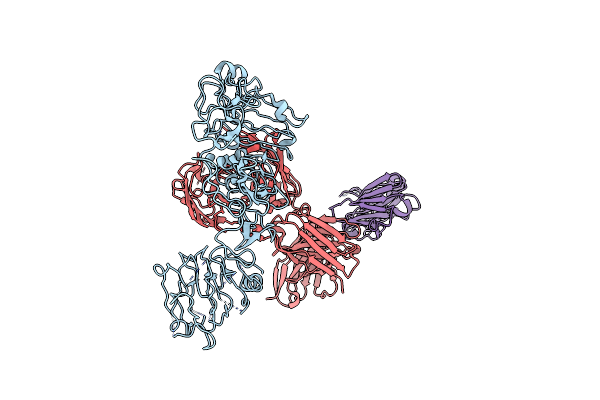
Local Refinement Structure Of Segfr And 528 Fv (From Hl-Type Bispecific Diabody Ex3) Complex
Organism: Homo sapiens, Synthetic construct
Method: ELECTRON MICROSCOPY Resolution:3.21 Å Release Date: 2025-05-28 Classification: ANTITUMOR PROTEIN/IMMUNE SYSTEM Ligands: NAG |
Organism: Homo sapiens, Synthetic construct
Method: ELECTRON MICROSCOPY
Release Date: 2025-05-28
Ligands: NAG
 |
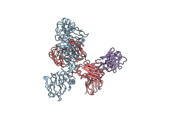
Poly-Alanine Model For Hl-Type Bispecific Diabody Ex3 Composed Of 528 And Okt3 Fvs In Ternary Complex With Segfr And Cd3Gamma-Epsilon (Closed Conformation)
Organism: Homo sapiens, Synthetic construct
Method: ELECTRON MICROSCOPY Release Date: 2025-05-28 Classification: ANTITUMOR PROTEIN/IMMUNE SYSTEM |
Organism: Homo sapiens, Synthetic construct
Method: ELECTRON MICROSCOPY
Release Date: 2025-05-28
 |
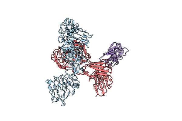
Poly-Alanine Model For Hl-Type Bispecific Diabody Ex3 Composed Of 528 And Okt3 Fvs In Ternary Complex With Segfr And Cd3Gamma-Epsilon (Middle Conformation)
Organism: Homo sapiens, Synthetic construct
Method: ELECTRON MICROSCOPY Release Date: 2025-05-28 Classification: ANTITUMOR PROTEIN/IMMUNE SYSTEM |
Organism: Homo sapiens, Synthetic construct
Method: ELECTRON MICROSCOPY
Release Date: 2025-05-28
 |
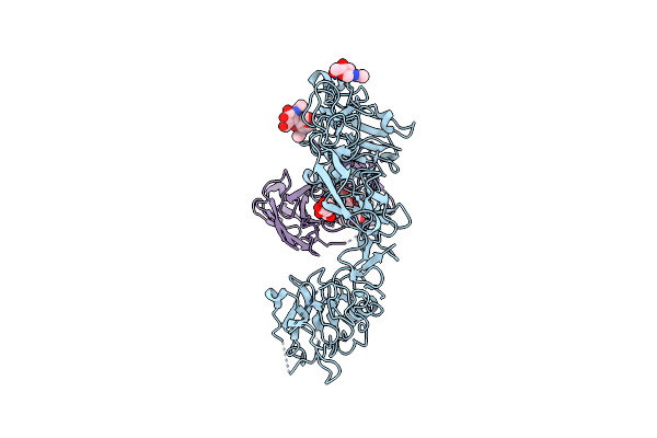
Poly-Alanine Model For Hl-Type Bispecific Diabody Ex3 Composed Of 528 And Okt3 Fvs In Ternary Complex With Segfr And Cd3Gamma-Epsilon (Open Conformation)
Organism: Homo sapiens, Synthetic construct
Method: ELECTRON MICROSCOPY Release Date: 2025-05-28 Classification: ANTITUMOR PROTEIN/IMMUNE SYSTEM |
Organism: Homo sapiens, Synthetic construct
Method: ELECTRON MICROSCOPY
Release Date: 2025-05-28
 |
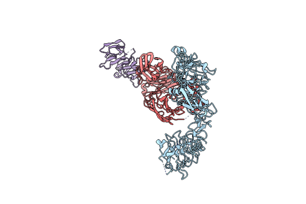
Local Refinement Structure Of Segfr And 528 Fv (From Lh-Type Bispecific Diabody Ex3) Complex
Organism: Homo sapiens, Synthetic construct
Method: ELECTRON MICROSCOPY Resolution:2.93 Å Release Date: 2025-05-28 Classification: ANTITUMOR PROTEIN/IMMUNE SYSTEM Ligands: NAG |
Organism: Homo sapiens, Synthetic construct
Method: ELECTRON MICROSCOPY
Release Date: 2025-05-28
Ligands: NAG
 |
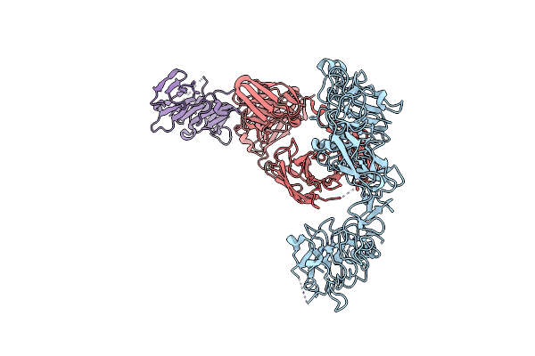
Poly-Alanine Model For Lh-Type Bispecific Diabody Ex3 Composed Of 528 And Okt3 Fvs In Ternary Complex With Segfr And Cd3Gamma-Epsilon (Closed Conformation)
Organism: Homo sapiens, Synthetic construct
Method: ELECTRON MICROSCOPY Release Date: 2025-05-28 Classification: ANTITUMOR PROTEIN/IMMUNE SYSTEM |
Organism: Homo sapiens, Synthetic construct
Method: ELECTRON MICROSCOPY
Release Date: 2025-05-28
 |
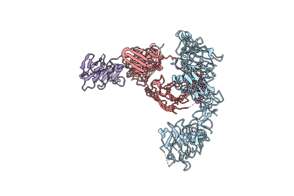
Poly-Alanine Model For Lh-Type Bispecific Diabody Ex3 Composed Of 528 And Okt3 Fvs In Ternary Complex With Segfr And Cd3Gamma-Epsilon (Middle Conformation)
Organism: Homo sapiens, Synthetic construct
Method: ELECTRON MICROSCOPY Release Date: 2025-05-28 Classification: ANTITUMOR PROTEIN/IMMUNE SYSTEM |
Organism: Homo sapiens, Synthetic construct
Method: ELECTRON MICROSCOPY
Release Date: 2025-05-28
 |
Poly-Alanine Model For Lh-Type Bispecific Diabody Ex3 Composed Of 528 And Okt3 Fvs In Ternary Complex With Segfr And Cd3Gamma-Epsilon (Open Conformation)
Organism: Homo sapiens, Synthetic construct
Method: ELECTRON MICROSCOPY Release Date: 2025-05-28 Classification: ANTITUMOR PROTEIN/IMMUNE SYSTEM |
Organism: Homo sapiens, Synthetic construct
Method: ELECTRON MICROSCOPY
Release Date: 2025-05-28
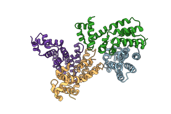 |
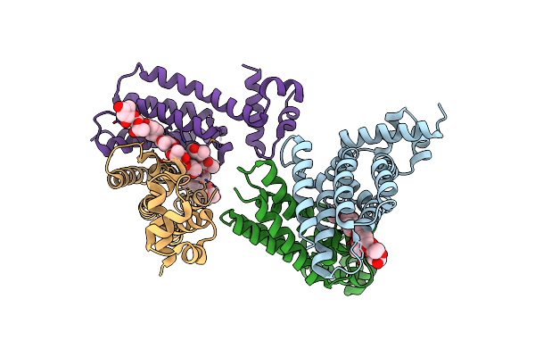
Crystal Structure Of A Transcription Factor
Organism: Streptomyces griseoluteus
Method: X-RAY DIFFRACTION Resolution:2.40 Å Release Date: 2022-04-27 Classification: TRANSCRIPTION |
Organism: Streptomyces griseoluteus
Method: X-RAY DIFFRACTION
Release Date: 2022-04-27
 |
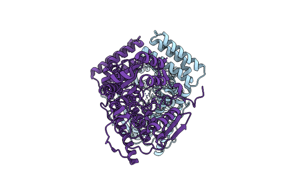
Crystal Structure Of A Transcription Factor In Complex With Ligand
Organism: Streptomyces griseoluteus
Method: X-RAY DIFFRACTION Resolution:2.91 Å Release Date: 2022-04-27 Classification: TRANSCRIPTION Ligands: JB0 |
Organism: Streptomyces griseoluteus
Method: X-RAY DIFFRACTION
Release Date: 2022-04-27
Ligands: JB0
 |
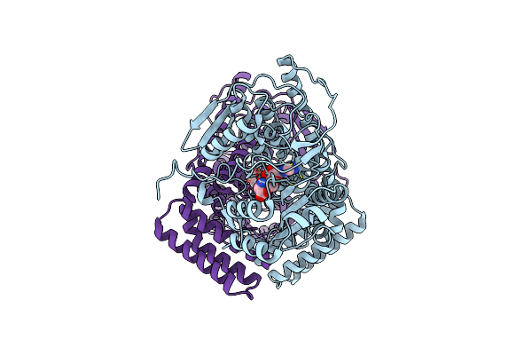
Crystal Structure Of The Human Cap-Specific Adenosine Methyltransferase
Organism: Homo sapiens
Method: X-RAY DIFFRACTION Resolution:2.70 Å Release Date: 2018-12-05 Classification: TRANSFERASE |
Organism: Homo sapiens
Method: X-RAY DIFFRACTION
Release Date: 2018-12-05
 |
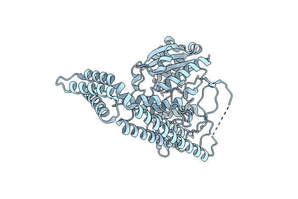
Crystal Structure Of The Human Cap-Specific Adenosine Methyltransferase Bound To Sah
Organism: Homo sapiens
Method: X-RAY DIFFRACTION Resolution:2.90 Å Release Date: 2018-12-05 Classification: TRANSFERASE Ligands: SAH |
Organism: Homo sapiens
Method: X-RAY DIFFRACTION
Release Date: 2018-12-05
Ligands: SAH
 |
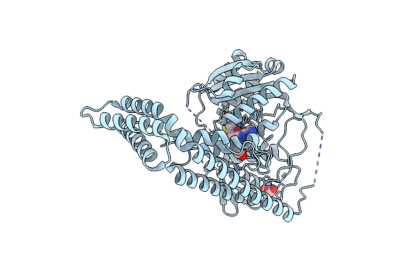
Crystal Structure Of The Zebrafish Cap-Specific Adenosine Methyltransferase
Organism: Danio rerio
Method: X-RAY DIFFRACTION Resolution:2.00 Å Release Date: 2018-12-05 Classification: TRANSFERASE |
Organism: Danio rerio
Method: X-RAY DIFFRACTION
Release Date: 2018-12-05
 |
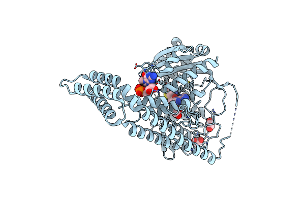
Crystal Structure Of The Zebrafish Cap-Specific Adenosine Methyltransferase Bound To Sah
Organism: Danio rerio
Method: X-RAY DIFFRACTION Resolution:1.80 Å Release Date: 2018-12-05 Classification: TRANSFERASE Ligands: SAH, EDO |
Organism: Danio rerio
Method: X-RAY DIFFRACTION
Release Date: 2018-12-05
Ligands: SAH, EDO
 |
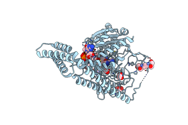
Crystal Structure Of The Zebrafish Cap-Specific Adenosine Methyltransferase Bound To Sah And M7G-Capped Rna
Organism: Danio rerio
Method: X-RAY DIFFRACTION Resolution:2.00 Å Release Date: 2018-12-05 Classification: TRANSFERASE Ligands: SAH, EDO, M7G |
Organism: Danio rerio
Method: X-RAY DIFFRACTION
Release Date: 2018-12-05
Ligands: SAH, EDO, M7G
 |
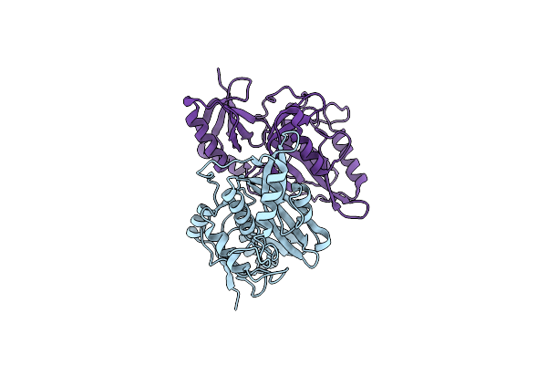
Crystal Structure Of The Zebrafish Cap-Specific Adenosine Methyltransferase Bound To Sah And M7G-Capped Rna
Organism: Danio rerio, Synthetic construct
Method: X-RAY DIFFRACTION Resolution:1.80 Å Release Date: 2018-12-05 Classification: TRANSFERASE/RNA Ligands: SAH, M7G, EDO, B3P |
Organism: Danio rerio, Synthetic construct
Method: X-RAY DIFFRACTION
Release Date: 2018-12-05
Ligands: SAH, M7G, EDO, B3P
 |
Crystal Structure Of The A47Q1 Mutant Of Pro-Protein-Glutaminase
Organism: Chryseobacterium proteolyticum
Method: X-RAY DIFFRACTION Resolution:1.50 Å Release Date: 2010-08-11 Classification: HYDROLASE |
Organism: Chryseobacterium proteolyticum
Method: X-RAY DIFFRACTION
Release Date: 2010-08-11